ISSN 0378-6471 (Print)⋅ISSN 2092-9374 (Online)
https://doi.org/10.3341/jkos.2019.60.11.1072
Original Article
중심망막정맥폐쇄에 동반된 황반부종에서 유리체내 서로 다른 세 가지 약물의 단기 효과 비교
Comparison of Short-term Effects of Intravitreal Injection of Three Modalities on Central Retinal Vein Occlusion
정지성1,2⋅이동우1,2⋅김병선1,2⋅유웅선1,2,3⋅정인영1,2,3⋅박종문1,2,3
Ji-Seong Jeong, MD1,2, Dong-Woo Lee, MD1,2, Byoung-Seon Kim, MD1,2, Woong-Sun Yoo, MD1,2,3, In Young Chung, MD, PhD1,2,3, Jong-Moon Park, MD, PhD1,2,3
경상대학교 의과대학 안과학교실1, 경상대학교병원 안과2, 경상대학교 건강과학연구원3 Department of Ophthalmology, Gyeongsang National University College of Medicine1, Jinju, Korea
Department of Ophthalmology, Gyeongsang National University Hospital2, Jinju, Korea Institute of Health Sciences, Gyeongsang National University3, Jinju, Korea
Purpose: To report the short-term effects of intravitreal bevacizumab alone, low-dose bevacizumab combined with low-dose tri- amcinolone injection, and intravitreal dexamethasone implant (Ozurdex®, Allergan, Irvine, CA, USA) injection in patients with macular edema following central retinal vein occlusion (CRVO).
Methods: The medical records of 70 patients (70 eyes) with macular edema secondary to CRVO were reviewed retrospectively.
Of these, 25 eyes (IVB group) were injected with intravitreal bevacizumab, 23 eyes (intravitreal low-dose bevacizumab and tri- amcinolone injection [IVB+IVTA] group) were injected with low-dose bevacizumab (0.625 mg/0.025 mL) combined with low-dose triamcinolone (1 mg/0.025 mL), and 20 eyes (intravitreal dexamethasone implant [IVD] group) were injected with an intravitreal dexamethasone implant. The best-corrected visual acuity (BCVA), central macular thickness (CMT), and intraocular pressure (IOP) of treated eyes were measured before injection and at 1 month and 3 months after injection.
Results: Groups were similar in age and gender distribution. At 1 month, the CMT of all groups was significantly lower, and the BCVA of all groups had increased significantly in patients with CRVO; there were no significant differences among the three groups (p = 0.246, p = 0.974). At 3 months, the CMT and BCVA had improved significantly only in the IVD and IVB+IVTA groups;
the short-term effect was comparable to the IVD group. IOP showed no significant change at 3 months after injection for all groups.
Conclusions: Considering various clinical variables in the treatment of macular edema associated with CRVO, intravitreal in- jection of bevacizumab, low-dose bevacizumab combined with triamcinolone, and dexamethasone implants may be used selectively.
J Korean Ophthalmol Soc 2019;60(11):1072-1079
Keywords: Bevacizumab, Dexamethasone, Intravitreal injections, Retinal vein occlusion, Triamcinolone
■Received: 2019. 5. 22. ■ Revised: 2019. 7. 11.
■Accepted: 2019. 10. 24.
■Address reprint requests to Jong-Moon Park, MD, PhD Department of Ophthalmology, Gyeongsang National University Hospital, #79 Gangnam-ro, Jinju 52727, Korea Tel: 82-55-750-8171, Fax: 82-55-758-4158
E-mail: [email protected]
*Conflicts of Interest: The authors have no conflicts to disclose.
ⓒ2019 The Korean Ophthalmological Society
This is an Open Access article distributed under the terms of the Creative Commons Attribution Non-Commercial License (http://creativecommons.org/licenses/by-nc/3.0/) which permits unrestricted non-commercial use, distribution, and reproduction in any medium, provided the original work is properly cited.
망막중심정맥폐쇄는 망막분지정맥폐쇄보다 드물게 발생 하지만, 상대적으로 양호한 자연경과를 보이는 망막분지정 맥폐쇄보다 황반부종, 망막허혈, 유리체출혈 및 신생혈관녹 내장 등의 합병증으로 인해 시력예후가 불량한 질환이다.1-3 망막정맥폐쇄에 동반된 황반부종은 혈관의 폐쇄로 인한 정
맥압의 증가에 따른 혈류학적인 요인과 혈관내피성장인자 (vascular endothelial growth factor, VEGF) 및 인터루킨 6 (interleukin [IL]-6), 인터루킨 8 (IL-8) 등과 같은 염증성 전 구물질의 증가로 인한 대사적인 요인에 의해 혈액망막장벽 이 파괴하며 발생한다.4 황반부종의 치료에 있어 망막분지 정맥폐쇄와는 달리 망막중심정맥폐쇄에서의 황반부 격자 레이저치료의 효과는 제한적이며,5 여러 대규모 연구를 통해 유리체내 트리암시놀론 아세토나이드(Triamcinolone acetonide, Allergan, Irvine, CA, USA),6 항혈관내피성장인 자(anti-VEGF),7-11 덱사메타손삽입물(Ozurdex®, Allergan)의 효과와 안정성이 보고되었다.12,13 한편, Ehrlich et al14는 한 가지 치료 약물에 반응이 없는 경우, 베바시주맙과 트리암 시놀론의 병합요법의 효과에 대해 보고하였으며, 황반부종 을 동반할 수 있는 여러 가지 망막혈관질환에서 유리체내 저용량의 베바시주맙-트리암시놀론 혼합주입술의 단기간 효과 및 안정성에 대한 연구가 대한안과학회지에 보고된 바 있다.14-16 아직 국내에서는 망막중심정맥폐쇄에 동반된 황반부종에서 서로 다른 세 가지 약물의 유리체내주입술의 단기간 효과 및 안정성에 대한 비교 연구가 없었기에, 저자 들은 유리체내 베바시주맙, 저용량 베바시주맙-트리암시놀 론 혼합물, 덱사메타손삽입물의 단기간 효과 및 안정성에 대한 비교를 통해 다양한 인자를 고려해야 하는 실제 임상 환경에서 중심망막정맥폐쇄에 동반된 황반부종을 치료하 는 데 있어 약제 선택에 도움이 되고자 하였다.
대상과 방법
2015년 3월부터 2018년 3월까지 본원에서 중심망막정맥 폐쇄에 동반된 황반부종으로 진단한 후 유리체내 주입술을 시행 받고, 3개월간 추적 관찰이 가능했던 총 70명 70안을 대상으로 의무기록을 통한 후향적 분석 연구를 시행하였다.
본 연구는 피험자 동의 면제 연구이며, 경상대학교병원 생 명의학연구윤리심의위원회의 승인을 받았으며, 헬싱키선언 을 준수하였다(승인 번호: 2019-05-006). 유리체내 베바시 주맙주입술을 시행한 25안, 저용량의 베바시주맙-트리암시 놀론을 혼합하여 유리체 내로 주입한 23안, 유리체내 덱사 메타손삽입물주입술을 시행한 22안의 세 군으로 분류하였 고, 세 군 간의 특별한 분류 기준은 없었다. 모든 환자에서 최대교정시력, 안압, 세극등현미경검사, 안저검사, 빛간 섭단층촬영(Spectralis SD-OCT, Heidelberg Engineering, Heidelberg, Germany)을 시행하였다. 당뇨병성 망막질환과 같은 기타 망막혈관질환, 나이관련황반변성, 이전에 다른 안내 질환으로 유리체내 베바시주맙 혹은 트리암시놀론주
, 각막혼탁을 포함한 각막질환, 녹내장, 시
축을 침범하는 백내장, 포도막염, 백내장수술을 제외한 안 구 내 수술을 시행한 병력 및 최근 3개월 내 유리체내 약물 주입술 및 기타 안과적 시술을 받은 환자는 제외하였다.
유리체내 주입술은 다음과 같이 시행되었다.15-17 시술 전 0.5% proparacaine (Paracaine®, Hanmi Pharm., Seoul, Korea) 으로 점안 마취한 후 5% povidone iodine으로 속눈썹을 포 함한 눈 주위를 닦았다. 이후 개검기를 끼우고 5% povidone iodine과 생리식염수로 충분히 세척한 뒤 주사기의 바늘 끝 이 눈꺼풀 가장자리나 속눈썹에 닿지 않도록 주의하며 30게 이지 바늘을 이용하여 유리체내 베바시주맙주입술을 시행 하였다. 유리체내 저용량 베바시주맙(Avastin® 25 mg/mL, Genentech, San Francisco, CA, USA)과 트리암시놀론 (MAQAID®, Hanmi Pharm.) 혼합주입술은 베바시주맙 0.625 mg/0.025 mL와 트리암시놀론 1 mg/0.025 mL를 주 사기 내에 혼합하여 0.05 mL로 만들어 한 번에 주입하였 다. 마지막으로 덱사메타손삽입물 그룹은 유리체내 덱사메 타손임플란트(OZURDEX®, Allergan)를 각막윤부에서 3.5 mm 떨어진 섬모체평면부를 통해 유리체 내로 삽입하였다. 주 입 후에는 멸균 면봉을 사용해 1분간 주사 부위를 압박하 여 약물의 역류를 예방하였고, 역류가 없음을 확인 후 mox- ifloxacin hydrochloride 0.5% (Moxifloxacin®, Hanmi Pharm.) 를 점안하고 시술을 마쳤다. 시술 후 1개월, 3개월째 시력, 안압 및 빛간섭단층촬영을 이용한 중심황반두께를 측정하 여 세 군에서 시술 후의 효과를 비교 분석하였다. 시력은 최대교정시력으로 하였으며 logMAR로 환산하여 분석하였 다. 통계적인 분석은 SPSS 21.0 (IBM Corp., Armonk, NY, USA)을 이용하였다. Wilcoxon signed rank test을 이용하여 수술 전후를 비교 분석하였으며, 세 군 간의 비교는 Kruskal Wallis test을 이용하였으며, 유의미한 차이를 보이는 변수 에 대해 Bonferroni’s method를 이용한 사후분석을 진행하 였다. p값이 0.05 미만인 경우에 통계적으로 유의하다고 판 단하였다.
결 과
총 70명 70안의 환자 중 유리체내 베바시주맙 단독 주입 군은 총 25명 25안이었으며, 평균 나이는 70.1 ± 7.8세였으 며, 유리체내 저용량 베바시주맙-트리암시놀론 혼합주입군 은 총 23명 23안, 평균 나이는 73.7 ± 8.8세였으며, 유리체 내 덱사메타손삽입물 주입군은 총 22명 22안, 평균 나이는 74.5 ± 8.9세였다. 덱사메타손삽입물군에서 상대적으로 고 령인 경향이 있었으나, 세 군 사이의 유의한 차이는 없었다.
전체 환자 중 고혈압은 65.7%, 당뇨는 31.4%였으며, 수정 체안은 58.6%였다. 초진 시 허혈성과 비허혈성의 비율, 발
Mean CMT (μm) IVB IVB + IVTA IVD p-value*
Baseline 682.24 ± 112.61 671.82 ± 250.93 701.18 ± 194.18 0.775
Post-op 1 month 363.18 ± 176.94 317.00 ± 96.04 324.24 ± 75.34 0.246
p-value† 0.001 0.001 0.001
Post-op 3 months 598.45 ± 82.58 456.67 ± 183.54 387.57 ± 121.82 0.003
p-value† 0.041 0.012 0.001
Values are presented as mean ± standard deviation.
IVB = intravitreal bevacizumab injection; IVB + IVTA = intravitreal low-dose bevacizumab and triamcinolone injection; IVD = intravitreal dexamethasone implant; CMT = central macular thickness; Post-op = postoperative.
*Kruskal-Wallis test; †p-value compared with baseline, Wilcoxon signed rank test.
Table 2. Comparison of CMT in the three groups
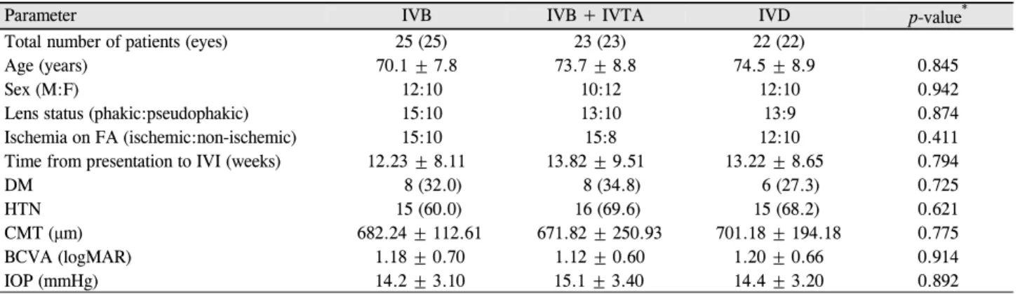
Parameter IVB IVB + IVTA IVD p-value*
Total number of patients (eyes) 25 (25) 23 (23) 22 (22)
Age (years) 70.1 ± 7.8 73.7 ± 8.8 74.5 ± 8.9 0.845
Sex (M:F) 12:10 10:12 12:10 0.942
Lens status (phakic:pseudophakic) 15:10 13:10 13:9 0.874
Ischemia on FA (ischemic:non-ischemic) 15:10 15:8 12:10 0.411
Time from presentation to IVI (weeks) 12.23 ± 8.11 13.82 ± 9.51 13.22 ± 8.65 0.794
DM 8 (32.0) 8 (34.8) 6 (27.3) 0.725
HTN 15 (60.0) 16 (69.6) 15 (68.2) 0.621
CMT (μm) 682.24 ± 112.61 671.82 ± 250.93 701.18 ± 194.18 0.775
BCVA (logMAR) 1.18 ± 0.70 1.12 ± 0.60 1.20 ± 0.66 0.914
IOP (mmHg) 14.2 ± 3.10 15.1 ± 3.40 14.4 ± 3.20 0.892
Values are presented as mean ± standard deviation or number (%) unless otherwise indicated.
IVB = intravitreal bevacizumab injection; IVB+IVTA = intravitreal low-dose bevacizumab and triamcinolone injection; IVD = intravitreal dexamethasone implant; M:F = male:female; FA = fluorescein angiography; IVI = intravitreal injection; DM = diabetes mellitus; HTN = hypertension; CMT = central macular thickness; BCVA = best-corrected visual acuity; logMAR= logarithm of the minimal angle of reso- lution; IOP = intraocular pressure.
*Kruskal-Wallis test.
Table 1. Baseline and demographic characteristics
병 후 유리체내 주사까지의 기간, 최대교정시력, 중심망막 두께 및 안압 등은 세 군 사이의 유의한 차이는 없었다 (Table 1).
빛간섭단층촬영을 이용하여 측정한 중심망막두께는 주 입술 전에 비해 주입술 후 1개월, 3개월까지 세 군에서 모 두 의미 있게 감소하였다(모두 p=0.001). 주입술 후 1개월 째 황반두께는 베바시주맙군은 평균 363.18 ± 176.94 µm, 저용량 베바시주맙-트리암시놀론 혼합군은 평균 317.00 ± 96.04 µm, 덱사메타손삽입물군은 평균 324.21 ± 75.34 µm 로 세 군 사이의 유의한 차이는 없었다(p=0.246). 주입술 3개 월째 중심망막두께의 결과를 보면, 유리체내 베바시주맙 단독 주입군의 경우 평균 598.45 ± 82.58 µm, 저용량 베바 시주맙-트리암시놀론 혼합군은 평균 456.67 ± 183.54 µm, 덱사메타손삽입물군은 평균 387.57 ± 121.82 µm로 세 군 모두 시간이 흐를수록 두께가 증가하는 경향을 보였다 (Table 2, Fig. 1). 세 군 사이의 유의한 차이(p=0.003)를 보 여 시행한 사후분석에서 베바시주맙 단독주입군에서 나머
지 두 군에 비해 의미 있는 두께의 증가 소견을 보였으며 (p=0.0007, Fig. 2), 저용량 베바시주맙-트리암시놀론 혼합 군과 덱사메타손 삽입물군 사이에는 통계학적으로 유의한 차이를 보이지 않았다(p=0.2702, Fig. 2).
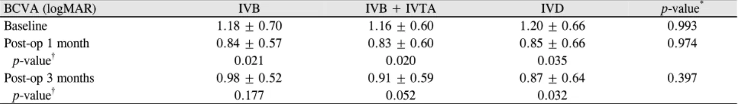
각 치료군에서 최대교정시력을 비교한 결과, 주입술 전 최대교정시력은 logMAR 시력으로 유리체내 베바시주맙 단독 주입군에서 평균 1.18 ± 0.70, 저용량 베밥시주맙-트 리암시놀론 혼합군에서 평균 1.16 ± 0.60, 덱사메타손삽입 물군에서 평균 1.20 ± 0.60으로 각 군 사이의 유의한 차이 는 없었다(p=0.993) (Table 3). 주입술 시행 1개월째 최대교 정시력은 베바시맙 단독군에서 평균 0.84 ± 0.57, 저용량 베바시주맙-트리암시놀론 혼합군에서 평균 0.83 ± 0.60, 덱 사메타손삽입물군에서 평균 0.85 ± 0.66으로, 모든 군에서 통계적으로 유의한 시력호전을 보였으며(p=0.021, p=0.020, p=0.035). 주입술 3개월째에는 베바시주맙 단독군과 저용 량 베바시주맙-트리암시놀론 혼합군에서는 시력개선의 효 과가 감소하면서 주입술 시행 전과 통계학적인 차이를 보
Figure 2. Comparison of central macular thickness (CMT)
among study groups at postoperative 3 months. Note that the CMT of IVB group was increased significantly more than IVD and IVB+IVTA groups. IVB = intravitreal bevacizumab in- jection; IVB + IVTA = intravitreal low-dose bevacizumab and triamcinolone injection; IVD = intravitreal dexamethasone implant; Mo = month(s).Figure 1. Changes of central macular thickness (CMT) among
study groups during 3 months of follow up period. The CMT of all groups was decreased at postoperative 1 month and 3 months compared with baseline. IVB = intravitreal bevacizumab in- jection; IVB + IVTA = intravitreal low-dose bevacizumab and triamcinolone injection; IVD = intravitreal dexamethasone implant.BCVA (logMAR) IVB IVB + IVTA IVD p-value*
Baseline 1.18 ± 0.70 1.16 ± 0.60 1.20 ± 0.66 0.993
Post-op 1 month 0.84 ± 0.57 0.83 ± 0.60 0.85 ± 0.66 0.974
p-value† 0.021 0.020 0.035
Post-op 3 months 0.98 ± 0.52 0.91 ± 0.59 0.87 ± 0.64 0.397
p-value† 0.177 0.052 0.032
Values are presented as mean ± standard deviation.
IVB = intravitreal bevacizumab injection; IVB + IVTA = intravitreal low-dose bevacizumab and triamcinolone injection; IVD = intravitreal dexamethasone implant; BCVA = best corrected visual acuity; logMAR = logarithm of the minimal angle of resolution; Post-op = postoperative.
* †
Table 3. Comparison of visual acuity in the three groups
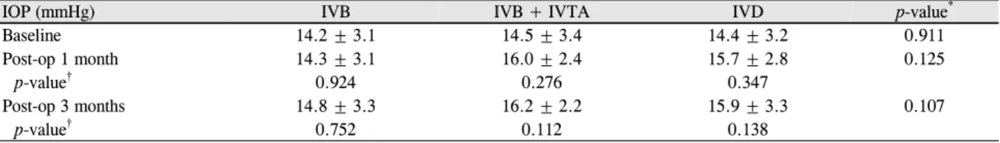
이지 않았다(p=0.177, p=0.052). 하지만 덱사메타손삽입물군 에서는 유일하게 시력호전이 3개월까지 유지되었다(p=0.032) (Table 3, Fig. 3).각 군에서 안압을 비교한 결과 주입술 전 안압은 세 군 간의 유의한 차이를 보이지 않았으며(p=0.524), 세 군 모두 에서 주입술 전과 비교하여 치료 후 1개월, 3개월째 모두 유의한 변화를 보이지 않았다(Table 4, Fig. 4). 연구 기간 중 세 군 모두에서 유리체내 주사와 연관된 심각한 합병증, 부작용 등은 나타나지 않았다.
고 찰
중심망막정맥폐쇄에 동반된 황반부종은 그 발병기전이 복잡하며, 환자의 방수 및 유리체 내에는 혈관내피성장인자 뿐만 아니라 다양한 inflammatory cytokine, angiopoiten-2, platelet-derived growth factor-AA, transforming growth fac- tor-β1, matrix metalloproteinase, soluble intercellular adhe- sion molecule 등이 증가되어 있음이 보고되었다.18-22 이러 한 황반부종을 치료하는 데 있어 여러 연구들을 통해 다양
한 방법이 발표되었고, 전통적으로 유리체내 항혈관내피성 장인자와 스테로이드가 치료의 중심에 있으나, 어떤 약물 을 선택해야 할지 정확한 기준은 정해져 있지 않은 상태이
다.3,6-13 본 연구에서는 중심망막정맥폐쇄에 동반된 황반부
종을 개선시키기 위한 유리체내 주사의 선택 약물로 망막 혈관질환에 동반된 부종의 치료에 이미 효과가 입증되고 광범위하게 사용 중인 항혈관내피성장인자인 베바시주맙
과23-26 최근 여러 연구를 통해 망막정맥폐쇄로 인한 황반부
종에서 그 효용성이 밝혀진 후로 임상에서 널리 사용되고 있는 덱사메타손삽입물12,13 그리고 저용량의 베바시주맙-트 리암시놀론 혼합물의 단기 효과 및 안정성을 비교하였다. Ehrlich et al14은 병합요법의 이론적 근거로, 질환 자체의 복잡한 병태생리의 해결을 위해 혈관형성 및 염증 반응을 동시에 억제하며, 트리암시놀론의 용량 및 안압상승의 부
Figure 3. Changes of visual acuity among study groups during
3 months of follow up period. At postoperative 1 month, the best corrected visual acuity (BCVA) of all groups was in- creased significantly. At postoperative 3 months, the BCVA was significantly improved only in the intravitreal dex- amethasone implant group compared with baseline. logMAR= logarithm of the minimal angle of resolution; IVB = intra- vitreal bevacizumab injection; IVB + IVTA = intravitreal low-dose bevacizumab and triamcinolone injection.
Figure 4. Changes of intraocular pressure (IOP) among study
groups during 3 months of follow up period. The IOP showed no significant change at postoperative 3 months after injection in all three groups. IVB = intravitreal bevacizumab injection;IVB + IVTA = intravitreal low-dose bevacizumab and tri- amcinolone injection; IVD = intravitreal dexamethasone implant.
IOP (mmHg) IVB IVB + IVTA IVD p-value*
Baseline 14.2 ± 3.1 14.5 ± 3.4 14.4 ± 3.2 0.911
Post-op 1 month 14.3 ± 3.1 16.0 ± 2.4 15.7 ± 2.8 0.125
p-value† 0.924 0.276 0.347
Post-op 3 months 14.8 ± 3.3 16.2 ± 2.2 15.9 ± 3.3 0.107
p-value† 0.752 0.112 0.138
Values are presented as mean ± standard deviation.
IOP = intraocular pressure; IVB = intravitreal bevacizumab injection; IVB + IVTA = intravitreal low-dose bevacizumab and triamcinolone injection; IVD = intravitreal dexamethasone implant; Post-op = postoperative.
*Kruskal-Wallis test; †p-value compared with baseline, Wilcoxon signed rank test.
Table 4. Comparison of IOP in the three groups
작용을 줄일 수 있음을 제시했다. 또한, Ip et al6의 보고에 의하면 트리암시놀론 1 mg과 4 mg 간에는 결과의 차이가 없으며, 오히려 부작용 면에서 4 mg이 불리하다고 하였으 며, 스테로이드의 안압상승의 부작용은 용량 의존적임을 제시한 연구 결과도 있었다.27 망막혈관질환에 따른 부종의 치료에 병합 치료의 효과를 보고했던 이전의 연구들은 1.25 mg/
0.05 mL 베바시주맙과 2 mg/0.05 mL 트리암시놀론 혼합 물을 이용했으나,14,28-30 본 연구에서는 이전에 본 교실에서 국내에 보고한 0.625 mg/0.025 mL와 트리암시놀론 1 mg/
0.025 mL를 혼합하여 사용함으로써 시술 후의 안구압박감 등의 불편감 및 추가적인 전방천자의 가능성을 줄이고자 하였다.15-17
본 연구에서는 세 군 모두에서 3개월의 경과 관찰기간 동안 시간이 지남에 따라 모든 군에서 중심황반두께가 다 시 증가하는 양상을 보였지만, 치료 전과 비교했을 때 중심 망막두께의 유의한 호전을 보였다. 주입술 1개월째, 각 치 료군 사이에 통계적으로 유의한 차이가 없이 비슷한 중심 망막두께의 감소를 보였다. Cekiç et al31은 망막정맥폐쇄에
서 유리체내 베바시주맙, 트리암시놀론, 베바시주맙-트리암 시놀론 혼합물을 비교하였을 때 1개월째 세 군에서 모두 비슷한 부종의 감소 효과를 보였다고 하여 본 연구와 비슷 한 결과는 보였다. 하지만, 주입술 3개월째에는 덱사메타손 삽입물군과 저용량 베바시주맙-트리암시놀론 혼합군은 베 바시주맙 단독군에 비해 통계적으로 유의한 해부학적 개선 효과를 보였다. 한편, 기능적인 면을 살펴보면 베바시주맙 단독군과 저용량 베바시주맙-트리암시놀론 혼합군에서는 주사 1개월째에만 유의한 시력호전을 보였으며, 덱사메타 손삽입물군은 1개월, 3개월째 모두 유의한 시력호전을 보 였다. 추가적으로 저용량 베바시주맙-트리암시놀론 혼합군 의 경우 치료 전에 비해 주입술 3개월째에는 통계적으로 유의하진 않았지만(p=0.052), 부분적인 시력호전을 보이는 것을 확인할 수 있었다.
이전의 연구에 의하면 당뇨황반부종에서 유리체내 베바 시주맙의 지속 시간은 평균 6주로 알려져 있으며,32 유리체 내 트리암시놀론의 반감기는 18.6일이며, 체내에서 3개월 (93 ± 28일)까지 측정될 수 있고,27,33 덱사메타손삽입물은 60일째 최대 효과를 보이며 약 90일까지 그 효과가 지속된
다고 하였다.12,34 이를 참고하여, 본 연구에서 각 약물의 단 기 효과에 대한 결과를 분석해 보면, 유지 치료를 위해 유 리체내 베바시주맙은 1-2개월, 저용량 베바시주맙-트리암 시놀론 혼합물의 경우에는 2-3개월, 덱사메타손삽입물의 경우 4개월 정도의 간격으로 주입술을 시행하는 것을 고려 해 볼 수 있을 것으로 사료된다.
본 연구에서 트리암시놀론-베바시주맙 혼합군과 덱사메 타손삽입물군에서 시간이 경과함에 안압이 상승하는 경향 을 보였으나, 통계적으로 유의하진 않았다. 이전의 연구들 이 다양한 빈도로 백내장 및 안압상승의 부작용을 보고한 결과와는 다르게,6,12,13,31 본 연구에서는 모든 환자에서 21 mg 이상의 안압상승은 보이지 않았으며, 백내장의 진행으로 수술받은 환자는 없었다. 하지만, 이는 3개월간의 단기간 임상연구임을 감안하면, 장기간의 추적 관찰 연구가 필요 할 것으로 사료된다.
본 연구에는 몇 가지 제한점이 있다. 첫째, 비교적 적은 수를 대상으로 이루어진 후향적 연구로 전향적 연구 방법과 같은 무작위 배정이 이루어지지 않아 선택 비뚤림(selection bias)의 가능성이 있다. 둘째, 단기간의 임상연구로 재발과 이에 따른 재주입술 여부에 관해서는 포함되지 않아서 차 후 많은 환자를 대상으로 긴 추적 관찰을 통한 추가적인 연 구가 필요하리라 생각된다. 셋째, 유리체내 트리암시놀론을 단독으로 사용한 군을 비교 대상으로 설정하지 못한 점을 들 수 있겠다. 하지만 재치료의 영향을 받지 않은 각각의 약물 고유의 단기 효과를 비교함으로써, 이를 통해 여러 가 지 변수가 있는 실제 임상에서 최소한의 주사 횟수와 경과 관찰 간격, 주사 약제의 선택 등 중심망막정맥폐쇄에 동반 된 황반부종의 표준화된 진료지침 설정에 도움이 될 것으 로 생각된다. 실제로 임상에서 주입술 시행 전 약물의 선택 에는 다양한 변수가 존재한다. 예를 들면 항혈관내피성장 인자는 심근경색, 뇌경색 같은 혈전색전성 질환의 발생 가 능성이 있으며, 짧은 반감기로 인해 잦은 주사가 필요하며, 스테로이드는 안압상승과 백내장 등의 합병증이 있다. 그 중 트리암시놀론의 경우는 약물 입자가 유리체 내에서 수 시간에서 수일간의 시야흐림 증상을 유발할 수 있어 치료 받은 눈이 유일한 눈인 환자에게서 주의 깊게 사용해야 한 다. 덱사메타손삽입물의 경우에는 공막고정술을 받은 눈이 나, 수정체 후낭손상이 동반된 경우에는 전방이동의 가능 성이 있으므로 주의해야 한다.
결론적으로 중심망막정맥폐쇄에 동반된 황반부종을 치 료하는 데 있어 유리체내 베바시주맙, 저용량 베바시주맙- 트리암시놀론 혼합물, 덱사메타손삽입물주입술 시행 후 모 든 군에서 1개월째 해부학적 및 기능적 개선을 보여주었다.
3개월까지 시력개선
이 유지되는 결과를 보였으나, 저용량 베바시주맙-트리암 시놀론 혼합물 군에서도 해부학적, 기능적 면에서 단기간 의 효과는 덱사메타손삽입물에 비교할 만한 결과를 보였다.
REFERENCES
1) Rogers SL, McIntosh RL, Lim L, et al. Natural history of branch retinal vein occlusion: an evidence-based systematic review.
Ophthalmology 2010;117:1094-101.e5.
2) Klein R, Klein BE, Moss SE, Meuer SM. The epidemiology of reti- nal vein occlusion: the Beaver Dam Eye Study. Trans Am Ophthalmol Soc 2000;98:133-43.
3) McAllister IL. Central retinal vein occlusion: a review. Clin Exp Ophthalmol 2012;40:48-58.
4) Yoshimura T, Sonoda KH, Sugahara M, et al. Comprehensive anal- ysis of inflammatory immune mediators in vitreoretinal diseases.
PLoS One 2009;4:e8158.
5) Evaluation of grid pattern photocoagulation for macular edema in central vein occlusion. The Central Vein Occlusion Study Group M report. Ophthalmology 1995;102:1425-33.
6) Ip MS, Scott IU, VanVeldhuisen PC, et al. A randomized trial com- paring the efficacy and safety of intravitreal triamcinolone with ob- servation to treat vision loss associated with macular edema secon- dary to central retinal vein occlusion: the Standard Care vs Corticosteroid for Retinal Vein Occlusion (SCORE) study report 5.
Arch Ophthalmol 2009;127:1101-14.
7) Prager F, Michels S, Kriechbaum K, et al. Intravitreal bevacizumab (Avastin) for macular oedema secondary to retinal vein occlusion:
12-month results of a prospective clinical trial. Br J Ophthalmol 2009;93:452-6.
8) Zhang H, Liu ZL, Sun P, Gu F. Intravitreal bevacizumab for treat- ment of macular edema secondary to central retinal vein occlusion:
eighteen-month results of a prospective trial. J Ocul Pharmacol Ther 2011;27:615-21.
9) Brown DM, Campochiaro PA, Singh RP, et al. Ranibizumab for macular edema following central retinal vein occlusion: six-month primary end point results of a phase III study. Ophthalmology 2010;117:1124-33.e1.
10) Korobelnik JF, Holz FG, Roider J, et al. Intravitreal aflibercept in- jection for macular edema resulting from central retinal vein occlu- sion: one-year results of the phase 3 GALILEO study. Ophthalmology 2014;121:202-8.
11) Heier JS, Clark WL, Boyer DS, et al. Intravitreal aflibercept in- jection for macular edema due to central retinal vein occlusion:
two-year results from the COPERNICUS study. Ophthalmology 2014;121:1414-20.e1.
12) Haller JA, Bandello F, Belfort R Jr, et al. Dexamethasone intra- vitreal implant in patients with macular edema related to branch or central retinal vein occlusion twelve-month study results.
Ophthalmology 2011;118:2453-60.
13) Yoon YH, Kim JW, Lee JY, et al. Dexamethasone intravitreal im- plant for early treatment and retreatment of macular edema related to branch retinal vein occlusion: the multicenter COBALT study.
Ophthalmologica 2018;240:81-9.
14) Ehrlich R, Ciulla TA, Moss AM, Harris A. Combined treatment of
intravitreal bevacizumab and intravitreal triamcinolone in patients with retinal vein occlusion: 6 months of follow-up. Graefes Arch Clin Exp Ophthalmol 2010;248:375-80.
15) Shin MH, Kang HJ, Seo JS, Chung IY. Combined low dose bev- acizumab-triamcinolone versus bevacizumab single intravitreal in- jection for branch retinal vein occlusion. J Korean Ophthalmol Soc 2018;59:650-6.
16) Kim BS, Chung IY, Park JM, Han YS. Comparison of intravitreal bevacizumab alone injection and intravitreal combination low-dose bevacizumab-triamcinolone injection or diabetic macular edema. J Korean Ophthalmol Soc 2014;55:1155-61.
17) Kim BJ, Kim HW, Han YS, et al. Comparison of bevacizumab and combined low-dose bevacizumab and triamcinolone in central reti- nal vein occlusion. J Korean Ophthalmol Soc 2016;57:438-44.
18) Kiire CA, Chong NV. Managing retinal vein occlusion. BMJ 2012;
344:e499.
19) Tuuminen R, Loukovaara S. Increased intravitreal angiopoietin-2 levels in patients with retinal vein occlusion. Acta Ophthalmol 2014;92:e164-5.
20) Jung SH, Kim KA, Sohn SW, Yang SJ. Association of aqueous hu- mor cytokines with the development of retinal ischemia and re- current macular edema in retinal vein occlusion. Invest Ophthalmol Vis Sci 2014;55:2290-6.
21) Tuuminen R, Loukovaara S. High intravitreal TGF-β1 and MMP-9 levels in eyes with retinal vein occlusion. Eye (Lond) 2014;28:1095-9.
22) Ehlken C, Grundel B, Michels D, et al. Increased expression of an- giogenic and inflammatory proteins in the vitreous of patients with ischemic central retinal vein occlusion. PLoS One 2015;10:e0126859.
23) Hsu J, Kaiser RS, Sivalingam A, et al. Intravitreal bevacizumab (avastin) in central retinal vein occlusion. Retina 2007;27:1013-9.
24) Iturralde D, Spaide RF, Meyerle CB, et al. Intravitreal bevacizumab (Avastin) treatment of macular edema in central retinal vein occlu-
sion: a short-term study. Retina 2006;26:279-84.
25) Matsumoto Y, Freund KB, Peiretti E, et al. Rebound macular ede- ma following bevacizumab (Avastin) therapy for retinal venous oc- clusive disease. Retina 2007;27:426-31.
26) Choi SW, Kim HW, Yun IH. Intravitreal bevacizumab treatment of macular edema in central retinal vein occlusion. J Korean Ophthalmol Soc 2010;51:707-15.
27) Beer PM, Bakri SJ, Singh RJ, et al. Intraocular concentration and pharmacokinetics of triamcinolone acetonide after a single intra- vitreal injection. Ophthalmology 2003;110:681-6.
28) Campochiaro PA, Nguyen QD, Hafiz G, et al. Aqueous levels of fluocinolone acetonide after administration of fluocinolone aceto- nide inserts or fluocinolone acetonide implants. Ophthalmology 2013;120:583-7.
29) Lee K, Jung H, Sohn J. Comparison of injection of intravitreal drugs with standard care in macular edema secondary to branch ret- inal vein occlusion. Korean J Ophthalmol 2014;28:19-25.
30) Ali RI, Kapoor KG, Khan AN, Gibran SK. Efficacy of combined intravitreal bevacizumab and triamcinolone for branch retinal vein occlusion. Indian J Ophthalmol 2014;62:396-9.
31) Cekiç O, Cakir M, Yazici AT, et al. A comparison of three different intravitreal treatment modalities of macular edema due to branch retinal vein occlusion. Curr Eye Res 2010;35:925-9.
32) Haritoglou C, Kook D, Neubauer A, et al. Intravitreal bevacizumab (Avastin) therapy for persistent diffuse diabetic macular edema.
Retina 2006;26:999-1005.
33) Tano Y, Sugita G, Abrams G, Machemer R. Inhibition of intra- ocular proliferations with intravitreal corticosteroids. Am J Ophthalmol 1980;89:131-6.
34) Chang-Lin JE, Attar M, Acheampong AA, et al. Pharmacokinetics and pharmacodynamics of a sustained-release dexamethasone in- travitreal implant. Invest Ophthalmol Vis Sci 2011;52:80-6.
= 국문초록 =
중심망막정맥폐쇄에 동반된 황반부종에서 유리체내 서로 다른 세 가지 약물의 단기 효과 비교
목적: 중심망막정맥폐쇄에 동반된 황반부종환자에서 유리체내 베바시주맙, 저용량 베바시주맙-트리암시놀론 혼합물, 덱사메타손삽입 물주입술의 단기간 치료 효과 및 안정성을 비교하고자 하였다.
대상과 방법: 중심망막정맥폐쇄환자 총 70명 70안 중, 유리체내 베바시주맙 단독 주입술을 시행 받은 환자 25안, 유리체내 저용량의 베바시주맙 0.625 mg/0.025 mL와 트리암시놀론 1 mg/0.025 mL를 혼합하여 유리체내 주입한 23안, 유리체내 덱사메타손삽입물을 시행 받은 환자 23안을 대상으로 시술 전, 시술 후 1개월, 3개월의 최대교정시력, 중심망막두께, 안압을 비교하였다.
결과: 유리체내 베바시주맙, 저용량 베바시주맙-트리암시놀론 혼합물, 덱사메타손삽입물 주입술 시행 후 모든 군에서 1개월째 중심망 막두께의 감소 및 시력개선을 보였으며, 세 군 사이에 유의한 차이는 없었다(p=0.246, p=0.974). 유리체내 덱사메타손삽입물군에서만 3개월까지 시력개선이 유지되는 결과를 보였으며(p=0.032), 저용량 베바시주맙-트리암시놀론 혼합물군에서도 단기간의 효과는 덱사 메타손삽입물에 비교할 만한 결과를 보였다(p=0.052). 안압은 모든 군에서 유의한 변화를 보이진 않았다.
결론: 중심망막정맥폐쇄에 동반된 황반부종의 치료에 있어서 다양한 임상적 변수를 고려하여 유리체내 베바시주맙, 저용량 베바시주 맙-트리암시놀론 혼합물, 덱사메타손삽입물을 선택적으로 사용해야 할 것으로 사료된다.
<대한안과학회지 2019;60(11):1072-1079>
정지성 / Ji-Seong Jeong
경상대학교 의과대학 안과학교실 Department of Ophthalmology, Gyeongsang National University College of Medicine