Betulin suppressed interleukin-1β-induced gene expression, secretion and proteolytic activity of matrix metalloproteinase in cultured articular chondrocytes and production of matrix metalloproteinase in the knee joint of rat
8
0
0
전체 글
(2) 20 proteoglycans and activates procollagenase in articular cartilage [7,8]. In addition to MMP-3, MMP-1 and MMP-13 were reported to play important roles in the destruction of cartilage in osteo arthritis. MMP-1 is a commonly detected metalloproteinase in synovial fluid from patients suffering from osteoarthritis [9-15]. Another degradative enzyme, ADAMTS-4, is a major aggre canase in cartilage of mouse and ADAMTS-5 has been known to be important in cartilage matrix destruction during osteoarthritis [16,17]. Therefore, we suggest it is valuable to find the potential activity of regulating (inhibiting) the expression and activity of MMPs by the compounds derived from various medicinal plants used as arthritis remedies in folk medicine, for development of new therapeutic strategies for osteoarthritis. We have tried to investigate the potential activity of some natural products on the expression and activity of MMP-3 in articular chondrocytes. As a result of our trial, we previously reported that several natural compounds affected the gene expression, secretion (production) and proteolytic activity of MMP-3, in vitro and in vivo [18-21]. According to a number of reports, betulin, a natural product derived from Betulae Cortex, a medicinal plant used for control ling various inflammatory diseases in folk medicine, showed the biological activities including anti-inflammatory effect [22-26]. Betulin showed an alleviating effect on alcoholic liver injury and renal injury through SIRT1/AMPK and TLR4/NF-kB signaling pathways, respectively [22,23]. It also attenuates lung injury in sepsis and mammary gland inflammatory injury [24,25]. Betulin suppresses the expression of pro-inflammatory cytokines in human cardiac cells [26]. However, to the best of our knowledge, there are no report about the effect of betulin on the gene expression, secretion, and enzyme activity of MMP-3, an articular cartilage-degradative enzyme that decomposes proteoglycans, in primary cultured rabbit articular chondrocytes, or on in vivo production of MMP-3 in the rat knee joint. Therefore, to evaluate the chondroprotective potential of betulin, we investigated its effects on IL-1b-induced gene expression, secretion, and enzyme activity of MMP-3 in vitro , and on production of MMP-3 in vivo.. Ra HJ et al. Primary cultures of chondrocytes from rabbit articular cartilage Male New Zealand White rabbits were obtained from Daehan Biolink (Seoul, South Korea) at 2 weeks of age. Animals were housed 1 animal per cage, provided with distilled water and food ad libitum, and kept under a 12 h light/dark cycle (lights on from 08:00~20:00) at constant temperature (22.5oC) and humidity (55%). Animals were cared for in accordance with the Guide for the Care and Use of Laboratory Animals, and care was regulated by Chungnam National University (the approval number of animal experiment: CNU-00555) (Daejeon, Korea). Rabbit articular chondrocytes were isolated from the tibial plateau and femoral condyle in cartilage of the knee joint. Cartilage was washed in phosphate-buffered saline (PBS) and minced into pieces measuring 2 mm3, approximately. Cartilage tissue was digested for 4 h with 0.2% type II collagenase at 37oC. After collection of individual cells by brief centrifugation, the cells were transferred to 100 mm culture dishes (seeding density: 105 cells/cm2) in 12 mL DMEM supplemented with 10% fetal bovine serum (FBS), in the presence of penicillin (100 units/ mL) and streptomycin (100 mg/mL). Cells were cultured at 37oC in a humidified, 5% CO2/95% air, water-jacketed incubator, and medium was replaced every other day [27].. Treatment of cells with betulin. methods. Chondrocytes were seeded on 6-well culture plates (for RTPCR) or 60 mm culture dishes (for western blotting) at a density of 105 cells/cm2. After 2 days in monolayer culture, the cells were incubated for 2 h in growth medium with 1, 10, 50, or 100 mM of betulin followed by incubation in the presence or absence of IL-1b (10 ng/mL) for 24 h. Betulin was dissolved in dimethylsulfoxide, diluted in PBS, and administered in culture medium (final concentrations of dimethylsulfoxide were 0.5%). The final pH values of these solutions were between 7.0 and 7.4. Culture medium and 0.5% dimethylsulfoxide in medium did not affect the gene expression, secretion, or proteolytic activity of MMP-3 in primary cultured chondrocytes. The supernatant was collected and centrifuged, and cell and supernatant fractions were stored at –80oC until use.. Materials. Cytotoxicity assay. All chemicals and reagents used in this experiment, including betulin (purity: 98.0%), were purchased from Sigma-Aldrich (St. Louis, MO, USA) unless otherwise specified. Dulbecco’s Modified Eagle’s Medium (DMEM) was purchased from Gibco-BRL (Grand Island, NY, USA) and recombinant human IL-1b was purchased from R&D Systems (Minneapolis, MN, USA).. Chondrocytes were seeded at a density of 2×10⁵/mL (0.1 mL/ well) in a 96-well microtiter plate, and allowed to attach for 24 h to keep the log phase growth at the time of drug treatment. Betulin was dissolved in DMSO, and administered in DMEM supplemented with 10% FBS (final concentrations of DMSO were under 0.5%). 0.5% DMSO alone did not affect the proliferation of chondrocytes. After incubation with the indicated drug concentrations for 72 h, cell proliferation was determined using. Korean J Physiol Pharmacol 2017;21(1):19-26. https://doi.org/10.4196/kjpp.2017.21.1.19.
(3) 21. Betulin and osteoarthritis. the sulforhodamine B (SRB) assay [28].. Isolation of total RNA and RT-PCR Total RNA was isolated from chondrocytes using the EasyBLUE Extraction Kit (INTRON Biotechnology, Inc. Kyung-ki-do, South Korea), and reverse transcribed using AccuPower RT Premix (BIONEER Corporation, Daejeon, South Korea) according to the manufacturer's instructions. About 2 mg of total RNA was primed with 1 mg of oligo (dT) in a final reaction volume of 30 mL. 2 mL of RT reaction product was amplified in 20 mL using Thermoprime Plus DNA Polymerase (ABgene, Rochester, NY, USA). PCR was performed with the following primers: MMP-3 (5’ATG GAC CTT CTT CAG CAA 3’, 5’TCA TTA TGT CAG CCT CTC 3’), MMP13 (5’AGG AGC ATG GCG ACT TCT AC 3’, 5’TAA AAA CAG CTC CGC ATC AA 3’), MMP-1 (5’TCA GTT CGT CCT CAC TCC AG 3’, 5’TTG GTC CAC CTG TCA TCT TC 3’), ADAMTS-4 (5’CAA GGT CCC ATG TGC AAC GT 3’, 5’CAT CTG CCA CCA CCA GTG TCT 3’), ADAMTS-5 (5’TGT CCT GCCAGC GGATGT 3’; 5’ACG GAA TTA CTG TAC GGC CTA CA 3’), and type II collagen (5’AAC ACT GCC AAC GTC CAG AT 3’, 5’CTG ACG CAC GGT ATA GGT GA 3’). GAPDH (5’ACT GGC GTC TTC ACC ACC AT 3’; 5’AAG GCC ATG CCA GTG AGC TT 3’) was used as a quantitative control. The PCR products increased as the concentration of RNA increased. The amplified fragment sizes were 350 base pairs (bp) for MMP-3, 458 bp for MMP-13, 300 bp for MMP-1, 90 bp for ADAMTS-4, 110 bp for ADAMTS-5, 220 bp for type II collagen, and 400 bp for GAPDH. After PCR, 15 mL of PCR products were subjected to 2% agarose gel electrophoresis and visualized with ethidium bromide under a transilluminator [27]. The signal intensity of each band was analyzed by GelQuant software (DNR Bio-Imaging Systems Ltd., Jerusalem, Israel).. Western blot analysis for measuring secretion level of MMP-3 in culture supernatant Chondrocytes (confluent in 60 mm culture dish) were incubated for 2 h in growth medium with 1, 10, 50, or 100 mM of betulin followed by incubation in the presence or absence of IL-1b (10 ng/ mL) for 24 h. After the treatment, the supernatant was collected and the cells were harvested using 3 x trypsin-EDTA solution and then centrifuged in a microcentrifuge (1,200 rpm, 3 min, 4oC). The Bradford assay was used to measure protein concentrations in culture supernatants to ensure consistent weight of protein samples subjected to electrophoresis. Culture supernatant samples containing MMP-3 protein (50 mg each) were subjected to 10% sodium dodecyl sulfate–polyacrylamide gel electrophoresis (SDSPAGE), and then transferred onto a polyvinylidene difluoride (PVDF) membrane. Blots were blocked using 5% skim milk in Tris-buffered saline/Tween 20 (TBS-T), and probed overnight with MMP-3 antibody in blocking buffer at 4oC. Antibody against MMP-3 was purchased from Santa Cruz Biotechnology (Santa www.kjpp.net. Cruz, CA, USA). Membranes were washed with TBS-T and probed for 1 h with a secondary antibody conjugated with horseradish peroxidase (Calbiochem, La Jolla, CA, USA). After 4 washes with TBS-T, immunoreactive bands were detected using an enhanced chemiluminescence kit (Pierce ECL western blotting substrate, Thermo Scientific, Waltham, MA, USA). The signal intensity of each band was analyzed by GelQuant software (DNR Bio-Imaging Systems Ltd., Jerusalem, Israel).. Casein zymography to measure the proteolytic activity of MMP-3 A modified casein-substrate zymography was carried out using culture supernatants from chondrocytes pretreated for 2 h with betulin and stimulated for 24 h with IL-1b in DMEM containing 0.5% FBS. The Bradford assay was used to measure protein con centrations in culture media to ensure consistency across samples. Samples were electrophoresed at 4oC in a 10% SDS gel containing 0.1% casein. After electrophoresis, gels were washed with 10 mM Tris-HCl (pH 8.0) containing 2.5% Triton X-100. Next, gels were incubated at 37oC for 48 h in 50 mM Tris-HCl (pH 8.0) containing 1% Triton X-100, 0.2 M NaCl, and 5 mM CaCl 2. Finally, gels were stained with 1% Coomassie Brilliant Blue, destained, and photographs were taken [27]. The signal intensity of each band was analyzed by GelQuant software (DNR Bio-Imaging Systems Ltd., Jerusalem, Israel).. In vivo experiments Male Sprague–Dawley rats (Daehan Biolink, Seoul, South Korea) weighing 200~210 g were used to investigate the effect of betulin on production of MMP-3 in articular cartilage in vivo . Animals were housed 5 per cage, provided with distilled water and food ad libitum , and kept under a 12 h light/dark cycle (lights on from 08:00~20:00) at constant temperature (22.5oC) and humidity (55%). Animals were cared for in accordance with the Guide for the Care and Use of Laboratory Animals, and care was regulated by Chungnam National University (the approval number of animal experiment: CNU-00555) (Daejeon, South Korea). Rats were randomly divided into 4 groups: control, IL1b only, 50 mM betulin plus IL-1b, or 100 mM betulin plus IL1b. Rats were anesthetized with vaporized diethyl ether, and those from the 50 mM betulin plus IL-1b and 100 mM betulin plus IL-1b treatment groups received a 30 mL injection of 50 mM or 100 mM betulin, respectively, into the right knee joint. After 3 h, rats from the IL-1b only group, the 50 mM betulin plus IL1b group, and the 100 mM betulin plus IL-1b group received a 30 mL injection of 20 ng IL-1b in sterile PBS into the right knee joint. Rats from the control group were injected with 30 mL of sterile PBS. Rats were euthanized via CO2 asphyxiation 72 h after injections. Articular cartilage (tibial plateau and femoral condyle) was isolated from each animal, homogenized, and prepared for Korean J Physiol Pharmacol 2017;21(1):19-26.
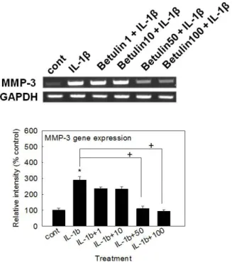
(4) 22 measurement of MMP-3 protein by western blot analysis. Tissue lysates from articular cartilage homogenates containing MMP3 protein (50 mg each) were subjected to 10% SDS-PAGE, and transferred onto a PVDF membrane. Blots were blocked with 5% skim milk in TBS-T, and probed with MMP-3 antibody (Santa Cruz Biotechnology, Santa Cruz, CA, USA) in blocking buffer overnight at 4oC. Membranes were washed with TBS-T, and probed for 1 h with a secondary antibody conjugated with horseradish peroxidase (Calbiochem, La Jolla, CA, USA). After 4 washes with TBS-T, immunoreactive bands were detected using an enhanced chemiluminescence kit (Pierce ECL western blotting substrate, Thermo Scientific, Waltham, MA, USA). The signal intensity of each band was analyzed by GelQuant software (DNR Bio-Imaging Systems Ltd., Jerusalem, Israel).. Statistics Means of individual group were converted to percent control. Ra HJ et al. and expressed as mean±S.E.M. The difference between groups was assessed using one-way ANOVA and Holm-Sidak test as a post-hoc test. p<0.05 was considered as significantly different.. Results Effect of betulin on MMP-3 gene expression in rabbit chondrocytes To examine the potential activity of betulin on the gene expression of MMP-3, the key matrix metalloproteinase involved in destruction of articular cartilage, MMP-3 gene expression was measured after pretreatment of betulin. As shown in Fig. 1, betulin inhibited IL-1b-induced MMP-3 gene expression.. Cytotoxicity of betulin to rabbit chondrocytes To investigate the potential cytotoxicity of betulin to cultured rabbit chondrocytes, effect of betulin on proliferation of rabbit chondrocytes using SRB assay was tested. As can be seen in Fig. 2, betulin showed no significant cytotoxicity at the concentrations of 1, 10, 50, and 100 mM. The numbers of cells in betulin-treated cultures were 100±10%, 91±18%, 102±13%, 107±9%, and 95±11% for control, 1, 10, 50, and 100 mM betulin, respectively.. Effect of betulin on the gene expression of MMP-1, MMP-13, ADAMTS-4, ADAMTS-5 or type II collagen in rabbit chondrocytes If betulin can affect the gene expression of MMP-3, the key matrix metalloproteinase involved in destruction of articular. Fig. 1. Effect of betulin on MMP-3 gene expression in rabbit chondrocytes. Primary cultured rabbit articular chondrocytes were pretreated with varying concentrations (1, 10, 50, and 100 mM) of betulin for 2 h and then stimulated with IL-1b (10 ng/mL) for 24 h. MMP-3 gene expression level was measured by RT-PCR. Three independent experiments were performed and the representative data were shown. The upper figure is a representative image data. The signal intensity of each band in images was analyzed by GelQuant software and means of individual group from the three independent experiments were converted to percent control and expressed as mean±S.E.M. Each bar in the lower figure (graph) represents a mean±S. E.M. of three independent experiments in comparison with that of the control set at 100% (Fig. 1, 3, 4 and 5). *significantly different from control (p<0.05). +significantly different from IL-1b alone (p<0.05) (cont: control, concentration unit is mM). Korean J Physiol Pharmacol 2017;21(1):19-26. Fig. 2. Effect of betulin on proliferation of rabbit chondrocytes. Chondrocytes were incubated for 72 h in the presence of varying concentrations of betulin. Cell viability was determined using SRB assay as described in Materials and Methods. Each bar represents a mean±S. E.M. of three independent experiments in comparison with that of the control set at 100%. https://doi.org/10.4196/kjpp.2017.21.1.19.
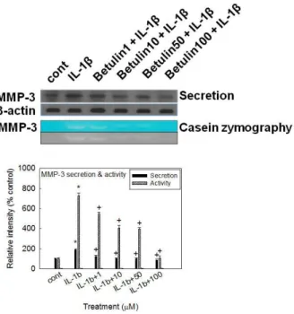
(5) 23. Betulin and osteoarthritis. cartilage, it should be investigated whether betulin affects the gene expression of MMP-1, MMP-13, ADAMTS-4 or ADAMTS-5, the other degradative enzymes related to destruction of articular cartilage, and type II collagen, in rabbit chondrocytes. As shown in Fig. 3, betulin showed the suppression of IL-1b-induced gene expression of MMP-1, MMP-13, ADAMTS-4, and ADAMTS-5, in rabbit chondrocytes. Furthermore, betulin showed an additional chondroprotective effect by restoring the compromised gene expression of type II collagen by IL-1b, in rabbit chondrocytes.. Effect of betulin on IL-1b-induced secretion of MMP-3 from rabbit articular chondrocytes If betulin can affect the MMP-3 gene expression at the transcriptional level, it should be investigated whether betulin affects IL-1b-induced secretion of MMP-3 proteins from rabbit articular chondrocytes. As shown in Fig. 4, stimulation with IL1b (10 ng/mL) increased secretion of MMP-3 from chondrocytes. However, betulin inhibited the effects of IL-1b on MMP-3 secretion.. secreted MMP-3, which is known to degrade proteoglycans, one of the two major matrix components of cartilage, culture supernatants from rabbit articular chondrocytes were analyzed for caseinolytic activity by casein zymography, after treatment with IL-1b for 24 h. As can be seen in Fig. 4, IL-1b increased the caseinolytic activity of MMP-3 in rabbit articular chondrocytes, and this effect was inhibited by pretreatment with betulin.. Effect of betulin on MMP-3 production in vivo To investigate whether betulin shows the potential effect in vivo , we examined the effect of intraarticular injection of betulin into the knee joint of rats on IL-1b-stimulated production of MMP-3 from articular cartilage tissues. As shown in Fig. 5, treatment with IL-1b (20 ng/30 mL) increased MMP-3 production in articular cartilage tissues. However, betulin inhibited IL-1binduced MMP-3 production, in vivo.. Discussion. To investigate the effect of betulin on the enzyme activity of. To restore the broken equilibrium between physiological synthesis and degradation of articular cartilage during the progression of osteoarthritis, discovering a useful and specific pharmacological tool can be a promising approach to the effective. Fig. 3. Effect of betulin on the gene expression of MMP-1, MMP-13, ADAMTS-4, ADAMTS-5, or collagen type II in rabbit chondrocytes. Primary cultured rabbit articular chondrocytes were pretreated with varying concentrations (1, 10, 50, and 100 mM) of betulin for 2 h and then stimulated with IL-1b (10 ng/mL) for 24 h. The gene expression level of MMP-1, MMP-13, ADAMTS-4, ADAMTS-5, or collagen type II was measured by RT-PCR.. Fig. 4. Effects of betulin on IL-1β-induced secretion of MMP-3 and caseinolytic activity of MMP-3 in rabbit articular chondrocytes. Primary cultured rabbit articular chondrocytes were pretreated with varying concentrations (1, 10, 50, and 100 mM) of betulin for 2 h and then stimulated with IL-1b (10 ng/mL) for 24 h. Culture supernatants were collected for measurement of both the levels of produced and secreted MMP-3 by western blot analysis and the proteolytic activity of MMP-3 by casein zymography.. Effect of betulin on proteolytic activity of MMP-3 in rabbit articular chondrocytes. www.kjpp.net. Korean J Physiol Pharmacol 2017;21(1):19-26.
(6) 24. Fig. 5. Effect of betulin on production of MMP-3 in vivo . The knee joint of rats were pretreated with 50 or 100 mM of betulin for 3 h and then stimulated with IL-1b (20 ng/30 mL) for 72 h, by intraarticular injection. Tissue lysates from articular cartilage homogenates containing MMP-3 proteins were collected for measurement of the level of produced MMP-3 in vivo , by western blot analysis. Equal protein loading was evaluated by b-actin levels.. control of this condition. Although osteoarthritis can be defined as a non-inflammatory disease, its development and progression have been attributed to low-grade inflammation in intraarticular sites, as well as to various inflammatory cytokines in articular tissues and fluids that are produced by chondrocytes and/or interact with chondrocytes [29-32]. IL-1b is an inflammatory cytokine that is produced by cells in articular tissues, including chondrocytes, and which can increase expression of MMPs and stimulate the progression of osteoarthritis. IL-1b plays an important role in the initiation and progression of destruction of articular cartilage by suppressing synthesis of collagen and stimulating MMP expression [31,33,34]. Particularly, MMP-3 has been reported to play a pivotal patho physiological role in osteoarthritis by degrading components of the extracellular matrix, such as proteoglycans. MMP-3 levels were increased more than MMP-1 levels in patients suffering from osteoarthritis in knee joints compared to the control group [7,35]. According to several reports, IL-1b-stimulated expression of MMPs is associated with the suppression of the NF-kB signa ling pathway [36-39]. Jung and his colleagues reported that Schisandrae Fructus, an anti-inflammatory medicinal plant used in folk medicine, inhibited IL-1b-induced expression and activity Korean J Physiol Pharmacol 2017;21(1):19-26. Ra HJ et al. of MMP-1, MMP-3, and MMP-13 and markedly suppressed the nuclear translocation of NF- k B by blocking Ik B-alpha degradation in SW1353 human chondrocytes [36]. Taraxasterol, a natural product with anti-inflammatory effect, suppressed IL-1binduced expression of MMP-1, MMP-3, MMP-13 and activation of NF-kB [37]. Another natural product, matrine, inhibited IL1b-induced expression of MMPs by suppressing the activation NF-kB in human chondrocytes in vitro [38]. Zhou et al. reported that coptisine, an isoquinoline alkaloid extracted from an antiinflammatory medicinal plant, also inhibited the expression of MMP-3 and MMP-13 through inhibition of NF-kB activation in human chondrocytes [39]. IL-1b activates several intracellular signal transduction cascades among which the NF-kB pathway is pivotal. NF-kB is a heterodimer composed of p65, p50 and IkBα subunits present in the cytoplasm as an inactive state. In response to various stimuli, the Ik Bα subunit is phosphorylated and degraded, thereby facilitating the translocation of p50-p65 heterodimer to the nucleus. The p50-p65 acts as a transcription factor regulating the expression of numerous genes including MMP-3 [36]. In the present study, betulin affected the gene expression, secretion, and proteolytic activity of MMP-3, by directly acting on articular chondrocytes. We found that betulin inhibited IL-1b-induced gene expression of MMP-3, MMP-1, MMP-13, ADAMTS-4, and ADAMTS-5 and restored the gene expression of type II collagen that had been inhibited by IL-1b, in rabbit articular chondrocytes (Fig. 1 and 3). Thus, the chondroprotective effect of betulin are supported by its regulation of the gene expression of diverse proteases involved in the destruction of articular cartilage in osteoarthritis, as well as by its promotion of the gene expression of type II collagen at the transcriptional level. Additionally, IL1b-stimulated secretion and the proteolytic activity of MMP-3 from articular chondrocytes were suppressed by betulin (Fig. 4). This result means that betulin can regulate the step of protein synthesis and secretion of MMP-3 and affects the proteolytic activity of overproduced and oversecreted MMP-3 in tissues of osteoarthritic articular cartilage. The underlying mechanism of action of betulin on the gene expression, secretion, and proteolytic activity of MMP-3 are not clear at present, although we are investigating whether betulin act as a potential regulator of NF-kB signaling pathway in articualr chondrocytes, based on a report on the inhibitory effect of betulin on lipopolysaccharide (LPS)-stimulated rat HBZY-1 renal mesan gial cells through inactivation of NF-kB signaling pathway [23]. Lastly, we investigated the effect of intraarticular injection of betulin into the knee joint of rats on IL-1b-stimulated production of MMP-3 in articular cartilage tissue. As can be seen in Fig. 5, betulin inhibited IL-1b-stimulated production of MMP-3 in articular cartilage tissue. This result shows that, in addition to its in vitro effects, betulin exerts chondroprotective effects in vivo when administered via intraarticular injection. Taken together, the inhibitory action of betulin on the gene https://doi.org/10.4196/kjpp.2017.21.1.19.
(7) Betulin and osteoarthritis. expression, secretion, and enzyme activity of MMP-3 in articular chondrocytes and production of MMP-3 in the knee joint of rats might explain, at least in part, the traditional use of Betulae Cortex as an anti-inflammatory agent for diverse inflammatory diseases, in folk medicine. We suggest it is valuable to find the natural products that have specific suppressive effects on the gene expression, secretion, and enzyme activity of MMP-3 - in view of both basic and clinical sciences - and the result from this study suggests a possibility of developing betulin as a candidate for novel agent controlling cartilage damage in osteoarthritis via intraarticular administration, although further studies are essentially required.. AcknowledgementS This research was supported by Basic Science Research Program through the National Research Foundation of Korea (NRF) funded by the Ministry of Education (NRF-2014R1A6A1029617).. Conflicts of interest The authors have no conflicts of interest to declare.. REFERENCES 1. Aigner T, McKenna L. Molecular pathology and pathobiology of osteoarthritic cartilage. Cell Mol Life Sci. 2002;59:5-18. 2. Mankin HJ. The response of articular cartilage to mechanical injury. J Bone Joint Surg Am. 1982;64:460-466. 3. Dean DD, Martel-Pelletier J, Pelletier JP, Howell DS, Woessner JF Jr. Evidence for metalloproteinase and metalloproteinase inhibitor imbalance in human osteoarthritic cartilage. J Clin Invest. 1989; 84:678-685. 4. Kullich W, Fagerer N, Schwann H. Effect of the NSAID nimesulide on the radical scavenger glutathione S-transferase in patients with osteoarthritis of the knee. Curr Med Res Opin. 2007;23:1981-1986. 5. Birkedal-Hansen H, Moore WG, Bodden MK, Windsor LJ, BirkedalHansen B, DeCarlo A, Engler JA. Matrix metalloproteinases: a review. Crit Rev Oral Biol Med. 1993;4:197-250. 6. Burrage PS, Mix KS, Brinckerhoff CE. Matrix metalloproteinases: role in arthritis. Front Biosci. 2006;11:529-543. 7. Garnero P, Rousseau JC, Delmas PD. Molecular basis and clinical use of biochemical markers of bone, cartilage, and synovium in joint diseases. Arthritis Rheum. 2000;43:953-968. 8. Lin PM, Chen CT, Torzilli PA. Increased stromelysin-1 (MMP-3), proteoglycan degradation (3B3- and 7D4) and collagen damage in cyclically load-injured articular cartilage. Osteoarthritis Cartilage. 2004;12:485-496. 9. Freemont AJ, Hampson V, Tilman R, Goupille P, Taiwo Y, Hoyland JA. Gene expression of matrix metalloproteinases 1, 3, and 9 by chondrocytes in osteoarthritic human knee articular cartilage is www.kjpp.net. 25 zone and grade specific. Ann Rheum Dis. 1997;56:542-549. 10. Goupille P, Jayson MI, Valat JP, Freemont AJ. Matrix metallo proteinases: the clue to intervertebral disc degeneration? Spine (Phila Pa 1976). 1998;23:1612-1626. 11. Kanyama M, Kuboki T, Kojima S, Fujisawa T, Hattori T, Takigawa M, Yamashita A. Matrix metalloproteinases and tissue inhibitors of metalloproteinases in synovial fluids of patients with temporo mandibular joint osteoarthritis . J Orofac Pain. 2000;14:20-30. 12. Jo H, Park JS, Kim EM, Jung MY, Lee SH, Seong SC, Park SC, Kim HJ, Lee MC. The in vitro effects of dehydroepiandrosterone on human osteoarthritic chondrocytes. Osteoarthritis Cartilage. 2003; 11:585-594. 13. Little CB, Barai A, Burkhardt D, Smith SM, Fosang AJ, Werb Z, Shah M, Thompson EW. Matrix metalloproteinase 13-deficient mice are resistant to osteoarthritic cartilage erosion but not chondrocyte hypertrophy or osteophyte development. Arthritis Rheum. 2009;60: 3723-3733. 14. Neuhold LA, Killar L, Zhao W, Sung ML, Warner L, Kulik J, Turner J, Wu W, Billinghurst C, Meijers T, Poole AR, Babij P, DeGennaro LJ. Postnatal expression in hyaline cartilage of constitutively active human collagenase-3 (MMP-13) induces osteoarthritis in mice. J Clin Invest. 2001;107:35-44. 15. Yoshihara Y, Nakamura H, Obata K, Yamada H, Hayakawa T, Fujikawa K, Okada Y. Matrix metalloproteinases and tissue inhi bitors of metalloproteinases in synovial fluids from patients with rheumatoid arthritis or osteoarthritis. Ann Rheum Dis. 2000;59: 455-461. 16. Echtermeyer F, Bertrand J, Dreier R, Meinecke I, Neugebauer K, Fuerst M, Lee YJ, Song YW, Herzog C, Theilmeier G, Pap T. Syndecan-4 regulates ADAMTS-5 activation and cartilage breakdown in osteo arthritis. Nat Med. 2009;15:1072-1076. 17. Stanton H, Rogerson FM, East CJ, Golub SB, Lawlor KE, Meeker CT, Little CB, Last K, Farmer PJ, Campbell IK, Fourie AM, Fosang AJ. ADAMTS5 is the major aggrecanase in mouse cartilage in vivo and in vitro. Nature. 2005;434:648-652. 18. Nam DC, Kim BK, Lee HJ, Shin HD, Lee CJ, Hwang SC. Effects of prunetin on the proteolytic activity, secretion and gene expression of MMP-3 in vitro and production of MMP-3 in vivo. KoreanJ Physiol Pharmacol. 2016;20:221-228. 19. Park JS, Kim DK, Shin HD, Lee HJ, Jo HS, Jeong JH, Choi YL, Lee CJ, Hwang SC. Apigenin regulates interleukin-1b -induced production of matrix metalloproteinase both in the knee joint of rat and in primary cultured articular chondrocytes. Biomol Ther (Seoul). 2016;24:163-170. 20. Park JS, Lee HJ, Lee DY, Jo HS, Jeong JH, Kim DH, Nam DC, Lee CJ, Hwang SC. Chondroprotective effects of wogonin in experimental models of osteoarthritis in vitro and in vivo. Biomol Ther (Seoul). 2015;23:442-448. 21. Kang BJ, Ryu J, Lee CJ, Hwang SC. Luteolin inhibits the activity, secretion and gene expression of MMP-3 in cultured articular chondrocytes and production of MMP-3 in the rat knee. Biomol Ther (Seoul). 2014;22:239-245. 22. Bai T, Yang Y, Yao YL, Sun P, Lian LH, Wu YL, Nan JX. Betulin alleviated ethanol-induced alcoholic liver injury via SIRT1/AMPK signaling pathway. Pharmacol Res. 2016;105:1-12. 23. Zhao H, Zheng Q, Hu X, Shen H, Li F. Betulin attenuates kidney injury in septic rats through inhibiting TLR4/NF-k B signaling Korean J Physiol Pharmacol 2017;21(1):19-26.
(8) 26 pathway. Life Sci. 2016;144:185-193. 24. Zhao H, Liu Z, Liu W, Han X, Zhao M. Betulin attenuates lung and liver injuries in sepsis. Int Immunopharmacol. 2016;30:50-56. 25. Guo MY, Li WY, Zhang Z, Qiu C, Li C, Deng G. Betulin suppresses S. aureus-induced mammary gland inflammatory injury by regulating PPAR-g in mice. Int Immunopharmacol. 2015;29:824-831. 26. Zhang SY, Zhao QF, Fang NN, Yu JG. Betulin inhibits pro-inflam matory cytokines expression through activation STAT3 signaling pathway in human cardiac cells. Eur Rev Med Pharmacol Sci. 2015; 19:455-460. 27. Moon PD, Jeong HS, Chun CS, Kim HM. Baekjeolyusin-tang and its active component berberine block the release of collagen and pro teoglycan from IL-1b-stimulated rabbit cartilage and down-regulate matrix metalloproteinases in rabbit chondrocytes. Phytother Res. 2011;25:844-850. 28. Skehan P, Storeng R, Scudiero D, Monks A, McMahon J, Vistica D, Warren JT, Bokesch H, Kenney S, Boyd MR. New colorimetric cytotoxicity assay for anticancer-drug screening. J Natl Cancer Inst. 1990;82:1107-1112. 29. Bonnet CS, Walsh DA. Osteoarthritis, angiogenesis and inflam mation. Rheumatology (Oxford). 2005;44:7-16. 30. Goldring MB, Otero M, Tsuchimochi K, Ijiri K, Li Y. Defining the roles of inflammatory and anabolic cytokines in cartilage metabolism. Ann Rheum Dis. 2008;67 Suppl 3:iii75-82. 31. Kobayashi M, Squires GR, Mousa A, Tanzer M, Zukor DJ, Antoniou J, Feige U, Poole AR. Role of interleukin-1 and tumor necrosis factor alpha in matrix degradation of human osteoarthritic cartilage. Arthritis Rheum. 2005;52:128-135.. Korean J Physiol Pharmacol 2017;21(1):19-26. Ra HJ et al 32. Loeser RF. Molecular mechanisms of cartilage destruction: mecha nics, inflammatory mediators, and aging collide. Arthritis Rheum. 2006;54:1357-1360. 33. Aida Y, Maeno M, Suzuki N, Shiratsuchi H, Motohashi M, Matsumura H. The effect of IL-1beta on the expression of matrix metalloproteinases and tissue inhibitors of matrix metalloproteinases in human chondrocytes. Life Sci. 2005;77:3210-3221. 34. Pantsulaia I, Kalichman L, Kobyliansky E. Association between radiographic hand osteoarthritis and RANKL, OPG and inflam matory markers. Osteoarthritis Cartilage. 2010;18:1448-1453. 35. Lijnen HR. Matrix metalloproteinases and cellular fibrinolytic activity. Biochemistry (Mosc). 2002;67:92-98. 36. Jeong JW, Lee HH, Choi EO, Lee KW, Kim KY, Kim SG, Hong SH, Kim GY, Park C, Kim HK, Choi YW, Choi YH. Schisandrae fructus inhibits IL-1b -induced matrix metalloproteinases and inflam matory mediators production in SW1353 human chondrocytes by suppressing NF-k B and MAPK activation. Drug Dev Res. 2015; 76:474-483. 37. Piao T, Ma Z, Li X, Liu J. Taraxasterol inhibits IL-1b -induced inflammatory response in human osteoarthritic chondrocytes. Eur J Pharmacol. 2015;756:38-42. 38. Lu S, Xiao X, Cheng M. Matrine inhibits IL-1b-induced expression of matrix metalloproteinases by suppressing the activation of MAPK and NF-kB in human chondrocytes in vitro. Int J Clin Exp Pathol. 2015;8:4764-4772. 39. Zhou K, Hu L, Liao W, Yin D, Rui F. Coptisine prevented IL-binduced expression of inflammatory mediators in chondrocytes. In flammation. 2016;39:1558-1565.. https://doi.org/10.4196/kjpp.2017.21.1.19.
(9)
수치

관련 문서
In this paper, we consider the texture background of rail track images and the sparse foreground of the defects to construct a low-rank matrix decomposition model
Gene expression signature as Predictor Of Survival in Breast Cancer. -Inkjet-synthesized oligonucleotide microarrays, 25000 oligos -70-gene
1 John Owen, Justification by Faith Alone, in The Works of John Owen, ed. John Bolt, trans. Scott Clark, "Do This and Live: Christ's Active Obedience as the
Pituitary tumor transforming gene (PTTG) expression in pituitary adenomas.. Pathophysiology of
This study tried to find out the effect of physical expression activity in the childlcare classroom influencing to the aggressions and the adjusting to school of the
The matrix A show the cost per computer (in thousands of dollars) and B the production figures for the year 2005 (in multiples of 1000 units).. Find a matrix C that
This study suggested diversity in the configuration of expression through expression of abstract images, the style of modern painting, which are expressed
Effects of phase I periodontal treatment on gingival crevicular fluid levels of matrix metalloproteinase-3 and tissue inhibitor of metalloproteinase-1.
