J Korean Soc Coloproctol Vol. 19, No. 1, 2003
13
Clinicopathological Characteristics of Spo- radic Colorectal Cancer with DNA Micro- satellite Instability
N am Kyu Kim , M.D., H oguen K im , M.D.
1, Jae K un Park, M.D., K ang Y ong Lee, M.D., Seung K ook Sohn, M.D., Jin Sik M in, M.D.
Departments of Surgery, 1Pathology, Yonsei University College of Medicine, Seoul, Korea
P urpose: Sporadic colorectal cancers, with DNA mi- crosatellite instability (MSI), have been characterized by a predilection area of proximal colon, younger age onset, exophytic growth and larger tumor size. MSI colorectal cancers have recently been had a good survival rate. The aim of this study is to determine the MSI status in sporadic colorectal cancers, and compare their clinical and path- ological characteristics with those of MSS (Microsatellite Stable) cancers.
M eth ods: Between March 1995 and December 1997, deep frozen fresh tissue of 107 eligible colorectal cancer patients, who underwent surgical resections, were used for analysis. Hereditary nonpolyposis colorectal cancer, and fam ilial adenom atous polyposis, patients were ex- cluded. All the patients were registered on a colorectal cancer database, and followed up completely with regular visits for a potential recurrence. Genom ic DN A was prepared by the SDS-proteinase K and phenol chloroform extraction m ethods. The DN A was am plified by PCR at five microsatellite loci (BAT26, BAT25, D2S123, D5S346, and D 17S250) to evaluate the M SI. The PCR products were separated in 6% polyacrylamide gels, containing 5.6 M urea, followed by autoradiography. The M SI was defined as being over 2 m arker positive, and the M SS as 1 m arker positive, all m arker negatives were classed as M SS. The survival rates were calculated by the Kaplan-
Meier methods.
R esults: MSI was noted at 16/107 (15%), with mean ages for the patients of 51.8 vs. 58.6 years old for MSI and MSS, respectively. For the patients under 40 years old 5 (31.3%) vs. 6 (6.6%) had MSI and MSS, respectively (p<0.01). The cancer was located in the right colon in 12 of each of the MSI and MSS (p<0.01). There were no MSI rectal cancer tumors. The average tumor sizes were 7.6±3.6 cm vs. 5.3
±2.2 cm (p<0.01) for MSI and MSS, respectively, but there were no correlations with the frequency of asso- ciated polyps, recurrence and distant metastasis between MSI and MSS. The cells were well differentiated (12.5%
vs. 17.6% ), moderately differentiated (68.8% vs. 76.9%), poorly differentiated (6.2% vs. 3.3%), and mucinous type (12.5% vs. 2.2% ), with MSI and MSS, respectively. The overall survival rates were 93.8% vs. 73.8% for MSI and MSS (P=0.07), respectively.
C onclusions: Sporadic colorectal cancer, with DNA micro- satellite instability (MSI), was located predominantly in the proximal colon, in the younger age onset, and larger size of tumor. The survival rate of the patients with MSI tumors were good, but with no statistical significance.
J K orean S oc C oloproctol 2003;19:13-19K e y W o rd s: Sporadic colorectal cancer, M icrosatellite instability
산발성 대장암, DNA 현미부수체 ꠏꠏꠏꠏꠏꠏꠏꠏꠏꠏꠏꠏꠏꠏꠏꠏꠏꠏꠏꠏꠏꠏꠏꠏꠏꠏꠏꠏꠏꠏꠏꠏꠏꠏꠏꠏꠏꠏꠏꠏꠏꠏꠏꠏꠏꠏꠏꠏꠏ
서 론
현미 부수체 불안정성(microsatellite instability, 이하 MSI)은 DNA 복제오류를 교정해주는 DNA 부정합 교 정 유전자(mismatch repair gene)의 돌연변이로 발생하 는 것으로 알려져 있다. 관련된 유전자는 hMSH2, hMLH1, hMSH6, hPMS1, hPMS2 등이다. 이 중에서도 hMSH2, hMLH1의 돌연변이가 주로 관여한다고 알려 져 있고 특히 hMLH1 유전자 촉진자(promoter)의 변화 된 메칠화가 주로 원인이 된다고 하였다. 이러한 현상 은 대장암 외에도 자궁 내막암, 위암, 췌장암 등에서 나타난다고 하는데1,2 현미 부수체 불안전성(MSI)은 가
DNA 현미 부수체 불안전성 특성을 가진 산발성 대장암의 임상병리학적 특징
연세대학교 의과대학 외과학교실, 1병리학교실
김남규․김호근1․박재균․이강영․손승국․민진식
책임저자: 김남규, 서울시 서대문구 신촌동 134 연세대학교 의과대학 외과학교실 (우편번호: 120-752)
Tel: 361-5562, Fax: 313-8289 E-mail: [email protected]
2002년 대한대장항문학회 춘계 학술대회에서 구연발표하였음.
보건복지부, 정보 통신부, IMT-2000 출연금 기술 개발지원 사업비 후원. BK 21 의과학 사업단의 연구비 일부 후원.
족성 비용종성 대장암에서 약 90% 이상 관찰되며 산 발성 대장암에서도 약 15%에서 발견된다고 한다.3,4 이 러한 유전자 돌연변이의 표현형으로 MSI를 보이는 대 장암은 DNA 현미 부수체 즉 짧게, 나란히 반복되는 DNA sequence에 삽입 혹은 결손으로 돌연변이가 관찰 되며 여러 가지 특징이 관찰되어 관심의 대상이 되어 왔다. 즉 일반적인 선종-암 연속(adenoma-carcinoma sequence)에서 보이는 일련의 종양 억제 유전자 및 암 유전자 즉 K-ras, p53 유전자 등의 돌연변이는 관찰이 안 되고 TGF-β receptor type II, β-catenin 등의 유전자 돌연변이가 주로 관찰되며5 임상병리학적 특징으로는 우측 대장에 호발하고, 세포의 미분화 및 점액성이 많 은 것이 특징이다. 아울러 종양 주위에 림프구 침윤이 많고, 덜 공격적이어서 예후가 좋은 것으로 보고되고 있다.6 MSI의 특징을 가진 종양의 임상 병리학적 특징 은 이미 많이 보고되어 있지만 보고자에 따라 약간의 차이가 있었다. 본 연구는 결장 및 직장암 신선 조직을 대상으로 미국 국립 암연구소에서 추천하는 표준 현 미 부수체 5개 유전자 자리(locus)를 조사하여 MSI의 빈도를 조사하고 임상 병리학적 특징과 비교 분석 하 고자 하였다.
방 법
1995년 3월부터 1997년 12월까지 연세대학교 의과대 학 세브란스 병원 외과에서 결장 및 직장암으로 수술 받은 107명을 대상으로 하였다. 수술실에서 암조직이 절제되는 즉시 암 조직과 정상 조직을 조직은행에 보 내 -80oC에 냉동 보관하였다. 연구 대상에서 가족력이
있는 비용종증 대장암이나 가족성 용종증의 환자는 제외하였다. 모든 환자의 임상 및 병리학적인 정보는 대장 및 직장암 데이터베이스에 기록되었고 수술 후 환자는 정기적으로 외래에서 검사하였고 재발 및 사 망 유무를 추적 조사하였다.
1) DNA 분리 및 MSI 분석
모든 조직은 조직학적으로 선암으로 확진된 상태이 고 종양조직은 cryostat으로 microdissection 하여 geno- mic DNA를 SDS-proteinase K와 phenolchloroform ex- traction method로 준비하였다. 정상과 종양 조직의 DNA를 5개 현미 부수체 유전자 자리(microsatellite loci;BAT26, BAT25, D2S123, D5S346, D17S250)에서 PCR 증폭을 시행하였다. 1.5 mM MgCl2 20μL, primer 20 pM, dATP, dGTP, dTTP 각각 0.2 mM, 5μM dCTP 및 1μCi of [α-32P] dCTP (3,000 Ci/mM; Dupont New England Nuclear, Boston, MA)의 혼합물과 조직에서 추 출한 DNA 50 ng과 PCR buffer, 1.25 units of Taq DNA polymerase (Life Technologie, Inc., Grand Island, NY)를 첨가하였다. 반응은 5분간 95oC에서 denaturation 후, DNA 증폭은 30초간 95oC에서 denaturation, 30초간 55∼
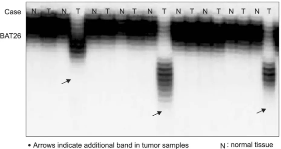
60oC에서 primer annealing, 15초간 72oC에서 elongation 의 cycle을 25∼30 cycle을 시행하였다. PCR product는 5.6 M urea를 함유한 6% polyacrylamide gels에서 분리하 였고 autoradiography를 시행하였다. MSI는 normal allele 부위에서 부가적인 band가 관찰되었다.
2) 분석 방법
2개 이상 marker의 양성인 경우를 MSI (microsatellite
Fig. 1. Example of MSI analysis at BAT26.
instability)로 하였고 1개 marker가 양성인 경우와 전부 음성인 경우는 MSS (microsatellite stability)로 분류하 였다. MSI 종양은 normal allele 부위에 추가 band가 나 타났다(Fig. 1).
임상병리학적 특징의 비교는 두 군 간의 chi-square test를 시행하였고 생존율 분석은 Kaplan-Meier 방법으로 하였고 생존율의 차이는 Log-rank test로 검증하였다.
결 과
1) 전체 107예 중 MSI는 16예로 15%에서 관찰되었다.
MSI 및 MSS의 평균 연령은 각각 51.8세, 58.6세였다 (P=0.03). 특히 40세 미만의 젊은 연령의 분포는 각각 5 예(31.3%), 6예(6.6%)로 MSI에서 40세 미만의 환자 분포 가 많았다(P<0.01). 남녀의 비는 MSI에서 10:6, MSS에 서 55:36으로 차이가 없었다. 암의 발생 부위는 우측
결장이 MSI에서 12예(75%), MSS에서 12예(13%)로 MSI 에서 우측 대장에 호발하였고 MSS의 경우는 좌측 결장 및 직장에 많았다(P<0.01)(Table 1).
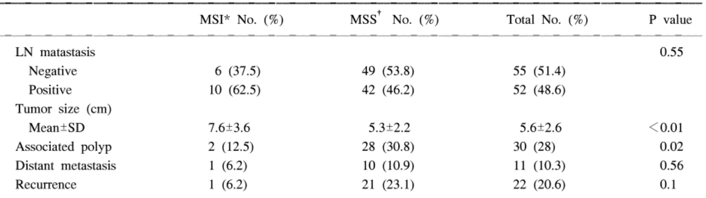
2) 림프절 전이가 없는 경우는 MSI, MSS 각각 6예 (37.5%), 49예(53.8%)이었고 림프절 전이가 있는 경우 는 각각 10예(62.5%), 42예(46.2%)로 차이가 없었다. 종 양의 평균 크기는 MSI에서 7.6 cm, MSS에서 5.3 cm 로 통계적으로 의의있게 차이가 있었다(P<0.01). 용종 이 동반된 경우는 MSI에서 2예(12.5%), MSS에서 28예 (30.8%)로 통계적으로 유의한 차이가 있었다(P=0.02). 원 격 전이는 MSI에서 1예(6%), MSS에서 10예(10.9%), 재발은 MSI에서 1예(6.2%), MSS에서 21예(23.1%)이었 다. 통계적으로 의의있는 차이는 없었다(Table 2).
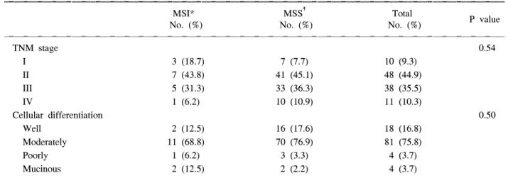
3) TNM 병기에 따른 분포는 1기에서 MSI 3예 (18.7%), MSS 7예(7.7%), 2기에서 MSI 7예(43.8%), MSS 41예(45.1%), 3기에서 MSI 5예(31.3%), MSS 33예
Table 2. Tumor characteristics and recurrence between MSI and MSS
ꠚꠚꠚꠚꠚꠚꠚꠚꠚꠚꠚꠚꠚꠚꠚꠚꠚꠚꠚꠚꠚꠚꠚꠚꠚꠚꠚꠚꠚꠚꠚꠚꠚꠚꠚꠚꠚꠚꠚꠚꠚꠚꠚꠚꠚꠚꠚꠚꠚꠚꠚꠚꠚꠚꠚꠚꠚꠚꠚꠚꠚꠚꠚꠚꠚꠚꠚꠚꠚꠚꠚꠚꠚꠚꠚꠚꠚꠚꠚꠚꠚꠚꠚꠚꠚꠚꠚꠚꠚꠚꠚꠚꠚꠚꠚꠚꠚꠚꠚꠚꠚꠚꠚꠚ
MSI* No. (%) MSS† No. (%) Total No. (%) P value
ꠏꠏꠏꠏꠏꠏꠏꠏꠏꠏꠏꠏꠏꠏꠏꠏꠏꠏꠏꠏꠏꠏꠏꠏꠏꠏꠏꠏꠏꠏꠏꠏꠏꠏꠏꠏꠏꠏꠏꠏꠏꠏꠏꠏꠏꠏꠏꠏꠏꠏꠏꠏꠏꠏꠏꠏꠏꠏꠏꠏꠏꠏꠏꠏꠏꠏꠏꠏꠏꠏꠏꠏꠏꠏꠏꠏꠏꠏꠏꠏꠏꠏꠏꠏꠏꠏꠏꠏꠏꠏꠏꠏꠏꠏꠏꠏꠏꠏꠏꠏꠏꠏꠏꠏ
LN matastasis 0.55
Negative 6 (37.5) 49 (53.8) 55 (51.4)
Positive 10 (62.5) 42 (46.2) 52 (48.6)
Tumor size (cm)
Mean±SD 7.6±3.6 5.3±2.2 5.6±2.6 <0.01
Associated polyp 2 (12.5) 28 (30.8) 30 (28) 0.02
Distant metastasis 1 (6.2) 10 (10.9) 11 (10.3) 0.56
Recurrence 1 (6.2) 21 (23.1) 22 (20.6) 0.1
ꠏꠏꠏꠏꠏꠏꠏꠏꠏꠏꠏꠏꠏꠏꠏꠏꠏꠏꠏꠏꠏꠏꠏꠏꠏꠏꠏꠏꠏꠏꠏꠏꠏꠏꠏꠏꠏꠏꠏꠏꠏꠏꠏꠏꠏꠏꠏꠏꠏꠏꠏꠏꠏꠏꠏꠏꠏꠏꠏꠏꠏꠏꠏꠏꠏꠏꠏꠏꠏꠏꠏꠏꠏꠏꠏꠏꠏꠏꠏꠏꠏꠏꠏꠏꠏꠏꠏꠏꠏꠏꠏꠏꠏꠏꠏꠏꠏꠏꠏꠏꠏꠏꠏꠏ
*MSI = Microsatellite Instability; †MSS = Microsatellite Stability.
Table 1. Age, sex and location of tumor between MSI and MSS
ꠚꠚꠚꠚꠚꠚꠚꠚꠚꠚꠚꠚꠚꠚꠚꠚꠚꠚꠚꠚꠚꠚꠚꠚꠚꠚꠚꠚꠚꠚꠚꠚꠚꠚꠚꠚꠚꠚꠚꠚꠚꠚꠚꠚꠚꠚꠚꠚꠚꠚꠚꠚꠚꠚꠚꠚꠚꠚꠚꠚꠚꠚꠚꠚꠚꠚꠚꠚꠚꠚꠚꠚꠚꠚꠚꠚꠚꠚꠚꠚꠚꠚꠚꠚꠚꠚꠚꠚꠚꠚꠚꠚꠚꠚꠚꠚꠚꠚꠚꠚꠚꠚꠚ
MSI* MSS† Total
No. (%) No. (%) No. (%) P value
16 (15) 91 (85) 107 (100)
ꠏꠏꠏꠏꠏꠏꠏꠏꠏꠏꠏꠏꠏꠏꠏꠏꠏꠏꠏꠏꠏꠏꠏꠏꠏꠏꠏꠏꠏꠏꠏꠏꠏꠏꠏꠏꠏꠏꠏꠏꠏꠏꠏꠏꠏꠏꠏꠏꠏꠏꠏꠏꠏꠏꠏꠏꠏꠏꠏꠏꠏꠏꠏꠏꠏꠏꠏꠏꠏꠏꠏꠏꠏꠏꠏꠏꠏꠏꠏꠏꠏꠏꠏꠏꠏꠏꠏꠏꠏꠏꠏꠏꠏꠏꠏꠏꠏꠏꠏꠏꠏꠏꠏ Age (year)
Mean±SD‡ 51.8±12.4 58.8±1.7 57.7±12.0 0.03
<40 year old 5 (31.3) 6 (6.6) 11 (10.3) <0.01
Sex
M:F 10:6 55:36 65:42 0.87
Location
Right 12 (75) 12 (13.2) 24 (22.4) <0.01
Left 4 (25) 41 (45.1) 46 (43)
Rectum 0 (0) 38 (41.8) 38 (35.6)
ꠏꠏꠏꠏꠏꠏꠏꠏꠏꠏꠏꠏꠏꠏꠏꠏꠏꠏꠏꠏꠏꠏꠏꠏꠏꠏꠏꠏꠏꠏꠏꠏꠏꠏꠏꠏꠏꠏꠏꠏꠏꠏꠏꠏꠏꠏꠏꠏꠏꠏꠏꠏꠏꠏꠏꠏꠏꠏꠏꠏꠏꠏꠏꠏꠏꠏꠏꠏꠏꠏꠏꠏꠏꠏꠏꠏꠏꠏꠏꠏꠏꠏꠏꠏꠏꠏꠏꠏꠏꠏꠏꠏꠏꠏꠏꠏꠏꠏꠏꠏꠏꠏꠏꠏ
*MSI = Microsatellite Instability; †MSS = Microsatellite Stability; ‡SD = standard deviation.
(36.3%), 4기에서 MSI 1예(6.2%), MSS 10예(10.9%) 이 었다. 양 군 간에 통계적으로 유의한 차이는 없었다 (P=0.54).
세포 분화도에 따른 분포는 고분화가 MSI 2예 (12.5%), MSS 16예(17.6%), 중등도 분화가 MSI 11예 (68.8%), MSS 70예(76.9%), 미분화가 MSI 1예(6.2%), MSS 3예(3.3%)이었고 점액성 암이 MSI 2예(12.5%), MSS 2예(2.2%)이었다(Table 3).
4) 전체 환자의 5년 생존율은 MSI 93.8%, MSS 73.8% (P=0.07)이었고 2기 및 3기 암만을 대상으로 분 석 하였을 때 MSI 100%, MSS는 76.5% (P=0.06)이었다 (Fig. 2).
고 찰
현미부수체 불안정성(MSI)은 DNA 부정합 교정 유 전자(hMSH2, hMLH1, hMSH6, hPMS1, hPMS2)의 돌연 변이로 인해 짧고 나란히 반복되는 DNA sequences의 길이 변화(length variation)가 초래되어 나타나는 것이 다. DNA 부정합 교정 유전자는 부정합된 DNA se- quences를 잡아내어 교정하는 역할을 하는데 이 기전 이 상실되면 세포 분열 시 많은 곳에서 염기의 삽입, 결손, 대치가 생기게 되며 MSI를 관찰할 수 있게 된다.
반복되는 현미부수체(repeatitive microsatellite sequen- ces)를 PCR 증폭시켜 이 부분에 현미 부수체의 길이 변화(length mutation in oligonucleotide repeats)가 나타 나는 것을 검색하는데 이 현미 부수체 불안전성에 대 한 의견 차이가 있다가 Boland 등이 international con-
sensus meeting을 통해 guideline을 만들었다.7,8 이러한 변화는 처음에 유전성 비용종성 대장암에서 주로 관 찰되었는데3 나중에는 산발성 대장암에서도 관찰된다 고 하였다.4 이 MSI 종양은 특징적으로 선종-암 연속 (adenoma-carcinoma sequence)에서 나타나는 염색체 불 안정성(chromosome instability) 즉 APC, p53, DCC, K-ras 유전자의 돌연변이는 보이지 않고 TGF-β 수용 체 돌연변이 등만이 관찰이 된다.9-11 따라서 이 특징적 인 암은 그 발생기전이 다르다고 추측하고 있다. 근래 에 대장암의 예후에 관계되는 분자 생물학적 표지자 의 연구가 많이 진행되면서 chromosome 18q DCC에 loss of heterozygosity가 2기, 3기 암에서 나타나면 예후 가 좋지 않다는 보고가 있었고,5,12 이외 p53 gene, chro- mosome 17p의 allelic loss, p21 waf1/cip1, TGF-β1 type Table 3. Cellular differentiation and TNM stage between MSI* and MSS†
ꠚꠚꠚꠚꠚꠚꠚꠚꠚꠚꠚꠚꠚꠚꠚꠚꠚꠚꠚꠚꠚꠚꠚꠚꠚꠚꠚꠚꠚꠚꠚꠚꠚꠚꠚꠚꠚꠚꠚꠚꠚꠚꠚꠚꠚꠚꠚꠚꠚꠚꠚꠚꠚꠚꠚꠚꠚꠚꠚꠚꠚꠚꠚꠚꠚꠚꠚꠚꠚꠚꠚꠚꠚꠚꠚꠚꠚꠚꠚꠚꠚꠚꠚꠚꠚꠚꠚꠚꠚꠚꠚꠚꠚꠚꠚꠚꠚꠚꠚꠚꠚꠚꠚꠚ
MSI* MSS† Total
P value
No. (%) No. (%) No. (%)
ꠏꠏꠏꠏꠏꠏꠏꠏꠏꠏꠏꠏꠏꠏꠏꠏꠏꠏꠏꠏꠏꠏꠏꠏꠏꠏꠏꠏꠏꠏꠏꠏꠏꠏꠏꠏꠏꠏꠏꠏꠏꠏꠏꠏꠏꠏꠏꠏꠏꠏꠏꠏꠏꠏꠏꠏꠏꠏꠏꠏꠏꠏꠏꠏꠏꠏꠏꠏꠏꠏꠏꠏꠏꠏꠏꠏꠏꠏꠏꠏꠏꠏꠏꠏꠏꠏꠏꠏꠏꠏꠏꠏꠏꠏꠏꠏꠏꠏꠏꠏꠏꠏꠏꠏ
TNM stage 0.54
I 3 (18.7) 7 (7.7) 10 (9.3)
II 7 (43.8) 41 (45.1) 48 (44.9)
III 5 (31.3) 33 (36.3) 38 (35.5)
IV 1 (6.2) 10 (10.9) 11 (10.3)
Cellular differentiation 0.50
Well 2 (12.5) 16 (17.6) 18 (16.8)
Moderately 11 (68.8) 70 (76.9) 81 (75.8)
Poorly 1 (6.2) 3 (3.3) 4 (3.7)
Mucinous 2 (12.5) 2 (2.2) 4 (3.7)
ꠏꠏꠏꠏꠏꠏꠏꠏꠏꠏꠏꠏꠏꠏꠏꠏꠏꠏꠏꠏꠏꠏꠏꠏꠏꠏꠏꠏꠏꠏꠏꠏꠏꠏꠏꠏꠏꠏꠏꠏꠏꠏꠏꠏꠏꠏꠏꠏꠏꠏꠏꠏꠏꠏꠏꠏꠏꠏꠏꠏꠏꠏꠏꠏꠏꠏꠏꠏꠏꠏꠏꠏꠏꠏꠏꠏꠏꠏꠏꠏꠏꠏꠏꠏꠏꠏꠏꠏꠏꠏꠏꠏꠏꠏꠏꠏꠏꠏꠏꠏꠏꠏꠏꠏ
* MSI = Microsatellite Instability; †MSS: Microsatellite Stability.
Fig. 2. Overall 5 year survival rate: MSI vs. MSS.
II receptor 등13,14이 분자 생물학적 예후인자로써 의미 있다는 보고가 있다. 예후인자로써 MSI 종양은 예후 가 좋고 약물치료 후 생존율도 높다는 보고가 있
다.4,15-22 산발성 대장 및 직장암에서 현미부수체 불안
전성이 관찰되는 빈도는 저자에 따라 15∼17%로 보고 되고 있다.3,4,16 MSI 종양은 문헌에 따라 다소 차이는 있지만 대개 우측 결장에 호발하며 세포 분화가 나쁘 고, 보다 젊은 연령층에 많고, 종양이 바깥으로 자라고 (exophytic), 종양의 크기가 크고, 미분화 세포 유형을 보이며, 점액형성이 특징이다. 아울러 크론병 때 나타 나는 림프구 침윤이 관찰되는 것이 특징이라고 하였 다.4,15 또한 MSI 상태는 이시성으로 다발성 대장암의 발생을 예견할 수도 있다고 하였다. 즉 Masubuchi 등 은 산발성 이시성 다발성 대장암에서 MSI의 소견이 89%가 양성인데 비하여 단독 대장암(single colorectal cancer)에서는 14%밖에 나오지 않아 이시성 다발성 대 장암 발견을 예측할 수 있다고 하였다.23 이외 Senguta 등24도 다발성 산발성 대장암인 경우 단독 대장암보다 MSI가 보다 높게 나타난다고 하였다. 저자들도 MSI 종 양에서 보이는 특징적인 소견을 관찰할 수 있었다. 본 연구에서도 우측 대장에 월등히 많고 직장은 없었으며 연령도 40세 미만이 보다 많았다. 종양의 평균크기도 MSI에서 보다 의의있게 컸으나 세포 분화도나 병기의 차이는 관찰할 수 없었다. Gryfee 등21은 607명 대장암 환자에서 17%에서 MSI 양성을 보고하였고 이것이 생존 율에 독립적인 영향을 미친다고 보고하였다. Aaltonen 등3은 12%에서 MSI을 보고하였고 암의 침윤 정도(T stage)에 관계없이 MSI 종양은 주위 림프절과 원격장 기 전이를 하지 않는다고 보고하였다. 저자들의 분석 결과 MSI 는 107명 대장암 중에서 16명(15%)에서 관 찰되었고 MSI 종양과 MSS 종양 사이에 림프절 전이 나 원격 전이에 대한 차이는 관찰되지 않았다. MSI high가 예후가 좋다는 보고가 많은데 Watanabe 등5은 5 FU 보조적 항암약물치료를 받은 대장암 환자의 약 20%에서 MSI high가 관찰되었다고 보고하였고 기존 의 알려진 여러 분자생물학적 표지자와 함께 MSI의 예후인자로써의 의의를 연구하였는데 3기 대장암에서 18q allelic loss가 있는 경우의 5년 생존율이 50%, 없는 경우가 69%로 의의있게 나왔으며, 특히 MSS 대장암 에서 18q allelic loss가 예후와 관계 있다고 하였다.
MSI 종양에서는 TGF-β1 type II receptor 변이가 관찰 되는 경우가 없는 경우보다 예후가 월등히 좋은 것으 로 보고하였다.5 그러나 MSI 상태가 생존율과 관계가 없다고 하는 저자들도 있어25-27 MSI 종양이 예후가 좋
다는 것은 아직 논란이 있다. 따라서 미국 국립 암연구 소 workshop에서도 MSI가 독립적인 예후인자라고 확 실히 결론을 내리고있지는 못하고 있으며 이 workshop 에서 5개의 현미부수체 표지자(marker)와 MSI의 high, low, MSS에 대한 정의를 하였다.8 요약하면 최소한 검 사해야 할 5개 MSI markers로 검사 결과 2개 이상 양성 이면 MSI high, 1개 양성이면 MSI low, 모두 음성이면 MSS로 분류할 것을 권하였다. MSI high가 좋은 예후 를 보이는 기전으로 설명하는 것은 APC, p53 유전자 의 돌연변이는 드물고 β-catenin (CTNNB1), trans- forming growth factor β receptor type II의 돌연변이가 많기 때문이라고 한다.9-11 이외 종양 주위에 림프구 침 윤이 많다는 사실이 예후가 보다 좋은 이유라고 설명 하였다.6 Lukish 등20은 40세 미만의 대장암 환자를 대 상으로 MSI를 조사한 결과 17명(47%)에서 발견되었고 5년 생존율이 MSI에서 68%, MSS 에서 32%로 MSI 종 양이 의의있게 높았다고 보고하였다. Halling 등17은 2 기 및 3기 대장암 508명을 대상으로 MSI, chromosome 8p allelic loss 조사에서 MSI 종양의 예후가 보다 좋았 고 재발까지의 시간도 길었다고 하였고, 8p allelic imbalance가 보이는 대장암은 예후가 나쁘다고 하였 다. Jernvall 등19은 255명의 대장암 환자를 대상으로 MSI를 조사한 결과 28명(12%)에서 나타났고, 대개 근 위부 대장암, 미분화, 종양의 크기가 컸으며 5년 생존 율이 MSI 종양은 100%, MSS 종양은 74%라고 보고하 였다. 그러나 원위부 대장 및 직장에서는 MSI 5년 생 존율이 21%, MSS 57%로 반대의 결과가 초래되었다.
따라서 직장암에서는 이런 원칙을 적용하지 말고 근 위부 대장에서만 적용 시 보다 의의 있는 예후 인자가 될 것이라고 주장하였다. MSI의 특징적 소견에 대해 서는 Wullenweber 등28이 MSI를 미국 국립 암 연구소 guidelines을 적용하여 연구하였는데 이들은 잘 알려진 MSI 종양 연구서 지침에 따라 조사한 결과 앞서 거론 된 임상적 특징이 잘 안 나타난다고 하였다. 즉 미분화 암인 경우 연령 제한이 없으면 MSI와 관련이 없고 대 장암의 근위부 위치의 경향은 연령 제한 없이도 MSI 와 관련이 있었다고 하였다. MSI 산발성 대장암의 예 후는 저자의 경우 증례가 적어 통계적으로 의의있는 5년 생존율의 차이를 관찰하지는 못하였지만 증례가 많아지면 다른 보고들처럼 의의있는 예후의 차이가 관찰되리라 생각한다. 그러나 MSI 종양의 예후는 병 기에 관계없이 93.8%로 월등히 좋은 결과를 보였다.
산발성 대장암에서 MSI 종양의 특징이 뚜렷하여 암의 발생에 관련된 기전 및 유전자의 이상도 다르리라고
추측이 된다. 산발성 대장암에서 MSI 분자 표지자의 임상적 의의는 현재까지의 연구 결과를 종합하면 향 후 다른 대장부위에 이시성 암이 발생할 확률도 높다 는 것을 예측할 수도 있고23,24 예후를 예측할 수 있는 의의 있는 분자 생물학적 표지자로써 사용 가능하다는
것이다.26,27
결 론
107명의 결장직장암의 신선조직을 대상으로 미국 국립 암연구소에서 추천한 5개 현미부수체 표지자로 검사한 결과 16명(15%)에서 MSI로 나왔다. MSI 대장 암은 특징적으로 종양의 크기가 크고, 40세 미만에서 많았으며 우측 대장에 월등히 많이 분포하였다. 5년 생존율도 MSS 종양보다 통계적으로 의의는 없지만 좋은 경향을 보였다. MSI 대장암은 특징적인 임상 및 병리학적 특징이 관찰되어 그 발생기전도 기존의 알 려진 것과 다를 것으로 추측이 되고 치료 후 예후인자 로써 의의는 향후 연구가 필요하다고 생각한다.
REFERENCES
1. Risinger JI, Berchuck A, Kohler MF, Watson P, Lynch HT, Boyd J. Genetic instability of microsatellites in endometrial carcinoma. Cancer Res 1993;53:5100-3.
2. Han HJ, Yanagisawa A, Kato Y, Park JG, Nakamura Y.
Genetic instability in pancreatic cancer and poorly dif- ferentiated type of gastric cancer. Cancer Res 1993;53:
5087-9.
3. Aaltonen LA, Peltomaki P, MecklinJP, Jarvinen H, Jass JR, Green JS, et al. A replication errors in benign and malignant tumors from hereditary nonpolyposis colorectal cancer patients. Cancer Res 1994;54:1645-8.
4. Thibodeau SN, Bren G, Schaid D. Microsatellite instability in cancer of the proximal colon. Science 1993;260:816-9.
5. Watanabe T, Wu TT, Catalano PJ, Ueki T, Satriano R, Haller DG, et al. Molecular predictors of survival after adjuvant chemotherapy for colon cancer. N Eng J Med 2001;344:1196-206.
6. Kim H, Jen J, Vogelstein B, Hamilton SR. Clinical and pathogical characteristics of sporadic colorectal cancer with DNA replication errors in microsatellite sequences. Am J Path 1994;145:148-56.
7. Rodriguez-Bigas MA, Boland CR, Hamilton SR. A National Cancer Institute Workshop on hereditary nonpolyposis co- lorectal cancer syndrome: meeting highlights and Bethesda guidelines. J Natl Cancer Inst 1997;89:1758-62.
8. Boland CR, Thibodeau SN, Hamilton SR, SIdransky D,
Eshleman JR, Burt RW, et al. National Cancer Institute Workshop on microsatellite instability for cancer detection and familial predisposition: development of international criteria for the determination of microsatellite instability in colorectal cancer. Cancer Res 1998;58;5248-57.
9. Morin PJ, Sparks AB, Korinek V, Barker N, Clever H, Vogelstein B, et al. Activation of beta-catenin Tcf signaling in colon cancer by mutations in β catenin or APC. Science 1997;275:1787-90.
10. Sparks AB, Morin PJ, Vogelstein B, Kinzler KW. Mu- tational analysis of the APC/beta-catenin/Tcf pathway in colorectal cancer. Cancer Res 1998;58:1130-42.
11. Markowitz S, Wang J, Myeroff L, Parsons R, Sun L, Lutter-baugh J, et al. Inactivation of the type II TGF-beta receptor in colon cancer cells with microsatellite instability.
Science 1995;268:1336-8.
12. Jen J, Kim H, Piantadosi S, Lin ZF, Levitt RC, Sistonen P, et al. Allelic loss of chromosome 18q and prognosis in colorectal cancer. N Eng J Med 1994;331:213-21.
13. Cheung JD, Werness BA, Meropol NJ. Paradoxical cor- relations of cyclin-dependent kinase inhibitors p21 waf1/cip1 and p27 kip1 in metastatic colorectal carcinoma. Clin Cancer Res 1999;5:1057-62.
14. Blobe GC, Schiemann WP, Lodish HF. Role of transforming growth factor β in human disease. N Engl J Med 2000;
342:1350-8.
15. Ionov Y, Peinado MA, Malkhosyan S, Shibata D, Perucho M. Ubiquitous somatic mutations in simple repeated se- quences reveal a new mechanism for colonic carcinogenesis.
Nature 1993;363:558-61.
16. Lothe RA, Peltomaki P, Meling GI, Aaltonen LA, Nystrom- Lahti M, Pylkkanen L, et al. Genomic instability in co- lorectal cancer: relationship to clinicopathological variables and family history. Cancer Res 1993;53:5849-52.
17. Halling KC, French AJ, McDonnell SK, Burgart LJ, Schaid DJ, Peterson BJ, et al. Microsatellite instability and 8p allelic imbalance in stage B2 and C colorectal cancers. J Natl Cancer Inst 1999;91:1295-303.
18. Johannsdottir JT, Bergthorsson JT, Gretarsdottir S, Krist- jansson AK, Ragnarsson G, Jonasson JG, et al. Replication error in colorectal carcinoma: association with loss of heterozygosity at mismatch repair loci and clinicopathologic variables. Anticancer Res 1999;19:1821-6.
19. Jernvall P, Makinen MJ, Karttunen TJ, Makela J, Vihko P.
Microsatellite instability: impact on cancer progression in proximal and distal colorectal cancer. Eur J Cancer 1999;
35:197-201.
20. Lukish JR, Muro K, DeNobile J, Katz R, Williams J, Cruess DF, et al. Prognostic significance of DNA replication errors in young patients with colorectal cancer. Ann Surg 1998;
227:51-6.
21. Gryfe R, Kim H, Hsiesh ETK, Aronson MD, Holowaty EJ,
Bull SB, et al. Tumor microsatellite instability and clinical outcome in young patients with colorectal cancer. N Engl J Med 2000;342:69-77.
22. Elsaleh H, Joseph D, Grieu F, Seps N, Spry N, Iacopetta B. Association of tumor site and sex with survival benefit from adjuvant chemotherapy in colorectal cancer. Lancet 2000;355:1745-50.
23. Masubuchi S, Konishi F, Togashi K, Okamoto T, Senba S, Shitoh K, et al. The significance of microsatellite instability in predicting the development of metachronous multiple colorectal carcinomas in patients with nonfamilial colorectal carcinoma. Cancer 1999;85:1917-24.
24. Sengupta SB, Yu CY, Boulos PB, Silva MD, Sams VR, Delhanty JDA. Genetic instability in patients with meta- chronous colorectal cancers. Br J Surg 1997;84:996-1000.
25. Ko LM, Cheung MH, Kwan MW, Wong CM, Lau KW,
Tang CM, et al. Genomic instability and alerations in APC, MCC and DCC in Hong Kong patients with colorectal cancer. Int J Cancer 1999;84:404-9.
26. Messerini L, Ciantelli M, Baglioni S, Palomba A, Zampi G, Papi L. Prognostic significance of microsatellite instability in sporadic mucinous colorectal cancer. Human Pathol 1999;30:629-34.
27. Salahshor S, Kressner U, Fisher H, Lindmark G, Glimelius B, Pahlman L, et al. Microsatellite instability in sporadic colorectal cancer is not an independent prognostic factor. Br J Cancer 1999;81:190-3.
28. Wullenweber HP, Sutter C, Autschbach F, Willeke F, Kienle P, Benner A, et al. Evaluation of Bethesda Guidelines in relation to microsatellite Instability. Dis Colon Rectum 2001;44:1281-9.