Received June 23, 2008, Accepted for publication August 25, 2008 Reprint request to: Kyu Kwang Whang, M.D., Serion Dermatology Clinic, 241-1, Nonhyeon-dong, Gangnam-gu, Seoul 135-830, Korea.
Tel: 82-2-515-5158, Fax: 82-2-757-5630, E-mail: skinewkk@hotmail.
com
ORIGINAL ARTICLE
A Combination of Dual-mode 2,940 nm Er:YAG Laser Ablation with Surgical Excision for Treating Medium- sized Congenital Melanocytic Nevus
Ji Yeon Lim, M.D., Yun Jeong, M.D.1, Kyu Kwang Whang, M.D.2
Department of Dermatology, School of Medicine, Ewha Womans University, Seoul, 1Maryknoll Hospital, Busan,
2Serion Dermatology Clinic, Seoul, Korea
Background: There are various treatment options for congenital melanocytic nevus (CMN), including surgical excision, dermabrasions, curettage, laser treatment, chemi- cal peels and cryosurgery. The proper choice of treatment depends on the size, location, thickness and clinical ap- pearance of the nevi, the risk for developing melanoma, the psychological effect and the cosmetic component.
Objective: The purpose of this study is to evaluate the outcome of a combination of surgical excision with Er: YAG laser ablation for treating CMNs. Methods: A total of 13 patients were included in this study. The nevus was excised as much as possible and only dermal suturing was performed, without epidermal suturing, for the primary closure. We then ablated the whole lesion, including the suture lines, by using a dual-mode 2,940 nm Er:YAG laser with three to five passes. All the lesions were followed up for 6 months and they were evaluated with respect to the healing status, infection, erythema, scarring, textural change and pigmentary change. Subject satisfaction was scored at the 16th week by the patients. Results: Eleven (83%) of the 13 patients were clinically rated as having a good to excellent result by the physicians’ Global Assessment Scale (GAS) scores for the lesions’ reduction of size, the degree of scarring and the pigmentary change with only a one stage procedure.
10 (77%) of the total 13 patients reported a good to excellent result at four months after treatment. Conclusion: A combination of surgical excision with Er:YAG laser ablation as a one stage procedure is a safe, effective modality and it should be considered as one of the options for treating
medium-sized CMNs. (Ann Dermatol 21(2) 120∼124, 2009)
-Keywords-
2,940 nm Er:YAG, Combined treatment, Congenital mela- nocytic nevus, Laser ablation, Surgical excision
INTRODUCTION
Congenital melanocytic nevus (CMN) is defined as a tissue malformation of the neuroectoderm that presents at birth or it appears within the first few months of life1,2. CMNs are usually classified according to their current size or the size that the CMN is predicted to attain at adulthood:
small (less than 1.5 cm in diameter), medium (1.5∼20 cm) and large (more than 20 cm)3. The classification system is important because it has an effect on the management of CMNs. In addition to size, several factors that are considered for the clinical management of CMN include the location, thickness, clinical appearance, the risk for developing melanoma, the psychological effect and the cosmetic component4. Thus, the management of CMNs needs to be individualized for each patient5. There are many treatment options for CMNs: surgical excision, dermabrasions, curettage, laser treatment, chemical peels and cryosurgery1,6. Surgical excision is the most common- ly used method because this completely removes the nevus cells. However, it requires two or more staged procedures in many cases, and physicians should consider the complications according to surgery, such as a post- operative long linear scar. In this background, lasers are now being increasingly used for treating large lesions and the CMNs located on cosmetically sensitive areas. But
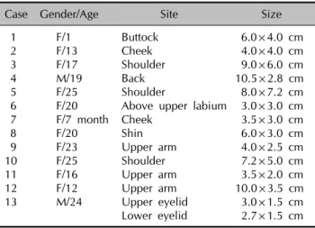
Table 1. Demographic data of the patients treated with a combination of surgical excision and Er:YAG laser ablation Case Gender/Age Site Size
1 F/1 Buttock 6.0×4.0 cm
2 F/13 Cheek 4.0×4.0 cm
3 F/17 Shoulder 9.0×6.0 cm
4 M/19 Back 10.5×2.8 cm
5 F/25 Shoulder 8.0×7.2 cm
6 F/20 Above upper labium 3.0×3.0 cm
7 F/7 month Cheek 3.5×3.0 cm
8 F/20 Shin 6.0×3.0 cm
9 F/23 Upper arm 4.0×2.5 cm
10 F/25 Shoulder 7.2×5.0 cm
11 F/16 Upper arm 3.5×2.0 cm
12 F/12 Upper arm 10.0×3.5 cm
13 M/24 Upper eyelid 3.0×1.5 cm
Lower eyelid 2.7×1.5 cm lasers are effective only for removing nevus cells of the upper dermis and they can not remove the deeper portion of the nevus cells. In addition, laser therapy requires multiple treatment sessions. Therefore, we introduce here a new method to treat medium-sized CMNs with the combination of surgical excision and Er:YAG laser abla- tion.
MATERIALS AND METHODS
Patients
Since 2004, we have treated 13 patients using a com- bination of surgical excision and Er:YAG laser ablation.
Informed consent was obtained from all the patients. The patients were two males and eleven females aged 7 months to 25 years. The CMN was located on the extre- mities in seven subjects, on the trunk in two subjects and on the face in four subjects (Table 1). All the patients had Fitzpatrick’s skin types III and IV.
Methods
First, we excised the nevus as much as possible, including the deeply pigmented or elevated portion, and especially the hair-bearing area, after administering local anesthesia (2% lidocaine with 1:80,000 epinephrine). Various de- signs of excision, including an elliptical shape, were used according to the size and location of the nevus. Second, only dermal suturing using vircryl was performed after the surgical dissection for achieving tension relief of the skin without suturing the epidermis. Third, we ablated the re- sidual pigmented areas, including the suture lines, with using a dual-mode 2,940 nm Er:YAG laser (Contour, Sciton, Inc., Palo Alto, CA, USA). On the average, three to five passes of ablation were done in 90∼100 μm ablation
mode and using a 4 mm laser beam spot at 25.0 J/cm2. Ablation was performed until the visible pigmented part was diminished and an even wound surface was created.
After the laser treatment, the wound was dressed with a chlorhexidine paraffin gauze-based dressing material (Bac- tigras, Smith & Nephew Co., Hull, UK) and topical anti- biotic ointment. The occlusive dressing was changed with using hydrocolloid dressing material at an interval of three days for two weeks until the re-epithelialization was completed.
Follow-up
All the lesions were followed up for 6 months. At each visit, the treated areas were evaluated with respect to the healing status, infection, postoperative hyperpigmentation and hypopigmentation, erythema, scarring and repigmen- tation. Photographs were taken prior to treatment and at each visit by using a Canon EOS 300 D digital camera (Canon Inc., Tokyo, Japan). The Global Assessment Scale (GAS) scores were checked to assess the overall results and the degree of the patient and physician satisfaction with the therapy at the 16th week according to the follow- ing scale: excellent, good, fair and poor. The postopera- tive changes, including erythema, pigmentary change and hypertrophic scarring, were also estimated by the physi- cians who were involved in the treatment; the postopera- tive changes were rated according to the following scale:
none, mild, moderate and severe.
RESULTS
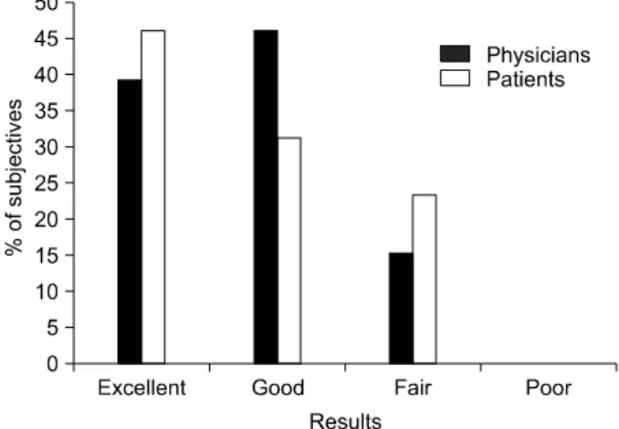
The treatment was well tolerated by all the patients and no major complications occurred. Re-epithelialization was complete within 10∼15 days in most cases. All the patients showed good to excellent results for a one-stage procedure (Fig. 1). With respect to the investigators’ GAS scores, five patients (37%) were rated as excellent and six patients (46%) were rated as good. Especially for the cosmetic outcome, this combination method showed excellent results and this was confirmed by the subjects’
GAS scores, which were checked at the 16th week follow up exam (Fig. 2). For the GAS scores by the patients, six patients (46%) were rated as excellent and four patients (31%) were rated as good. Infections did not occur in any of the patients. Erythema was observed in all patients, but this was rated as mild to moderate in 11 (83.6%) of 13 lesions and it gradually subsided. Pigmentary changes, including postoperative hyperpigmentation, hypopigmen- tation and repigmentation, were observed in 11 patients, and six patients (46.2%) showed mild changes and three patients (23.0%) showed moderate changes. Two patients
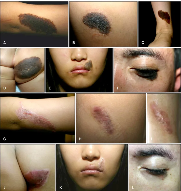
Fig. 1. Congenital melanocytic nevi before (A∼F) and after treatment (G∼L) with a combination of surgical excision and Er:YAG laser ablation (A, G, excellent/excellent; B, H, excellent/excellent; C, I, excellent/excellent; D, J, good/excellent; E, K, excellent/excellent;
F, L, excellent/excellent. Global Assessment Scale (GAS) scores by the physicians/GAS scores by the patients).
showed a severe degree of repigmentation and we further treated these lesions with pigment-specific Q-switched Nd:YAG lasers (Medlite IV, Medlite, USA). Then the pig- mentation became decreased. Three patients showed no signs of hypertrophic scarring, but most of the patients (61.6%) showed a mild degree of hypertrophic changes (Fig. 3). All the lesions were followed up for 6 months.
Although our cases showed excellent clinical results,
long-term follow up will be needed for assess the lesions for recurrence of the nevus or delayed scarring.
DISCUSSION
The management of CMN is a challenge to physicians because there are several factors to consider for deciding on the proper treatment modality, including the patient’s
Fig. 2. Global Assessment Scale (GAS) scores by the physicians
and patients at 16 weeks after treatment. Fig. 3. Assessment by physicians for erythema, the pigmentary changes and hypertrophic scarring at 16 weeks after treatment.
age and the lesion’s size, location, depth, shape and malignant potential. But no specific guidelines have been established for treating CMNs5. There are many treatment options for CMNs: surgical excision, dermabrasions, curet- tage, laser treatment, chemical peels and cryosurgery1,6,7. To decide on the modality of treatment, we should consi- der two main issues8. One is the potential for malignant transformation3,9,10 and the other is the cosmetic compon- ent, and particularly for large-sized CMNs or those CMNs located on cosmetically complex areas, including the face and genital area6,11. Surgical excision is the most com- monly used method and it requires full reconstructive me- thods such as direct excision with primary closure, including serial excision, skin grafting, tissue expansion, free tissue transfer etc1,12,13. Complete and full-thickness excision is an ideal method in terms of preventing the development of melanoma and for cosmetic reasons. But in most cases, except for the small CMNs, serial excision or tissue expansion will be needed so that multiple procedures are required for complete removal. Also, crea- ting a long linear scar and scar widening due to tension should be considered and there are many complications after tissue expansion, including deflation or disruption of the expanders, hematoma, severe infection and flap ische- mia1,5,12. On the other hand, laser treatment is useful for large-sized CMNs, it is effective for cosmetically sensitive areas and it is safe for children14. The lasers used for the treatment of CMN include pigment-specific lasers15-19, resurfacing lasers20-23 and a combination of the two14,17,24,25. In particular, the Er:YAG laser is precise and it can be used for controlled resurfacing. It causes significantly less deep thermal injury, there is a shorter re-epithelialization time and it is less painful compared with the CO2
laser20,23. But it is only effective with respect to the upper
levels of the dermis and multiple treatments at variable intervals can be required for achieving a cosmetically satisfactory outcome.
Therefore, we introduce a new method to treat medium- sized CMNs with using the combination of surgical ex- cision along with Er:YAG laser ablation. This method has some advantages over other modalities. First, it enables a much larger volume of lesion to be removed in a one stage procedure. It is the time-saving method and it can promote early recovery. Second, it can remove most of the deep dermal components by excision and this had been confirmed to prevent the development of melanoma, as compared with laser treatment only. Third, the suture line with this combined technique can be shorter as compared with radical excision of the whole nevus and it does not sacrifice the normal adjacent skin. Moreover, the formation of a surgical scar can be diminished because of the laser ablation over the suture line. Fourth, it is bene- ficial for treating CMNs located on cosmetically sensitive areas. There were no severe complications associated with the procedure, and only mild erythema and pigmentary change were observed. Particularly, the patients treated by this method reported high satisfaction scores. Ten (77%) of the total 13 patients in our study reported the cosmetic results to be good to excellent at four months after treat- ment.
We report here that the combination of surgical excision and Er:YAG laser resurfacing is a safe, effective method for managing medium-sized CMNs because it is an one-stage procedure that causes less scarring. Further, this combined treatment showed a good cosmetic outcome with a high degree of satisfaction, as reported by both the physicians and the patients. We suggest that this combined treatment will be a good option for treating medium-sized CMNs.
REFERENCES
1. Tromberg J, Bauer B, Benvenuto-Andrade C, Marghoob AA.
Congenital melanocytic nevi needing treatment. Dermatol Ther 2005;18:136-150.
2. Whang KK, Kim MJ, Song WK, Cho S. Comparative treat- ment of giant congenital melanocytic nevi with curettage or Er:YAG laser ablation alone versus with cultured epithelial autografts. Dermatol Surg 2005;31:1660-1667.
3. Kopf AW, Bart RS, Hennessey P. Congenital nevocytic nevi and malignant melanomas. J Am Acad Dermatol 1979;1:
123-130.
4. Hoffman D, Ratner D. Diagnosis and management of a cha- nging congenital melanocytic nevus. Skinmed 2006;5:242- 245.
5. Marghoob AA, Borrego JP, Halpern AC. Congenital melano- cytic nevi: treatment modalities and management options.
Semin Cutan Med Surg 2003;22:21-32.
6. Arneja JS, Gosain AK. Giant congenital melanocytic nevi.
Plast Reconstr Surg 2007;120:26e-40e.
7. Konz B. Therapy of congenital melanocytic nevi. Excision, dermabrasion, laser. Hautarzt 2007;58:659-660, 662-666, 668-670.
8. Kanzler MH. Management of large congenital melanocytic nevi: art versus science. J Am Acad Dermatol 2006;54:874- 876.
9. Watt AJ, Kotsis SV, Chung KC. Risk of melanoma arising in large congenital melanocytic nevi: a systematic review. Plast Reconstr Surg 2004;113:1968-1974.
10. Sahin S, Levin L, Kopf AW, Rao BK, Triola M, Koenig K, et al. Risk of melanoma in medium-sized congenital melanocy- tic nevi: a follow-up study. J Am Acad Dermatol 1998;39:
428-433.
11. Shipkov CD, Anastassov YK, Yonkov A. The place of laser treatment in the management of congenital melanocytic nevi. Ann Plast Surg 2006;56:222-224.
12. Lawrence CM. Treatment options for giant congenital naevi.
Clin Exp Dermatol 2000;25:7-11.
13. Bauer BS, Few JW, Chavez CD, Galiano RD. The role of tissue expansion in the management of large congenital pig- mented nevi of the forehead in the pediatric patient. Plast
Reconstr Surg 2001;107:668-675.
14. Burd A. Laser treatment of congenital melanocytic nevi. Plast Reconstr Surg 2004;113:2232-2233.
15. Helsing P, Mork G, Sveen B. Ruby laser treatment of conge- nital melanocytic naevi--a pessimistic view. Acta Derm Venereol 2006;86:235-237.
16. Kono T, Ercocen AR, Nozaki M. Treatment of congenital me- lanocytic nevi using the combined (normal-mode plus Q- switched) ruby laser in Asians: clinical response in relation to histological type. Ann Plast Surg 2005;54:494-501.
17. Kim S, Kang WH. Treatment of congenital nevi with the Q- switched Alexandrite laser. Eur J Dermatol 2005;15:92-96.
18. Noordzij MJ, van den Broecke DG, Alting MC, Kon M. Ruby laser treatment of congenital melanocytic nevi: a review of the literature and report of our own experience. Plast Recon- str Surg 2004;114:660-667.
19. Grevelink JM, van Leeuwen RL, Anderson RR, Byers HR. Cli- nical and histological responses of congenital melanocytic nevi after single treatment with Q-switched lasers. Arch Dermatol 1997;133:349-353.
20. Rajpar SF, Abdullah A, Lanigan SW. Er:YAG laser resurfacing for inoperable medium-sized facial congenital melanocytic naevi in children. Clin Exp Dermatol 2007;32:159-161.
21. Ostertag JU, Quaedvlieg PJ, Kerckhoffs FE, Vermeulen AH, Bertleff MJ, Venema AW, et al. Congenital naevi treated with erbium:YAG laser (Derma K) resurfacing in neonates: clinical results and review of the literature. Br J Dermatol 2006;154:
889-895.
22. Horner BM, El-Muttardi NS, Mayou BJ. Treatment of congeni- tal melanocytic naevi with CO2 laser. Ann Plast Surg 2005;
55:276-280.
23. Reynolds N, Kenealy J, Mercer N. Carbon dioxide laser der- mabrasion for giant congenital melanocytic nevi. Plast Re- constr Surg 2003;111:2209-2214.
24. Dave R, Mahaffey PJ. Combined early treatment of conge- nital melanocytic naevus with carbon dioxide and NdYag lasers. Br J Plast Surg 2004;57:720-724.
25. Park SH, Koo SH, Choi EO. Combined laser therapy for difficult dermal pigmentation: resurfacing and selective pho- tothermolysis. Ann Plast Surg 2001;47:31-36.