J Korean Surg Soc 2012;82:110-115 http://dx.doi.org/10.4174/jkss.2012.82.2.110
ORIGINAL ARTICLE
Journal of the Korean Surgical Society
JKSS
pISSN 2233-7903ㆍeISSN 2093-0488
Received August 9, 2011, Revised October 13, 2011, Accepted November 7, 2011 Correspondence to: Jeong Kyun Lee
Department of Surgery and Institute of Medical Science, Wonkwang University College of Medicine, 344-2 Sinyong-dong, Iksan 570-749, Korea
Tel: +82-63-859-1492, Fax: +82-63-855-2386, E-mail: [email protected]
cc Journal of the Korean Surgical Society is an Open Access Journal. All articles are distributed under the terms of the Creative Commons Attribution Non-Commercial License (http://creativecommons.org/licenses/by-nc/3.0/) which permits unrestricted non-commercial use, distribution, and reproduction in any medium, provided the original work is properly cited.
Application of single incision laparoscopic surgery for appendectomy in children
Dong Baek Kang, Seung Hyun Lee, Seok Youn Lee, Jung Taek Oh, Dong Eun Park, Cheol Lee
1, Duk Hwa Choi
1, Won Cheol Park, Jeong Kyun Lee
Departments of Surgery and 1Anesthesiology, Digestive Disease Research Institute and Institute of Medical Science, Wonkwang University College of Medicine, Iksan, Korea
Purpose: Recently, single incision laparoscopic surgery (SILS) has been popular in use with its progress studied for more minimally invasive surgery and cosmetic improvement. We investigated the feasibility and efficacy of SILS for appendec- tomy (SILS-A) in children and compare it with conventional laparoscopic appendectomy (C-LA). Methods: We studied, ret- rospectively, adolescent patients who underwent C-LA or SILS-A. There were 25 patients in the C-LA group and 30 patients in the SILS-A group. The clinical outcomes were compared between the groups. Results: The SILS-A procedures were per- formed successfully in adolescent patients . There were no significant difference between the C-LA and SILS-A group with respect to demographic data and post-operative outcomes. There was one complication (4%) in the C-LA group and two complications (6.6%) in the SILS-A group, but there was no significant difference. Conclusion: SILS-A was technically fea- sible and safe in children. Considering little postoperative scar and no difference in post-operative outcomes compared to C-LA, SILA could be applicable in adolescent patients. Larger studies and further technical implements will be necessary to assess the true benefit of this approach.
Key Words: Single incision, Laparoscopy, Child
INTRODUCTION
Acute appendicitis is the most common disease requir- ing emergency surgery in children. In numerous studies, when conventional laparoscopic appendectomy (C-LA) using 3 ports is compared with open appendectomy, it has advantages of reduced pain, reduced hospital stay, and en- hanced cosmetic effects [1-3]. Recently, as technology and innovation continue to advance the field of minimally in-
vasive surgery, single incision laparoscopic surgery (SILS) is being applied to diverse surgeries as a new technique for minimal invasive surgery [4-7].
In studies comparing single incision laparoscopic sur- gery for appendectomy (SILS-A) with a C-LA in adults, al- though early pain was observed, the former was superior from a cosmetic viewpoint, and the incidence of complica- tions was not different. Thus, recently, it was reported as a technique that could be performed safely in adults [8-10].
However, studies on the application of SILS-A in chil- dren are few, and recently, Oltmann et al. [11] showed that SILS-A operating times in patients with non-perforated appendicitis are somewhat longer than with C-LA, but should decrease with improved instrumentation and experience.
Therefore, we performed this study to examine the fea- sibility and efficacy of SILS-A in children by comparing SILS-A with C-LA.
METHODS
The study was performed on 55 cases of appendicitis in adolescent patients who underwent either a conventional 3-port laparoscopic appendectomy or single incision lapa- roscopic surgery for appendectomy (SILS-A) by the same surgeon from July 2009 to March 2011 at our hospital.
Patients receiving a C-LA were 25 cases, and SILS-A were 30 cases.
Among the patients who underwent SILS-A were 8 cas- es of complicated appendicitis due to perforation or gan- grenous appendicitis; 5 cases of which had one additional port inserted to facilitate the manipulation of laparoscopy or drainage.
This study was a retrospective review of medical records. The technique of SILS-A was approved by the Ethical Committee of our hospital.
Prior to surgery, abdominal ultrasonography or com- puted tomography was performed on all patients. In re- gard to surgical methods, C-LA, SILS-A, and laparotomy, were explained to the guardians, after which the method was selected by the patients themselves and their guar- dians.
Complicated appendicitis is defined as cases showing gangrene or perforation changes detected by surgical findings or histological findings, or cases with an abscess in the vicinity of the appendix.
General anesthesia was administered to all patients.
Simultaneously with the diagnosis of appendicitis, an an- tibiotic, 2nd generation cephalosporin, was administered;
and for cases diagnosed as appendicitis associated with complications, aminoglycoside and metronidazole were
also administered.
In the supine position, the surgeon stood at the left low- er area of the patient, leaning toward the lower ex- tremities, and the first assistant manipulated the laparo- scope on the right upper side of the surgeon. C-LA was performed using 3-trocar techniques, a 10-mm trocar was inserted through the vicinity of the umbilicus, a 5-mm tro- car was inserted between the pubic bone and the middle of the umbilicus, and another 5-mm trocar was inserted in the vicinity of the McBurney point. The mesoappendix was ligated and dissected by the application of a LigaSure (Valleylab, Boulder, CO, USA) and electric coagulation.
The appendiceal base was ligated by the use of one Endo loop (Ethicon Inc., Somerville, NJ, USA).
To prevent infection in the area of the trocar insertion, the surgeon removed the resected appendix to the ex- tracorporeal area using a Lap-bag (Sejong Medical, Paju, Korea) through the 10-mm trocar area. The abdominal cavity was washed with saline. Afterward, for cases show- ing perforation or severe inflammation, such as an abscess in the vicinity of the appendix, sufficient drainage after surgery was achieved by installing a Jackson-pratt drain through the 3rd trocar.
SILS-A was performed in the supine position under general anesthesia, and in the umbilical area, according to the open incision method, a 1.5- to 2-cm vertical incision was made. If the umbilical area was severely dirty or mal- odorous, avoiding the center of the umbilical area, in the area above the umbilical area or based on the umbilical area, a half-moon incision window 1.5- to 2-cm in size was made. When the insertion route to the abdominal cavity was secured, a wound retractor (Alexis, Applied Medical Resources Co., Rancho Santa Margarita, CA, USA) was inserted. One 5-mm trocar for use with a 30o, 5-mm lapa- roscopic camera (Karl-Storz, Tuttlingen, Germany) and the injection of CO2 gas, two homemade 5-mm trocars to reduce collisions of the tips of the trocars during SILS-A, and a three-way catheter to remove smoke generated dur- ing the use of the electric coagulator were fixed using silk in the finger area of the surgical gloves to prevent the leak- age of the air.
For cases difficult to resect because of perforation or se- vere inflammation, such as an abscess in the vicinity of the
appendix and requiring drainage, an additional 5-mm tro- car was inserted in the vicinity of the McBurney point.
Meso-appendectomy and appendectomy were per- formed by using identical conventional laparoscopic methods or extra-corporeal appendectomy when the cecal base can be mobilized to the midline.
In the 6 cases of intra-corporeal SILS-A, after the re- section of the appendix, the resected appendix was added to the finger of the glove that was no longer required and ligated with a forcep.
If the appendix was big or contamination was severe, it was removed to the extracorporeal area using the Lap-bag;
the abdominal cavity was washed, and the wound re- tractor was removed. If drainage was required, a 5-mm trocar was inserted in the vicinity of the McBurney point, and a Jackson-pratt drain was installed.
In the 24 cases of extra-corporeal SILS-A, the tip of the appendix or mesentery is grasped. The insufflation is re- leased and the appendix is extruded through the um- bilicus while removing the glove. The appendix is brought out of the wound until the cecal base can be grasped with a Babcock clamp and appendectomy is performed in the standard open fashion (Fig. 1).
In all patients, a patient-controlled analgesia (Accufu- sor, WooYoung Medical, Jincheon, Korea) was used. The patient-controlled analgesia, 18 μg/kg of fentanyl and 3 mg/kg of Keromin (Ketorolac Tromethamine, Hana Pharm Co., Hwasung, Korea) were diluted with metoclo- pramide and saline to a 100-mL volume and injected. For cases presenting with severe pain, higher than 5 points on a verbal numerical rating scale, despite the use of pa- tient-controlled analgesia, as additional analgesic, Kero- min was injected intravenously.
Statistical analysis was performed by using the Student’s t-test and the chi-square test with the SPSS ver.
17.0 (SPSS Inc., Chicago, IL, USA). P-values lower than 0.05 were considered to be statistically significant.
RESULTS
The ratio of males to females for the patients who under- went SILS-A was 17 : 13; their mean age was 9.3 ± 4.0 years.
In the group that underwent a C-LA, the ratio of males to females was 14 : 11; their mean age was 8.7 ± 3.5 years.
In patients who underwent SILS-A, 8 patients had com- plicated appendicitis; additional trocar had to be inserted for severe inflammation, abscess and drainage (5 patients), but none of those cases were converted to 3-port laparo- scopic appendectomy or a laparotomy. In the group that underwent a C-LA, 7 patients had complicated appendici- tis; none of those cases were converted to laparotomy.
The operation time of the group that underwent SILS-A was 46.2 ± 18.5 minutes; C-LA was 40.5 ± 15.2 minutes.
Although the time was longer for the group that under- went SILS-A, no statistically significant differences were detected (P = 0.067).
The hospitalization periods after surgery of the group that underwent SILS-A were 4.0 ± 1.5 days, and that of the group that underwent C-LA, 3.8 ± 2.0 days. The hospital- ization period showed no statistically significant diffe- rence.
The frequency of additional analgesics administered to SILS-A group was 1.2 ± 1.5 times, and that for C-LA group was 0.8 ± 0.5 times. The frequency of additional analgesics in SILS-A group was higher than C-LA but showed no statistically significant difference (P = 0.078) (Table 1).
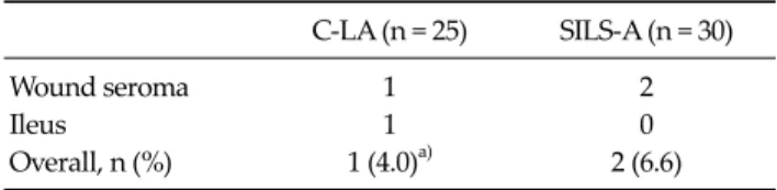
In regard to postoperative complications, in SILS-A group, seroma in the umbilical area developed in 2 pa- tients, and in C-LA group, seroma and ileus developed si- multaneously in 1 patient. They recovered after con- servative management (Table 2). Fig. 2 is immediate post- operative scar after SILS-A in a 9-year-old female patient with gangrenous type appendicitis.
DISCUSSION
Since the first laparoscopic appendectomy was re- ported by Semm [12] in Germany for an appendix without inflammation, it has been performed by numerous sur- geons. In comparison with open appendectomy, laparo- scopic appendectomy have the benefits of reduction of postsurgical pain, decreased operative trauma resulting in quicker recovery, shorter hospital stays, and improved cosmesis. As a result, it is now widely performed in adults as well as pediatric patients by many practicing surgeons
C-LA (n = 25) SILS-A (n = 30)
Wound seroma 1 2
Ileus 1 0
Overall, n (%) 1 (4.0)a) 2 (6.6)
C-LA, three-port conventional laparoscopic appendectomy; SILS- A, single incision laparoscopic surgery for appendectomy.
a)Wound seroma and ileus was in same patient.
Table 2. Postoperative complications in C-LA and SILS-A Fig. 2. Immediate post-operative scar after single incision laparo- scopic surgery for appendectomy in 9-year-old female patient with gangrenous type appendicitis.
Fig. 1. Extra-corporeal appendectomy in single incision laparo- scopic surgery for appendectomy.
C-LA (n = 25)
SILS-A
(n = 30) P-value
Gender (M/F) 14/11 17/13 0.220
Age (yr) 8.7 ± 3.5 9.3 ± 4.0 0.126
Complicated appendicitisa) 7 8 0.265
Mean OP time (min) 40.5 ± 15.2 46.2 ± 18.5 0.067 Hospital stay (day) 3.8 ± 2.0 4.0 ± 1.5 0.125 No. of IV pain control 0.8 ± 0.5 1.2 ± 1.5 0.078 Values are presented as number or mean ± SD.
OP, operation; C-LA, three ports conventional laparoscopic appendectomy; SILS-A, single incision laparoscopic surgery for appendectomy.
a)Perforated or gangrenous type appendicitis.
Table 1. Demographic data and operative comparison between C-LA and SILS-A in children
[1-3].
As laparoscopic minimal invasive surgery draws atten- tion, interest in no-scar surgical methods is on the rise.
Together with the development of equipment, Natural Orifice Transluminal Endoscopic Surgery, single-trocar or single incision surgical methods have been applied to di- verse diseases in the abdominal cavity [4-7,13]. Although it differs slightly depending on the surgeon, single in- cision laparoscopic surgery for appendectomy makes an incision window through the umbilicus in most cases. It is applied to appendectomy as a new technique of minimal invasive surgery because the umbilicus is located in the middle of the abdomen, so diverse intra-abdominal ap- proaches can be performed; blood vessels and nerves are absent, so incision windows can be readily created; even
after surgery, wounds become depressed within the um- bilicus and, thus, may be considered as an existing con- genital scar [8-10,14].
Reviewing the reports that compared single incision laparoscopic surgery with a conventional 3-port laparo- scopic appendectomy in adults, the former was found to reduce scars in addition to having the advantages of a 3-port laparoscopic appendectomy; thus, it is advanta- geous for cosmetic improvement. Nonetheless, short- comings, long operation time, and substantial early post- surgical pain, have been reported [8-10].
Oltmann et al. [11] reported that single incision laparo- scopic surgery for appendectomy is both feasible and safe across the pediatric age range. Although operating room times are somewhat longer than with conventional 3-port laparoscopic appendectomy, they concluded that it should decrease with improved instrumentation and ex- perience.
To overcome the longer operative time, we used a 30o,
5-mm laparoscopic camera to minimalize collisions with and interference between the laparoscopic surgical equip- ment and the laparoscopic camera. For cases in which col- lision and interference phenomenon between laparo- scopic surgical equipment and laparoscopic cameras oc- cur, in the view of 30° - 5-mm laparoscopic cameras, lapa- roscopic manipulation was made easy by using the flexi- ble laparoscopic Roticulator Grasper, Dissector, and Shear (Covidien, Norwalk, CT, USA).
Finally, we adapted extra-corporeal appendectomy when the cecal base could be mobilized to the midline. In 24 out of 30 patients (80%) extra-corporal appendectomy was applicable, but in 6 patients (20%) it was not appli- cable due to non-mobile cecum and adhesion. We think that it is an important point to choose intra or extra-cor- poreal appendectomy whether the cecum is mobile or not.
In the cases of mobile cecum, extra-corporeal appendec- tomy method in adolescent patients is a appropriate meth- od to avoid unnecessary manipulation and reduce oper- ation time.
Extra-corporeal laparoscopic appendectomy using sin- gle umbilical incision, initially published by Pelosi and Pelosi [15], may offer some advantage in terms of expense.
Removal of the appendix extracorporeally in the manner of conventional surgery eliminates need for expensive devices. Visnjic [16] reported that transumbilical ex- tra-corporeal laparoscopically assisted appendectomy op- erative time in children was shorter and cost less than con- ventional 3 port laparoscopic appendectomy. Hence, they called this method “High-tech low-budget surgery.”
Through such methods as those discussed above, single incision laparoscopic surgery for appendectomy in chil- dren can even be applied to appendicitis patients; and the operation time may not be significantly longer.
Kang et al. [9] reported that early pain was more severe in single incision laparoscopic surgery for appendectomy in adults than it was in a conventional 3-port laparoscopic appendectomy. This might be caused by the fact that al- though the skin incision in the umbilical area is small, the actual length of the fascia incision is longer, and through a small incision window, laparoscopic equipment is used si- multaneously, which irritates the incision window.
Visnjic [16] also reported that in transumbilical ex-
tra-corporeal laparoscopically assisted appendectomy in children, the administration of rescue analgesia was not statistically different than in conventional 3-port laparo- scopic appendectomy.
In our study, similarly, in the single incision laparo- scopic surgery for appendectomy in children, analgesic administration was significantly greater, and this is thought to be associated with shorter operation time and less fascial irritation by performing extra-corporeal lapa- roscopic appendectomy.
Postoperative complications in patients who under- went single incision laparoscopic surgery for appendec- tomy were treated without significant side effects or com- plications, except wound problems. A seroma in the um- bilical area developed in 2 patients (6.6%) and treated in outpatient clinics.
In the report of Oltmann et al. [11], they reported the in- cidence of wound complication was 5.2% (1/19), and Visnjic [16] was 13.7% (4/29). The higher incidence of port site infection could be expected in single incision laparo- scopic surgery for appendectomy than conventional 3-port laparoscopic appendectomy, especially in extra- corporeal appendectomy due to the exposure and manip- ulation of the appendix on the incision site. Therefore, an adjusted operative technique using minimal, gentle move- ments, and adequate wound protection is required. We routinely used a wound retractor for wound protection, and wound seroma, not wound infection, developed in only 2 cases.
In conclusion, single incision laparoscopic surgery for appendectomy in children is technically feasible and safe.
Considering little postoperative scar and no difference of post-operative outcomes compared to conventional 3-port laparoscopic appendectomy, single incision laparoscopic surgery for appendectomy in children could be appli- cable. Larger studies and further technical implements will be necessary to assess the true benefit of this approach.
CONFLICTS OF INTEREST
No potential conflict of interest relevant to this article
was reported.
ACKNOWLEDGEMENTS
This paper was supported by Wonkwang University 2010.
REFERENCES
1. Towfigh S, Chen F, Mason R, Katkhouda N, Chan L, Berne T. Laparoscopic appendectomy significantly reduces length of stay for perforated appendicitis. Surg Endosc 2006;20:495-9.
2. Khan MN, Fayyad T, Cecil TD, Moran BJ. Laparoscopic versus open appendectomy: the risk of postoperative in- fectious complications. JSLS 2007;11:363-7.
3. Golub R, Siddiqui F, Pohl D. Laparoscopic versus open ap- pendectomy: a metaanalysis. J Am Coll Surg 1998;186:
545-53.
4. White WM, Haber GP, Goel RK, Crouzet S, Stein RJ, Kaouk JH. Single-port urological surgery: single-center experi- ence with the first 100 cases. Urology 2009;74:801-4.
5. Bucher P, Pugin F, Morel P. Single port access laparoscopic right hemicolectomy. Int J Colorectal Dis 2008;23:1013-6.
6. Kuon Lee S, You YK, Park JH, Kim HJ, Lee KK, Kim DG.
Single-port transumbilical laparoscopic cholecystectomy:
a preliminary study in 37 patients with gallbladder
disease. J Laparoendosc Adv Surg Tech A 2009;19:495-9.
7. Oltmann SC, Rivas H, Varela E, Goova MT, Scott DJ.
Single-incision laparoscopic surgery: case report of SILS adjustable gastric banding. Surg Obes Relat Dis 2009;5:
362-4.
8. Hong TH, Kim HL, Lee YS, Kim JJ, Lee KH, You YK, et al.
Transumbilical single-port laparoscopic appendectomy (TUSPLA): scarless intracorporeal appendectomy. J Lapa- roendosc Adv Surg Tech A 2009;19:75-8.
9. Kang KC, Lee SY, Kang DB, Kim SH, Oh JT, Choi DH, et al.
Application of single incision laparoscopic surgery for ap- pendectomies in patients with complicated appendicitis. J Korean Soc Coloproctol 2010;26:388-94.
10. Vidal O, Valentini M, Ginestà C, Martí J, Espert JJ, Benarroch G, et al. Laparoendoscopic single-site surgery appendectomy. Surg Endosc 2010;24:686-91.
11. Oltmann SC, Garcia NM, Ventura B, Mitchell I, Fischer AC.
Single-incision laparoscopic surgery: feasibility for pedia- tric appendectomies. J Pediatr Surg 2010;45:1208-12.
12. Semm K. Endoscopic appendectomy. Endoscopy 1983;15:
59-64.
13. Horgan S, Cullen JP, Talamini MA, Mintz Y, Ferreres A, Jacobsen GR, et al. Natural orifice surgery: initial clinical experience. Surg Endosc 2009;23:1512-8.
14. Saber AA, Elgamal MH, El-Ghazaly TH, Dewoolkar AV, Akl A. Simple technique for single incision transumbilical laparoscopic appendectomy. Int J Surg 2010;8:128-30.
15. Pelosi MA, Pelosi MA 3rd. Laparoscopic appendectomy using a single umbilical puncture (minilaparoscopy). J Reprod Med 1992;37:588-94.
16. Visnjic S. Transumbilical laparoscopically assisted appen- dectomy in children: high-tech low-budget surgery. Surg Endosc 2008;22:1667-71.