http://www.jdapm.org 135
Case Report pISSN 2383-9309❚eISSN 2383-9317
J Dent Anesth Pain Med 2017;17(2):135-138❚https://doi.org/10.17245/jdapm.2017.17.2.135
Airway obstruction by dissection of the inner layer of a reinforced endotracheal tube in a patient with Ludwig’s angina: A case report
Sung-Min Shim, Jae-Ho Park, Dong-Min Hyun, Hwa-Mi Lee
Department of Anesthesiology and Pain Medicine, Gangneung Asan Hospital, University of Ulsan College of Medicine, Gangneung, Korea
Intraoperative airway obstruction is perplexing to anesthesiologists because the patient may fall into danger rapidly. A 74-year-old woman underwent an emergency incision and drainage for a deep neck infection of dental origin. She was orally intubated with a 6. 0 mm internal diameter reinforced endotracheal tube by video laryngoscope using volatile induction and maintenance anesthesia (VIMA) with sevoflurane, fentanyl (100 μg), and succinylcholine (75 mg). During surgery, peak inspiratory pressure increased from 22 to 38 cmH2O and plateau pressure increased from 20 to 28 cmH2O. We maintained anesthesia because we were unable to access the airway, which was covered with surgical drapes, and tidal volume was delivered. At the end of surgery, we found a longitudinal fold inside the tube with a fiberoptic bronchoscope. The patient was reintubated with another tube and ventilation immediately improved. We recognized that the tube was obstructed due to dissection of the inner layer.
Keywords: Intubation; Bronchoscopes; Ludwig’s Angina; Reinforced Endotracheal Tube.
This is an Open Access article distributed under the terms of the Creative Commons Attribution Non-Commercial License (http://creativecommons.org/licenses/by-nc/3.0/) which permits unrestricted non-commercial use, distribution, and reproduction in any medium, provided the original work is properly cited.
Received: 2017. April. 11.•Revised: 2017. May. 16.•Accepted: 2017. May. 21.
Corresponding Author: Hwa-Mi Lee, Department of Anesthesiology and Pain Medicine, Gangneung Asan Hospital, University of Ulsan College of Medicine, 38 Bangdong-gil, Sacheon-myeon, Gangneung-si, Gangwon-do, Korea
Tel: +82-33-610-3409 Fax: +82-33-641-8180 E-mail: [email protected] Copyrightⓒ 2017 Journal of Dental Anesthesia and Pain Medicine
Endotracheal intubation is performed routinely by anesthesiologists. The establishment of the endotracheal tube during both elective and emergency situations has allowed for immediate life-saving interventions during resuscitation, maintenance of oxygenation and ventila- tion, and delivery of inhaled anesthesia. However, the obstruction of an endotracheal tube is a potentially life- threatening complication [1]. Herein we report a case of obstruction of a reinforced endotracheal tube due to dissection of the inner layer in a patient who was difficult to intubate and was scheduled for an incision and drainage of a deep neck infection that was of dental origin.
CASE REPORT
A 74-year-old woman (weight = 48 kg; height = 151 cm) presented to the emergency room with complaints of pain on sublingual and tongue swelling. Neck com- puterized tomography confirmed a deep neck infection, and she was diagnosed with Ludwig’s angina. She was scheduled for emergency incision and drainage. Preopera- tive evaluation revealed that she had hypertension and bronchiectasis. An electrocardiogram (ECG) was per- formed and yielded no abnormal findings. Chest radio- graph showed underlying bronchiectasis in both lungs.
Results from laboratory tests showed an increased white blood cell count. An 18-gauge intravenous catheter was
Sung-Min Shim, et al
136 J Dent Anesth Pain Med 2017 June; 17(2): 135-138
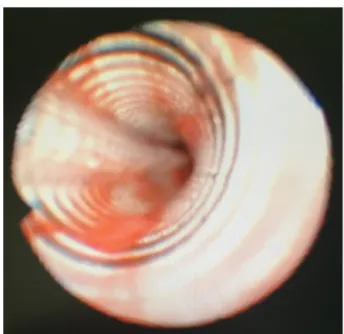
Fig. 1. Fiberoptic bronchoscopic view of the inside of the endotracheal tube. A longitudinal fold is seen inside the tube.
Fig. 2. Video laryngoscopic view for reintubation. A new endotracheal tube was prepared at the bedside for entrance of vocal cord.
inserted into the right arm and lactated Ringer’s solution was instilled. She entered the operating room without premedication. Oxygen saturation (SpO2), non-invasive blood pressure (NIBP), and ECG were monitored. The patient’s initial vital signs were as follows: NIBP = 120/68 mmHg, SpO2 = 100%, and heart rate = 85 beats/min. Preoxygenation was performed with 100%
oxygen. Volatile induction and maintenance anesthesia (VIMA) with sevoflurane, fentanyl (100 μg), and succinylcholine (75 mg) were used for induction. Direct laryngoscopy failed due to the severe tongue and neck swelling. After several attempts, she was orally intubated with a 6. 0 mm internal diameter reinforced endotracheal tube by video laryngoscope. Rocuronium 30 mg was then injected. Anesthesia had been maintained with 2 volume
% of sevoflurane, 2 L/min of N2O, and 2 L/min of O2
(FiO2 0. 5). Volume controlled ventilation mode was used. The tidal volume was 400 ml, positive end ex- piratory pressure (PEEP) was 4 cmH2O, and the respiratory rate was 11 breaths/min.
In 40 minutes, peak inspiratory pressure increased from 22 cmH2O to 38 cmH2O and plateau pressure increased from 20 cmH2O to 28 cmH2O. We were not able to examine the endotracheal tube because her face was covered with surgical drapes. Since tidal volume was delivered, we managed to maintain ventilation within the normal range for the subsequent 40 minutes. At the end of the operation, we inspected the tube, but there was no kicking. On auscultation, weak breath sounds were heard from both lungs. We tried to pass a suction catheter through the endotracheal tube but it did not pass beyond 24 cm from the inlet. To determine the cause for obstruction, a fiberoptic bronchoscopic examination was conducted, and we recognized a longitudinal fold in the inner layer of the endotracheal tube forming a partial occlusion (Fig. 1). Since prior direct laryngoscope had failed, we exchanged the reinforced tube with a 6. 0 mm internal diameter silastic tube using video laryngoscope to confirm visualization of the vocal cords (Fig. 2). After replacement with a new Silastic tube, the peak inspiratory pressure and plateau pressure decreased to 22 cmH2O and
20 cmH2O, respectively. She was transferred to the intensive care unit with a T-piece.
DISCUSSION
Similar cases of dissection between the inner and outer layers of a reinforced endotracheal tube were reported in the literature (Table 1). Most of them were related to repetitive resterilization with N2O [2,3] or even without N2O [4,5]. In some cases, the dissection was attributed to faulty manufacturing [6-9], while one case claimed that it was due to difficulty with stylet removal [10]. During production of the tube, a liquid polyvinyl chloride layer coats a rod. The spiral steel wire was lodged in the rod.
Reinforced endotracheal tube dissection
http://www.jdapm.org 137
Age/
Gender Procedure Anesthetic gas Management Problem Reference
49/F Transphenoidal hypophysectomy Sevoflurane Reintubation Resterilization Matthias Paul et al 2003 [5]
NA+ Laryngo-microsurgery Total intravenous
anesthesia Reintubation Resterilization Tose R et al 2003 [4]
39/F Lumbar discectomy Sevoflurane/N2O Reintubation Faulty Manufacture Isabel A. et al 2005 [6]
62/F Posterior lumbar fusion Sevoflurane/N2O Maintain anesthesia,
extubation after operation Resterilization Y. S. Jeon et al 2007 [2]
39/F Excision of meningioma Isoflurane Reintubation Faulty Manufacture Ashish Rajkumar et al 2011 [7]
52/M Thyroidectomy Sevoflurane/N2O Reintubation Resterilization Esra Mercanoglu et al 2013 [3]
25/F Laparoscopic cholecystectomy Sevoflurane Reintubation Resterilization
63/F Posterior lumbar fusion Sevoflurane Reintubation Faulty Manufacture Eunkyeong Choi et al 2013 [8]
18/F Occipital craniotomy Sevoflurane/N2O Reintubation Faulty Manufacture Omar Itani et al 2015 [9]
12/NA* Open appendectomy Sevoflurane Reintubation Stylet Aggression Fabricio Tavares et al 2015 [10]
74/F Incision and drainage for deep
neck infection Sevoflurane/N2O Maintain anesthesia,
reintubation after operation Resterilization Our case NA+, full text is not available, NA*, full text is not available. Article in Spanish.
Table 1. Summary of similar cases
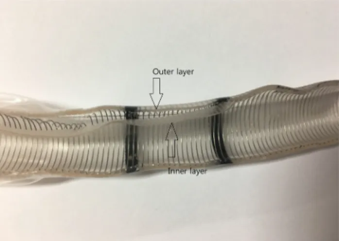
Fig. 3. Cut image of the dissected tube. The inner layer was detached from the outer layer and coil.
The coating process is repeated many times, creating the outer layer [5]. Formation of air bubbles in the wall of a reinforced endotracheal tube can occur during pro- duction. The exposure to heat, such as during autoclaving, could create a separation in the inner layer, also leading to the formation of an air bubble. Additionally, the use of N2O may expand it.
We assume that our case was related to the resteriliza- tion process. However, our center does not reuse tubes;
the tube used in this case had not been used by other patients and was only resterilized with ethylene oxide.
In addition to resterilization, our case had other com- plex problems. A metal stylet was used because of a difficult airway. N2O was used for general anesthesia. We
reasoned that the following contributed to the dissection of the reinforced endotracheal tube: first, the resteriliza- tion may have created an air bubble between the layers;
second, additional damage to the inner layer may have been inflicted by the metal stylet during its insertion or removal; and third, the air bubble increased during general anesthesia due to the use of N2O (Fig. 3).
There are several causes for an increase in peak inspiratory pressure, including the patient biting on the tube [11], kinking of the tube [12], internal obstruction by secretions, a pneumothorax, and bronchospasms.
These should be considered in the differential diagnosis.
Peak inspiratory pressure and plateau pressure are related to airway resistance and lung compliance, respectively.
The difference between the peak and plateau pressures represents the pressure needed to overcome the resistance to airflow [13]. We think that the resistant pressure of 10 cmH2O (38 cmH2O - 28 cmH2O) was airway resis- tance caused by the obstruction of the dissected tube.
If there is a sudden increase in peak inspiratory pressure, our recommendation is as follows: First, inspect for kink- ing of the tube. If there is no problem with the external tube, stop N2O and give the patient 100% oxygen. Then, pass a suction catheter through the tube. If it cannot pass through the tube, perform a fiberoptic bronchoscope for direct internal visualization of the tube [3,9].
In conclusion, ultimately, the use of a resterilized
Sung-Min Shim, et al
138 J Dent Anesth Pain Med 2017 June; 17(2): 135-138
reinforced endotracheal tube must be avoided. Due to the possibility of faulty manufacturing, strict examination of the tube prior to use is highly recommended. When using a metal stylet, insertion and removal of the stylet should be performed with care. Since there is a recent trend towards the use of inhalation anesthetics, such as sevoflurane and desflurane, but not N2O, combined with short-acting opioids for general anesthesia, remifentanil is recommended instead of N2O.
AUTHOR ORCIDs
Sung-Min Shim: http://orcid.org/0000-0001-7530-367x Jae-Ho Park: http://orcid.org/0000-0002-7778-6043 Dong-Min Hyun: http://orcid.org/0000-0002-3735-7383 Hwa-Mi Lee: http://orcid.org/0000-0002-1919-5528
NOTES: There are no financial or other issues that might lead to conflict of interest.
REFERENCES
1. Szekely S, Webb R, Williamson J, Russell W. The Australian Incident Monitoring Study. Problems related to the endo- tracheal tube: an analysis of 2000 incident reports. Anaesth Intensive Care 1993; 21: 611-6.
2. Jeon Y, Kim Y, Joo J, Kang E, In J, Choi J, et al. Partial airway obstruction caused by dissection of a reinforced endotracheal tube. Eur J Anaesthesiol 2007; 24: 983-4.
3. Mercanoglu E, Topuz D, Kaya N. The dissection of reinforced endotracheal tube internal wall causing intraoperative airway obstruction under general anesthesia.
Case report. Braz J Anesthesiol 2013; 63: 372-4.
4. Tose R, Kubota T, Hirota K, Sakai T, Ishihara H, Matsuki A. Obstruction of an reinforced endotracheal tube due to dissection of internal tube wall during total intravenous anesthesia. Masui 2003; 52: 1218-20.
5. Paul M, Dueck M, Kampe S, Petzke F. Failure to detect an unusual obstruction in a reinforced endotracheal tube with fiberoptic examination. Anesth Analg 2003; 97:
909-10.
6. Santos IA, Oliveira CA, Ferreira L. Life-threatening ventilatory obstruction due to a defective tracheal tube during spinal surgery in the prone position. Anesthesiology 2005; 103: 214-5; discussion 5.
7. Rajkumar A, Bajekal R. Intraoperative airway obstruction due to dissection of a reinforced endotracheal tube in a prone patient. J Neurosurg Anesthesiol 2011; 23: 377.
8. Choi E, Cho HS, Lee JW. Intraoperative airway obstruction from a whole dissection of the inner wall of a reinforced endotracheal tube. Korean J Anesthesiol 2013; 65: 585-6.
9. Itani O, Mallat C, Jazzar M, Hammoud R, Shaaban J.
Obstruction of a non-resterilized reinforced endotracheal tube during craniotomy under general anesthesia. Anesth Essays Res 2015; 9: 260-2.
10. Mendonça F, Martins L, Gazzi R, Palmieri J. Dissection of the wired endotracheal tube's lumen during general anesthesia: a case report. Rev Bras Anestesiol 2015; Epub ahead of print. Available from https://doi.org/10.1016/
j.bjan.2015.09.010.
11. Ball J, Platt S. Obstruction of a reinforced oral tracheal tube. Br J Anaesth 2010; 105: 699-700.
12. Peck MJ, Needleman SM. Reinforced endotracheal tube obstruction. Anesthe Analg 1994; 79: 193.
13. Marino PL. Marino's the ICU Book. 4 ed. Philadelpia (PA), Lippincott Williams & Wilkins. 2013, pp 487-93.