Case Report
J Gynecol Oncol Vol. 21, No. 1:62-64, March 2010 DOI:10.3802/jgo.2010.21.1.62
62
Lipoleiomyoma of broad ligament mimicking ovarian cancer in a postmenopausal patient:
case report and literature review
Mehmet Coskun Salman1, Zeliha Atak1, Alp Usubutun2, Kunter Yuce1
Departments of 1Obstetrics and Gynecology, 2Pathology, Hacettepe University Faculty of Medicine, Ankara, Turkey
Lipoleiomyoma is a very rare tumor which is composed of adipocytes and smooth muscle cells. It is most commonly located in uterine corpus although cervical, ovarian, and retroperitoneal locations were also reported. Lipoleiomyoma located in broad ligament is extremely uncommon and only five cases were reported to date. Here, we report the sixth case of lipoleiomyoma of broad ligament which was diagnosed in a postmenopausal woman who was subjected to exploratory laparotomy with a preoperative diagnosis of a solid adnexal mass suggesting an ovarian malignancy.
Key Words: Lipoleiomyoma, Adnexal mass, Ovarian neoplasms
Received June 14, 2009, Revised July 2, 2009, Accepted July 6, 2009
Correspondence to Mehmet Coskun Salman
Department of Obstetrics and Gynecology, Hacettepe University Faculty of Medicine, 06100 Sihhiye, Ankara, Turkey
Tel: 90-312-3053128, Fax: 90-312-3116372 E-mail: [email protected]
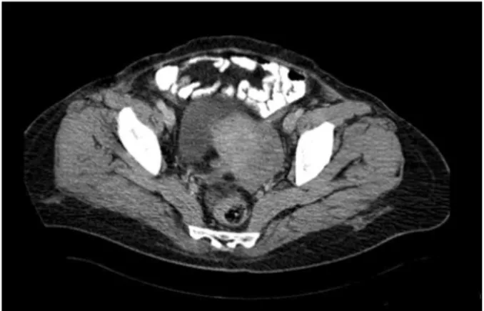
Fig. 1. Computerized tomography showing solid left adnexal mass.
INTRODUCTION
Lipoleiomyoma is a very rare tumor accounting for 0.35 to 2.1% of all uterine leiomyomas and is composed of adipocytes and smooth muscle cells.1,2 Lipoleiomyoma is most com- monly located in uterine corpus although cervical, ovarian, and retroperitoneal locations were also reported.1-3 However, lipoleiomyoma located in broad ligament is extremely un- common. A review of the English literature revealed only five cases of broad ligament lipoleiomyoma reported by this time.1,4-6
Here, we report the sixth case of lipoleiomyoma of broad lig- ament which was diagnosed in a postmenopausal woman who was subjected to exploratory laparotomy with a preoperative diagnosis of a solid adnexal mass suggesting an ovarian mali- gnancy. The English literature regarding lipoleiomyoma of uterus and lipoleiomyoma of broad ligament was reviewed as well.
CASE REPORT
A 54-year-old, gravida 2, parity 2 woman was admitted to our clinic for lower abdominal pain. Her past medical and sur-
gical histories were unremarkable and she experienced her last menstruation 8 years ago. Abdominal examination re- vealed some tenderness at left lower quadrant and a left ad- nexal mass was palpated on bimanual pelvic examination.
Routine laboratory tests including tumor markers were in normal limits. A transvaginal ultrasonography showed a solid mass of 75×40 mm at left adnexal site. Computerized tomog- raphy of abdomen and pelvis confirmed the heterogeneous solid mass of left adnexa measuring 70×35 mm (Fig. 1). An exploratory laparotomy was performed with a preoperative diagnosis of solid adnexal mass suggesting ovarian malig- nancy given the age of the patient and solid nature of the mass.
During laparotomy, a 7 to 8 cm mass was detected within the left broad ligament. Otherwise, the uterus and ovaries were atrophic and exploration of the whole abdomen was free of ad-
Broad ligament lipoleiomyoma in a postmenopausal patient
63
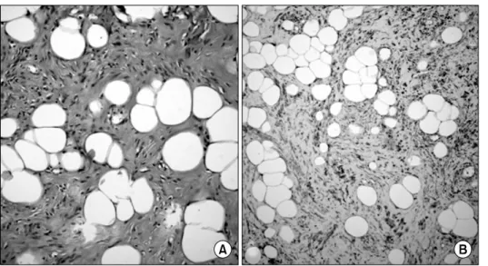
Fig. 2. Microscopic view of the tu- mor demonstrating an admixture of smooth muscle and adipose cells on hematoxylin-eosin (×200) (A) and positive staining with desmin on immunohistochemistry (×100) (B).
ditional abnormalities. A total abdominal hysterectomy with salpingo-oophorectomy was performed. Frozen section analy- sis did not suggest malignancy. Final pathologic examination showed a lipoleiomyoma without mitotic figures, nuclear aty- pia or pleomorphism which immunohistochemically showed reactivity with desmin (Fig. 2). The patient was discharged home 3 days after surgery following an uneventful post- operative course and she is asymptomatic clinically 8 months after the surgery.
DISCUSSION
Lipoleiomyoma of the uterus is an uncommon pelvic tumor with an incidence varying from 0.03% to 0.2%.7 Finding an ad- mixture of mature adipocytes and smooth muscle cells on mi- croscopy is required to be able to designate a neoplasm as lipoleiomyoma. The adipocytes may be evenly distributed throughout the tumor or they may be concentrated in only fo- cal areas. Also, adipocyte component in lipoleiomyoma may differ widely and a certain level of adipocytes was not defined to achieve the diagnosis of lipoleiomyoma. These tumors may contain microscopic foci of adipocytes resembling regular leiomyomas in gross appearance or high amounts of adipo- cytes may be detected resulting in yellow and lobulated cut surface.1 The tumor in our case had a yellow cut surface gross- ly and high amounts of adipocytes on microscopy.
The clinical features are uncertain due to its rarity. The exact histogenesis has not been explained clearly. Nevertheless, im- munohistochemical studies indicated a complex histogenesis of lipoleiomyoma which might arise from immature mesen- chymal cells or from transformation of smooth muscle cells into adipocytes.8 It was also demonstrated that lipoleiom- yomas may be associated with some metabolic disorders in- cluding hyperlipidemia, hypothyroidism and diabetes mellitus.
This suggests that changes in lipid metabolism after meno- pausal transition may play a role in the development of lip-
omatous change in leiomyomas.9 This hypothesis is con- sistent with advanced age of most of the patients at time of diagnosis. However, the current case did not have any of the aforementioned metabolic disorders.
By contrast with ordinary leiomyomas which tend to occur predominantly in women of reproductive age and regress after menopause, the lipoleiomyomas are frequently seen in older women. Actually, mean age of the patients in the largest two series so far was 55.4 years and almost 60% of patients were aged older than 50.1,2 Our patient was a 54-year-old post- menopausal woman as consistent with the literature.
Most patients are asymptomatic and are diagnosed in- cidentally, but among symptomatic ones pelvic pain, palpable mass or abnormal bleeding are the most common symptoms similar to those caused by leiomyomas. Patients with symp- toms usually undergo physical exam followed by imaging mo- dalities which reveal a solid pelvic mass.1 Although some fea- tures in different imaging modalities may suggest the possible diagnosis of these tumors, the precise diagnosis is based on pathologic examination.7
The presenting symptom was pain in our case who was found to have an adnexal mass on examination. Therefore, imaging was requested and a heterogenous solid mass located at left adnexal site which did not have an appearance of regular leiomyomas was reported on ultrasonography and computed tomography. Also, during surgery, the mass grossly did not resemble a leiomyoma which was fairly soft on palpation and located outside the uterus. The cut surface was reported to be yellow on gross pathological examination and it contained high amounts of adipocytes on microscopic evaluation. Cu- rrent patient was subjected to laparotomy with a suspicion of ovarian malignancy. The presence of a pelvic mass in a post- menopausal woman is certainly not enough to consider ovar- ian malignancy especially if it is not associated with ascites and elevated tumor markers. However, the heterogenous sol- id nature of the lesion on imaging modalities and its adnexal
J Gynecol Oncol Vol. 21, No. 1:62-64, 2010 Mehmet Coskun Salman, et al.
64 localization may suggest a malignant neoplasm that warrants surgical exploration with frozen section analysis where available.
Although the lipoleiomyoma is most commonly located in uterine corpus, it may be found elsewhere in pelvis. However, the extrauterine location including broad ligament is the rar- est site reported by this time.1-6 In fact, to best of our knowl- edge, only five cases of broad ligament lipoleiomyoma were reported previously.1,4-6
The long-term follow-up of patients with uterine lipoleio- myoma demonstrated that these lesions are benign without any recurrences or disease-related deaths if they are diag- nosed as the unique pelvic pathology.1 On the other hand, among patients with uterine lipoleiomyoma in two largest series, 18.8% of patients were reported to have associated gy- necologic malignancies which may originate from uterus, cer- vix or ovaries,1,2 Therefore, the patients with uterine lip- oleiomyoma should be subjected to detailed clinical and pathological evaluation in order not to overlook a coexistent gynecologic malignancy.
In conclusion, lipoleiomyoma of the broad ligament may ne- cessitate surgical intervention not only due to associated symptoms but also to be able to exclude an ovarian malig- nancy given its solid nature and the postmenopausal age of the affected individuals. The specimens should be examined care- fully to document whether or not a coexisting gynecologic ma- lignancy is present. The patients without an associated malig- nancy may be followed with routine annual gynecologic ex-
ams, because the tumor lacks the risk to recur.
CONFLICT OF INTEREST
No potential conflict of interest relevant to this article was reported.
REFERENCES
1. Wang X, Kumar D, Seidman JD. Uterine lipoleiomyomas: a clin- icopathologic study of 50 cases. Int J Gynecol Pathol 2006; 25:
239-42.
2. Aung T, Goto M, Nomoto M, Kitajima S, Douchi T, Yoshinaga M, et al. Uterine lipoleiomyoma: a histopathological review of 17 cases. Pathol Int 2004; 54: 751-8.
3. Mira JL. Lipoleiomyoma of the ovary: report of a case and review of the English literature. Int J Gynecol Pathol 1991; 10: 198-202.
4. Fernandez FA, Val-Bernal F, Garijo-Ayensa F. Mixed lipomas of the uterus and the broad ligament. Appl Pathol 1989; 7: 70-1.
5. Bajaj P, Kumar G, Agarwal K. Lipoleiomyoma of broad ligament:
a case report. Indian J Pathol Microbiol 2000; 43: 457-8.
6. Cinel L, Dusmez D, Nabaei SH, Taner D, Pata O. Two intra- ligamentary lipomatous tumors with immunohistochemical fea- tures. Acta Obstet Gynecol Scand 2002; 81: 786-7.
7. Prieto A, Crespo C, Pardo A, Docal I, Calzada J, Alonso P.
Uterine lipoleiomyomas: US and CT findings. Abdom Imaging 2000; 25: 655-7.
8. Dellacha A, Di Marco A, Foglia G, Fulcheri E. Lipoleiomyoma of the uterus. Pathologica 1997; 89: 737-41.
9. Lin KC, Sheu BC, Huang SC. Lipoleiomyoma of the uterus. Int J Gynaecol Obstet 1999; 67: 47-9.