103 Introduction
Stress-induced cardiomyopathy (SCMP) is a nonischemic reversible cardiomyopathy characterized uniquely by a left ventricular (LV) wall motion abnormality termed “apical ballooning” and the prognosis is generally excellent.1)2) How- ever, several critical complications are possible. The forma- tion of LV thrombus is one of these rare complications which can result in systemic embolism.3) Here, we report a case of SCMP complicated by LV mural thrombus which gradu- ally increased in mobility as LV contractility recovered and which eventually required surgical removal.
Case
A 66-year-old woman who presented with severe dys- phagia visited our emergency room. On admission her mental status was alert, and she had a blood pressure of 115/76 mm Hg, a respiratory rate of 20 per minute, a pulse rate of 96 per minute, and a body temperature of 36.4°C. She did not have any history of heart disease, hypertension or diabetes mellitus, but had undergone subtotal gastrectomy due to
pISSN 1975-4612/ eISSN 2005-9655 Copyright © 2015 Korean Society of Echocardiography www.kse-jcu.org http://dx.doi.org/10.4250/jcu.2015.23.2.103
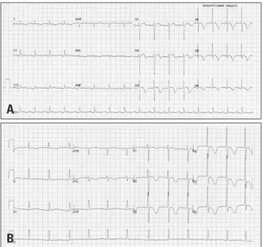
stomach cancer. Urgent gastrofibroscopy was performed and a piece of chicken trapped at the lower esophagus was removed. Additionally, a small polyp was found at the previ- ous surgical anastomosis site which subsequently confirmed pathologically as early gastric cancer. The patient also com- plained of mild dyspnea and diffuse chest discomfort on admission, and an electrocardiogram (ECG) showed ST eleva- tion with QT prolongation (QTc = 480 msec), and T wave inversion on leads V2–6 (Fig. 1A). Troponin T and creatinine kinase-MB (CK-MB) levels were elevated to 0.29 ng/mL and 20.46 ng/mL, respectively (reference range; < 0.01 ng/dL for troponin T, < 3.61 ng/mL for CK-MB). Transthoracic echo- cardiography (TTE) revealed mid and apical LV segmental wall motion abnormalities with apical ballooning that did not correspond to a coronary artery territory, as well as a left ventricular ejection fraction of 41% (LV end-diastolic volume/
LV end-systolic volume = 84/49 mL) (Fig. 2A). In addition, a 19 × 18-mm-sized non-mobile echogenic mass suspicious for a mural thrombus was found at the apex of the left ven- tricle (Fig. 2B). As the features were consistent with SCMP CASE REPORT J Cardiovasc Ultrasound 2015;23(2):103-106
• Received: September 23, 2014 • Revised: October 27, 2014 • Accepted: May 19, 2015
• Address for Correspondence: Jin Oh Na, Department of Cardiology, Korea University Guro Hospital, 148 Gurodong-ro, Guro-gu, Seoul 152-703, Korea Tel: +82-2-2626-3025, Fax: +82-2-863-1109, E-mail: [email protected]
• This is an Open Access article distributed under the terms of the Creative Commons Attribution Non-Commercial License (http://creativecommons.org/licenses/by-nc/3.0) which permits unrestricted non-commercial use, distribution, and reproduction in any medium, provided the original work is properly cited.
Surgical Removal of a Left Ventricular Thrombus Which Showed Morphologic Changes Over Time in a Patient with
Stress-Induced Cardiomyopathy
Jah Yeon Choi, MD1, Eun Jin Park, MD1, Sung Hun Park, MD1, Hee Dong Kim, MD1, Ji Young Song, MD1, Ji Bak Kim, MD1, Sun Ki Lee, MD1,
Yang Gi Ryu, MD2, Man Jong Baek, MD2, and Jin Oh Na, MD1
Departments of 1Cardiology, 2Thoracic and Cardiovascular Surgery, Korea University Guro Hospital, Seoul, Korea
Although stress-induced cardiomyopathy (SCMP) is a reversible disease and the prognosis is usually excellent, several complications can occur and can result in fatal adverse events. The formation of left ventricular (LV) thrombus is one of these critical complications of SCMP. This report describes a case of SCMP complicated by formation of a LV thrombus that became increasingly mobile as LV contractility recovered, and for which surgical removal was performed. Here, we report a case of SCMP complicated by LV thrombus and review the literature regarding this topic.
KEY WORDS: Stress cardiomyopathy · Left ventricular thrombus · Echocardiography.
Journal of Cardiovascular Ultrasound 23 | June 2015
104
complicated by LV mural thrombus, anticoagulation as well as conventional heart failure therapy was initiated. After one week, ST elevation disappeared on follow-up ECG (Fig. 1B) and follow-up TTE demonstrated resolution of the mid and apical LV segmental wall motion abnormalities and fully re- covered LV systolic function. However, the LV mural throm- bus had partially detached from the LV wall with recovery of LV contractility and was adherent to the ventricular wall by a narrow stalk. The remarkably increased mobility of the LV thrombus was thought to carry a very high thromboembolic risk (Fig. 2C). Thus, surgical removal of the thrombus was decided upon to prevent embolic complications.
Oblique aortotomy was performed under support of cardio- pulmonary bypass. The mass, which had its base attached to the trabecular endocardium of the apico-inferior wall of the LV cavity, was completely removed by suction tip through the aortic valve. The mass appeared to be an amorphous bizarre shape, measuring 1.7 × 1.2 × 0.7 cm in size (Fig. 3). On subse- quent pathohistological examination, the mass was confirmed to be a thrombus.
On postoperative day 7, follow-up TTE was performed. LV A
Fig. 1. Electrocardiogram on admission showing ST elevation and T wave inversion in the precordial leads and a prolonged QT interval (480 msec) (A). ST elevation disappeared after one week on follow-up electrocardiogram (B).
A B
C D
Fig. 2. Initial transthoracic echocardiography demonstrating apical ballooning (A) and a mural thrombi at the apex (B). Follow-up transthoracic echocardiography after 1 week showing a highly mobile and pedunculated thrombi (C). Transthoracic echocardiography after surgical removal of thrombus (D).
B
Left Ventricular Thrombus in SCMP | Jah Yeon Choi, et al.
105 systolic function was normal without any regional wall mo-
tion abnormalities (Fig. 2D). There was no evidence of resid- ual thrombus or signs of systemic embolism. The patient re- covered without any other complications and was discharged 18 days after the operation.
Discussion
SCMP was first described in Japan in 1991.1) It is charac- terized by a reversible LV systolic dysfunction with distinct
‘apical ballooning’ accompanied by slightly increased cardiac enzyme levels and electrocardiographic changes including QT prolongation, ST elevation, or T wave inversion. Despite many investigations, the mechanism of SCMP is not yet fully understood and many investigators have suggested a catecholamine-mediated mechanism as the cause of SCMP.2) Although the long term prognosis of patients with SCMP is usually excellent,3) well-known complications of SCMP are cardiogenic shock, congestive heart failure and sudden car- diac death. Besides these morbidity, ventricular tachycardia, LV rupture, apical thrombus formation and embolism have been reported as rare complications of SCMP.3) According to previous studies, the prevalence of LV thrombus in cases of SCMP is approximately 2.5%, and the occurrence rate of embolic complications in this condition is reported to be as high as 33% despite proper anticoagulation therapy.4)5) The pathogenesis of LV thrombus formation in SCMP is still not fully known and some theories have been suggested hemo- stasis,6) catecholamine induced platelet activation,7)8) and cat- echolamine induced cardiomyocyte damage9) as underlying mechanism.
Anterior acute myocardial infarction (AMI) can also be complicated by LV thrombus. The incidence of LV throm- bus formation in anterior AMI was reported to be 30–40%
before the era of reperfusion therapy,10)11) and the embolic rate of the condition was 10–15%.12) The relatively low in- cidence of LV thrombus formation (2.5% vs. 30–40%) and high embolic risk (33% vs. 10–15%) in SCMP compared to AMI might be related to faster and better recovery of LV
function. As LV wall motion recovers, the mobility of mural thrombus may increase rapidly by partial detachment from the ventricular wall, which can lead to increased embolic risk. In SCMP, the mural thrombus that was initially immo- bile can become highly mobile within a short time. And this phenomenon can explain why initially mobile LV thrombus in SCMP is not associated with a higher embolic event rate than initially immobile LV thrombus in previous studies.5) Of 11 previously reported cases of SCMP that were compli- cated by a mobile LV thrombus, LV thrombus was immobile or even absent in the initial examination of 6 cases.13-20) Thus, the embolic risk of LV thrombus in SCMP may not be deter- mined based on the initial mobility of the thrombus.
In all reported cases with SCMP, anticoagulation therapy was promptly initiated after the detection of the thrombus.
Despite appropriate anticoagulation therapy, however, car- diac embolic events occurred in one third of patients with SCMP complicated by LV thrombus. In contrast, when the thrombus was removed by surgery using a trans-apical ap- proach there was no embolic event in two reported cases.19)20) In the present case, a trans-aortic approach following oblique aortotomy was performed. Although the apical approach can provide good and easy access to the thrombus, myocardial injury is inevitable. Using a trans-aortic approach following aor- totomy, visualization or extraction of the thrombus through the aortic valve may be somewhat difficult, but myocardial dam- age can be avoided, and this approach may be more beneficial for the heart, especially in a clinical setting.
To the best of our knowledge, this is the first reported case of SCMP complicated by LV thrombus which was surgically removed by a trans-aortic approach after oblique aortotomy.
Although LV thrombus is a rare complication of SCMP, the embolic risk is relatively high because of rapid change of throm- bus mobility. Therefore, short-term TTE follow-up after an- ticoagulation therapy might be recommended in this clinical setting, and if the thrombus becomes highly mobile as LV sys- tolic function improves, surgical removal could be a reasonable therapeutic option.
References
1. Dote K, Sato H, Tateishi H, Uchida T, Ishihara M. [Myocardial stunning due to simultaneous multivessel coronary spasms: a review of 5 cas- es]. J Cardiol 1991;21:203-14.
2. Lyon AR, Rees PS, Prasad S, Poole-Wilson PA, Harding SE. Stress (Takotsubo) cardiomyopathy--a novel pathophysiological hypothesis to ex- plain catecholamine-induced acute myocardial stunning. Nat Clin Pract Cardiovasc Med 2008;5:22-9.
3. Bybee KA, Kara T, Prasad A, Lerman A, Barsness GW, Wright RS, Rihal CS. Systematic review: transient left ventricular apical ballooning: a syndrome that mimics ST-segment elevation myocardial infarction. Ann Intern Med 2004;141:858-65.
4. de Gregorio C, Grimaldi P, Lentini C. Left ventricular thrombus forma- tion and cardioembolic complications in patients with Takotsubo-like syn- drome: a systematic review. Int J Cardiol 2008;131:18-24.
5. de Gregorio C. Cardioembolic outcomes in stress-related cardiomyopathy com- Fig. 3. Removed thrombus measuring 1.7 × 1.2 × 0.7 cm in size.
Journal of Cardiovascular Ultrasound 23 | June 2015
106
plicated by ventricular thrombus: a systematic review of 26 clinical studies. Int J Cardiol 2010;141:11-7.
6. van Dantzig JM, Delemarre BJ, Bot H, Visser CA. Left ventricular thrombus in acute myocardial infarction. Eur Heart J 1996;17:1640-5.
7. Wittstein IS, Thiemann DR, Lima JA, Baughman KL, Schulman SP, Gerstenblith G, Wu KC, Rade JJ, Bivalacqua TJ, Champion HC. Neurohumoral features of myocardial stunning due to sudden emotional stress. N Engl J Med 2005;352:539-48.
8. Ardlie NG, Glew G, Schwartz CJ. Influence of catecholamines on nucleo- tide-induced platelet aggregation. Nature 1966;212:415-7.
9. Mann DL, Kent RL, Parsons B, Cooper G 4th. Adrenergic effects on the biology of the adult mammalian cardiocyte. Circulation 1992;85:790-804.
10. Nihoyannopoulos P, Smith GC, Maseri A, Foale RA. The natural history of left ventricular thrombus in myocardial infarction: a rationale in support of masterly inactivity. J Am Coll Cardiol 1989;14:903-11.
11. Weinreich DJ, Burke JF, Pauletto FJ. Left ventricular mural thrombi complicating acute myocardial infarction. Long-term follow-up with serial echocardiography. Ann Intern Med 1984;100:789-94.
12. Stratton JR, Resnick AD. Increased embolic risk in patients with left ventricular thrombi. Circulation 1987;75:1004-11.
13. Singh V, Mayer T, Salanitri J, Salinger MH. Cardiac MRI documented left ventricular thrombus complicating acute Takotsubo syndrome: an uncom- mon dilemma. Int J Cardiovasc Imaging 2007;23:591-3.
14. de Gregorio C, Cento D, Di Bella G, Coglitore S. Minor stroke in a Takotsubo-like syndrome: a rare clinical presentation due to transient left ventricular thrombus. Int J Cardiol 2008;130:e78-80.
15. Haghi D, Papavassiliu T, Heggemann F, Kaden JJ, Borggrefe M, Suselbeck T. Incidence and clinical significance of left ventricular thrombus in tako-tsubo cardiomyopathy assessed with echocardiography. QJM 2008;101:381-6.
16. Wakabayashi K, Dohi T, Daida H. Takotsubo cardiomyopathy associat- ed with epilepsy complicated with giant thrombus. Int J Cardiol 2011;
148:e28-30.
17. Kurisu S, Inoue I, Kawagoe T, Ishihara M, Shimatani Y, Nakama Y, Maruhashi T, Kagawa E, Dai K. Incidence and treatment of left ventricu- lar apical thrombosis in Tako-tsubo cardiomyopathy. Int J Cardiol 2011;
146:e58-60.
18. Matsuzono K, Ikeda Y, Deguchi S, Yamashita T, Kurata T, Deguchi K, Abe K. Cerebral embolic stroke after disappearing takotsubo cardiomy- opathy. J Stroke Cerebrovasc Dis 2013;22:e682-3.
19. Suzuki R, Kudo T, Kurazumi H, Takahashi M, Shirasawa B, Mika- mo A, Hamano K. Transapical extirpation of a left ventricular thrombus in Takotsubo cardiomyopathy. J Cardiothorac Surg 2013;8:135.
20. Seitz MJ, McLeod MK, O’Keefe MD, Seah PW. A rare cause of Ta- kotsubo cardiomyopathy related left ventricular apical thrombus requiring surgery. Heart Lung Circ 2012;21:245-6.