Fibrous dysplasia is a common benign disorder of bone in which normal bone marrow is replaced with fibro-osseous tissue. Patients are often asymptomatic, and fibrous dysplasia is often detected incidentally in patients with malignancy in whom 18F-fluoro-2- deoxy-glucose positron emission tomography (18F -FDG PET/
CT) is performed for metastatic disease. PET/CT findings of fibrous dysplasia have been reported on several papers.1-5) The level of FDG uptake in fibrous dysplasia can be either intense or be normal with normal metabolism without any increased FDG uptake. Fibrous dysplasia may show the glucose avidity of the lesion and may mimic metastasis on PET/CT. In this clinical situation, fibrous dysplasia should be considered in differential diagnosis based on other imaging findings using MRI as well as radiography.
Case Report
A 46-year-old woman underwent a wide excision and adjuvant
Case Report
J Korean Bone Joint Tumor Soc 2010; 16: 47-50 • DOI:10.5292/jkbjts.2010.16.1.47 www.kbjts.or.krRole of MRI and Plain Radiograph to Diagnose Fibrous Dysplasia Mimicking Metastasis on PET/CT in a Patient with Breast Cancer
Song-Mee Cho, M.D., Won-Hee Jee, M.D.*, Ie Ryung Yoo, M.D.
†, Ahwon Lee, M.D.
‡, and Yang-Guk Chung, M.D.
§Department of Radiology, St. Paul’s Hospital, College of Medicine, Th e Catholic University of Korea,
Departments of *Radiology and
†Nuclear Medicine,
‡Pathology and
§Orthopedic Surgery, Seoul St. Mary’s Hospital, College of Medicine, Th e Catholic University of Korea, Seoul, Korea
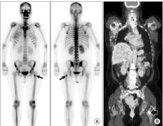
chemotherapy for breast cancer. At six months after the surgery, a focal hot uptake was found in left proximal femur on bone scintigraphy and was interpreted as a possible metastasis (Fig. 1A).
Three days later, the patient underwent PET/CT. A focal FDG uptake was observed in left proximal femur on PET/CT and was interpreted as a single metastasis (Fig. 1B). The surgeon concluded the femoral lesion as an osteoblastic metastasis based on the findings on bone scintigraphy and PET/CT in addition to the sclerotic lesion on plain radiographs (Fig. 2). The patient had radiation on the lesion of left proximal femur. In five months after radiation, she was admitted for intermittent left hip pain for one month. She underwent MRI. There was a well-defined lesion in left proximal femur. It was hypointense on T1- and T2-weighted images (Fig. 3A, B). Moderate perilesional hyperintense signal was seen on fat-suppressed T2-weighted images (Fig. 3C). The mass showed mild contrast enhancement on contrast-enhanced T1-weighted images (Fig. 3D, E). Mild post-radiation edema was observed in adjacent soft tissue.
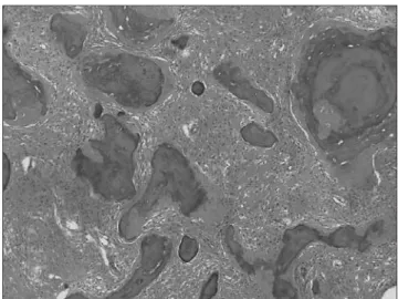
MR images were interpreted as fibrous dysplasia in the left proximal femur after reviewing the plain radiographs. Limb salvage operation including a wide excision of left proximal femur was performed one week later. A pathologic examination revealed that fibrous tissue blended into the bone spicules, which had irregular shapes and Received April 26, 2010 Revised June 1, 2010 Accepted June 11, 2010
Correspondence to: Won-Hee Jee, M.D.
Department of Radiology, Seoul St. Mary’s Hospital, College of Medicine, The Catholic University of Korea, 505, Banpo-dong, Seocho-gu, Seoul 137-701, Korea TEL: +82-2-2258-6238 FAX: +82-2-599-6771 E-mail: [email protected]
Fibrous dysplasia is a common benign disorder of bone in which normal bone marrow is replaced with fi bro-osseous tissue. As PET/CT is increasingly used for the staging of different malignant disease, incidentally found fi brous dysplasia with increased FDG uptake may mimic metastasis. We report on a 46-year-old woman with fi brous dysplasia who underwent PET/CT because of suspected recurrence of breast cancer and was misdiagnosed as a bony metastasis with a focal FDG uptake on left proximal femur. This lesion was interpreted as fi brous dysplasia based on MRI in addition to the plain radiographs. We conclude that MRI in addition to radiography may help to differentiate fi brous dysplasia mimicking metastasis on PET/CT in the patients with malignancy.
Key words: fi brous dysplasia, PET/CT, MRI
Copyrights © 2010 by The Korean Bone and Joint Tumor Society
“This is an Open Access article distributed under the terms of the Creative Commons Attribution Non-Commercial License (http://creativecommons.org/licenses/by-nc/3.0/) which permits unrestricted non-commercial use, distribution, and reproduction in any medium, provided the original work is properly cited.”
대한골관절종양학회지:제16권 제1호 2010
48
Song-Mee Cho, et al.
imperceptible osteoblast rimming. Final diagnosis was made as a fibrous dysplasia (Fig. 4).
Discussion
PET/CT is essential for the staging and localization of the metastatic lesion in the management of the patients with breast cancer. The skeleton is the most common site of distant metastases in patients
treated with mastectomy and adjuvant chemotherapy. PET/CT and bone scintigraphy have been shown to be complementary in the detection of skeletal metastases. PET/CT is more sensitive than bone scintigraphy for the detection of lytic metastases or lesions predominantly involving the bone marrow, whereas bone scintigraphy is more sensitive than PET/CT for the detection of osteoblastic metastases.6)
PET/CT findings of fibrous dysplasia are less well known since PET/CT does not have a role in the assessment of benign disease.
However, as PET/CT is increasingly used for the staging of different malignant disease incidentally found fibrous dysplasia on PET/
CT is reported on several papers.1-5) In our case fibrous dysplasia in left femur had a focal FDG uptake on PET/CT and this lesion was misdiagnosed as single metastasis from breast cancer following primary surgery and adjuvant chemotherapy.
Fibrous dysplasia is a benign disorder of unknown cause in which the normal bone structure is replaced by fibrous connective tissue. Radiography and CT can reveal the characteristic sclerotic and hyperplastic change in bone in fibrous dysplasia. MRI has been reported to identify specific findings of fibrous dysplasia.7-9) The characteristic MR findings of fibrous dysplasia are hypointense signal intensity on T2-weighted images in the substantial number of cases.7) Signal intensity on T1- and T2-weighted images and the degree of contrast enhancement on T1-weighted images depend on Figure 1. A 46-year-old woman with breast cancer. Bone scintigraphy (A) shows a focal hot uptake (arrow) in left proximal femur. PET/CT (B) shows a focal FDG uptake (arrow) in left proximal femur and was mis-interpreted as a single metastasis from breast cancer.
Figure 2. Radiograph in a 46-year-old woman shows an intramedullary lesion with a diffuse sclerotic lesion (arrow) on left proximal femur.
49
MRI of Fibrous Dysplasia
Figure 3. A 46-year-old woman who underwent MRI for intermittent left hip pain after radiation for one month. Left proximal femoral lesion was interpreted as a fibrous dysplasia based on MR findings. T1-weighted (A) and T2-weighted (B) coronal images show a well-defined hy pointense lesion (arrows in A and B) in left proximal femur. Fat-suppressed T2-weighted coronal image (C) shows moderate perilesional hyperintense signal (arrowheads) on the left proximal femur suggesting post-radiation edema. Contrast-enhanced T1-weighted coronal image (D) shows mild contrast enhance ment in the mass (arrow). Axial fat-sup pres sed con trast-enhanced T1-weighted image (E) also shows mild contrast enhance ment in the mass.
Adjacent muscles in the medial compartment of the thigh shows relatively well-demarcated contrast enhancement, suggesting post-radiation edema.
Figure 4. In microscopic examination, fibrous tissue blended into the bone spicules, which had irregular shapes and imperceptible osteoblast rimming (H&E ×100).
the amount of bony trabeculae, cellularity, collagen, and cystic and hemorrhagic changes.7) Fibrous dysplasia was interpreted based on MRI in addition to plain radiographs in our case. We conclude that MRI may help to differentiate fibrous dysplasia from metastasis in addition to the radiographic findings, which may mimic metastasis on PET/CT in a patient with primary malignancy.
References
1. Kao CH, Sun SS, Shen YY, Chen YK. Misdiagnosis of multiple bone metastases due to increased FDG uptake in polyostotic fi brous dysplasia. Clin Nucl Med. 2007;32:409-10.
2. Stegger L, Juergens KU, Kliesch S, Wormanns D, Weckesser M. Unexpected fi nding of elevated glucose uptake in fi brous dysplasia mimicking malignancy: contradicting metabolism
50
Song-Mee Cho, et al.
and morphology in combined PET/CT. Eur Radiol. 2007;17:
1784-6.
3. Strobel K, Bode B, Lardinois D, Exner U. PET-positive fi brous dysplasia--a potentially misleading incidental finding in a patient with intimal sarcoma of the pulmonary artery. Skeletal Radiol. 2007;36(Suppl 1):24-8.
4. Shigesawa T, Sugawara Y, Shinohara I, Fujii T, Mochizuki T, Morishige I. Bone metastasis detected by FDG PET in a patient with breast cancer and fibrous dysplasia. Clin Nucl Med. 2005;30:571-3.
5. Tsuyuguchi N, Ohata K, Morino M, et al. Magnetic resonance imaging and [11C]methyl-L-methionine positron emission tomography of fibrous dysplasia--two case reports. Neurol Med Chir (Tokyo). 2002;42:341-5.
6. Cook GJ, Houston S, Rubens R, Maisey MN, Fogelman I.
De tec tion of bone metastases in breast cancer by 18F-FDG PET: diff ering metabolic activity in osteoblastic and osteolytic lesions. J Clin Oncol. 1998;16:3375-9.
7. Jee WH, Choi KH, Choe BY, Park JM, Shinn KS. Fibrous dysplasia: MR imaging characteristics with radiopathologic correlation. Am J Roentgenol. 1996;167:1523-7.
8. Maeda M, Kimura H, Tsuchida C, Ishii Y, Kubota T. MR imaging of monostotic fi brous dysplasia of the clivus. A case report. Acta Radiol. 199334:527-8.
9. Utz JA, Kransdorf MJ, Jelinek JS, Moser RP Jr, Berrey BH.
MR appearance of fi brous dysplasia. J Comput Assist Tomogr.
1989;13:845-51.
유방암환자의 양전자방출단층촬영술에서 암 전이로 오인된 섬유형성이상 진단의 자기공명영상과 단순촬영의 역할
조송미 • 지원희* • 유이령† • 이아원‡ • 정양국§
가톨릭대학교 의과대학 성바오로병원 영상의학과, 가톨릭대학교 의과대학 서울성모병원 *영상의학과,
†핵의학과, ‡병리과, §정형외과
섬유형성이상은 골수가 섬유-골성 조직으로 치환되는 흔한 양성 골 질환이다. 암환자에서 병기의 결정과 추적 검사 시 암 전이의 발견에 PET/CT의 역할이 증가됨에 따라 우연히 발견된 FDG 섭취 증가를 보이는 섬유형성이상에서 암 전이와의 감별이 중요하다. PET/CT를 시행한 46세의 유방암 환자에서 좌측 대퇴골에 국소 FDG 섭취를 보여 암 전이로 의심되었으나 단순촬영과 자기공명 영상 소견에서 섬유 형성이상으로 진단한 환자의 증례를 보고하고자 한다. 이 환자에서 단순활영과 자기공명영상 소견은 PET/CT에서 유방암 전이로 오인된 섬유형성이상의 감별에 도움이 되었다.
색인단어: 섬유형성이상, PET/CT, 자기공명영상
접수일 2010년 4월 26일 심사수정일 2010년 6월 1일 게재확정일 2010년 6월 11일 교신저자 지원희, 서울시 서초구 반포동 505, 가톨릭대학교 의과대학 서울성모병원 영상의학과 TEL 02-2258-6238, FAX 02-599-6771, E-mail [email protected]