Warthin’s tumor of the parotid gland: a case report
Chul-Hwan Kim, Sung-Il Han, Moon-Young Kim
Department of Oral and Maxillofacial Surgery, College of Dentistry, Dankook University, Cheonan, Korea
Abstract(J Korean Assoc Oral Maxillofac Surg 2012;38:366-70)
Initially described in 1929, Warthin’s tumor is a benign neoplasm predominantly found in the parotid gland of the salivary glands. Warthin’s tumor is synonymous for cystadenoma lymphomatosum, adenolymphoma, and cystadenolymphoma. This tumor usually causes minimal pain, but the patient may complain of tinnitus, pain in the ear, or deafness. The tumor is treated with surgical excision, which is easily performed due to the superficial location of the tumor. In our case, a 69-year-old man visited our clinic primarily complaining of swelling in the left parotid gland area. Computed tomography revealed well-defined margins of the tumor in the superficial lobe of the left parotid gland and heterogeneous contrast enhancement. We performed superficial parotidectomy, with the pathological examination confirming Warthin’s tumor. We report a typical case of Warthin’s tumor and a literature review.
Key words: Salivary gland, Neoplasms, Adenolymphoma, Oral surgery
[paper submitted 2012. 3. 2 / revised 1st 2012. 5. 22, 2nd 2012. 6. 12 / accepted 2012. 6. 13]
from it than women at a ratio of 5 : 12,3. Unlike benign mixed tumors, it recurs in less than 2%, and only 1% develops into malignant tumor. Therefore, the treatment principle is that it is tracked after conservative excision4.
There are several hypotheses concerning Warthin’s tumor, but the widely accepted theory explains it as a condition that develops from the hyperplasia of salivary duct cells isolated in the lymph node of the parotid gland during embryogenesis.
Other theories suggest that Warthin’s tumor is caused by the infiltration of lymphocyte inside the existing adenoma or interaction of hyperplasia of grandular epithelium and excessive lymphal tissues inside the stroma5,6.
This case report concerns the successful excision of a typical case of Warthin’s tumor found in the parotid gland, with its clinical and radiological features and surgical pro- cedures presented including studies in the references.
II. Case Report
A 69-year-old male patient visited the dental clinic attached to Dankook University, complaining of swelling on his left cheek. He had been aware of this swelling for 2 years but had not sought medical help since it had not caused any pain or discomfort. Recently, however, he found that the swelling was worsening and consequently visited the clinic. His medical history revealed that he had been treated with drugs
I. Introduction
As a benign neoplasm of salivary glands and named after pathologist Aldred Scott Warthin, Warthin’s tumor has synonyms such as adenolymphoma, cystadenolymphoma, and papillary cystadenoma lymphomatosum1. Among these, papillary cystadenoma lymphomatosum is widely recognized by pathologists since it reflects the tumor’s characteristic tissue shape. Papillary cystademona refers to the double-layer epithelium with papillary bumps inside the cystoma, whereas lymphomatosum pertains to the lymphal tissues surroun- ding and supporting the tumor. Warthin became the first to substantiate this tumor, which has been widely known as Warthin’s tumor - a designation that is easy to use - since then2.
This type of tumor is the second most common after pleomorphic adenoma among other benign tumors found in the salivary glands. It is most frequently found in the parotid gland among middle-aged patients, with more men suffering
Chul-Hwan Kim
Department of Oral and Maxillofacial Surgery, College of Dentistry, Dankook University, 119, Dandae-ro, Dongnam-gu, Cheonan 330-714, Korea
TEL: +82-41-550-1991 FAX: +82-41-551-8988 E-mail: [email protected]
This is an open-access article distributed under the terms of the Creative Commons Attribution Non-Commercial License (http://creativecommons.org/licenses/by-nc/3.0/), which permits unrestricted non-commercial use, distribution, and reproduction in any medium, provided the original work is properly cited.
CC
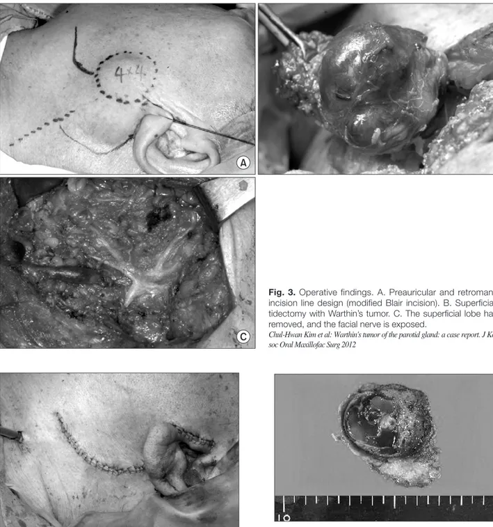
The skin incision upon surgery started from the tragus and extended backward along the curve of the ear to the bottom of the mandibular angie as a variant Blair incision. A vasoconstrictor was injected, with skin incision and platysma incision done followed by tissue dissection to expose the investing layer of the parotid gland and well-encapsulated tumor. The main trunk was first identified, and the branch was dissected; the tumor and the investing layer of the parotid gland were then carefully removed.(Fig. 3) Due to time and cost constraints, no other test was conducted aside from CT;
thus, to rule out the possibility of a malignant tumor, some tissues were removed and subjected to a frozen section test.
The result showed that the tumor was benign, so a drainage tube was inserted and suturing was done by layer.(Fig. 4) The excised tumor measured 3.5×3.5 cm and consisted of grayish, yellowish, and gelatinous material.(Figs. 5, 6)
After surgery, the patient did not experience any particular symptom such as facial nerve palsy; he was discharged in 6 days after the removal of suture material.
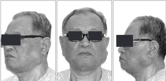
for hyper tension for 20 years and that he used to smoke half a pack of cigarettes a day for 30 years but had given up smoking 2 years before. A hard, immobile tumor with diameter of 4 cm was found around his left mandibular angie but with no tenderness, heat sensation, or difficulty of mouth opening by palpation involved. The patient complained of a recent amblyacousia. No intraoral abnormality was found, and no infection in his lymph node and other areas was palpated on his head and neck.(Fig. 1)
No abnormality was found in his panoramic radiograph. He was subjected to computed tomography (CT), which identified a well-defined, contrast-enhanced round lesion with diameter of 3.5×3.5 cm and which included multiple cystomas and small multiple lymph nodes in Levels II and III.(Fig. 2)
After considering the combined clinical and radiological test results, the patient was assumed to have a benign tumor like pleomorphic adenoma or Warthin’s tumor rather than mucoepidermoid carcinoma since the patient did not feel pain and the lesion was well-defined.
Fig. 1. Preoperative extraoral findings:
tumor of the left parotid gland.
Chul-Hwan Kim et al: Warthin's tumor of the parotid gland: a case report. J Korean Assoc Oral Maxillofac Surg 2012
Fig. 2. Preoperative computed tomo- graphy (CT) findings. Enhanced CT image of Warthin’s tumor of the super- ficial lobe of the left parotid gland.
3.5×3.5 cm well-defined margins of the tumor and heterogeneous contrast enhancing with multifocal cystic por- tion. A. Axial view. B. Coronal view.
Chul-Hwan Kim et al: Warthin's tumor of the parotid gland: a case report. J Korean Assoc Oral Maxillofac Surg 2012
parotid gland was parotid tail, followed by the anterior margin of the ear lobe. It is at times found in the mouth, mostly in the palate or buccal mucosa2,9. It is often multicentric and bilateral (10-30% and 5-9%, respectively). Most cases are found in population aged 58-70 with rare cases (below 6%) of below 40 years of age10. Five men for every woman are known to suffer from this condition; according to a more recently published paper, however, the male and female
III. Discussion
As the second most frequent type of tumor found in the salivary gland, Warthin’s tumor accounts for 14-30% of all tumors found in the parotid gland7. This tumor is at times found in the submaxillary gland, but is more frequently found in the parotid gland, mostly in the investing layer, but rarely in the deep lobe (10%)8. The most frequent location in the
Fig. 3. Operative findings. A. Preauricular and retromandibular incision line design (modified Blair incision). B. Superficial paro- tidectomy with Warthin’s tumor. C. The superficial lobe has been removed, and the facial nerve is exposed.
Chul-Hwan Kim et al: Warthin's tumor of the parotid gland: a case report. J Korean As- soc Oral Maxillofac Surg 2012
Fig. 4. Postoperative findings.
Chul-Hwan Kim et al: Warthin's tumor of the parotid gland: a case report. J Korean As- soc Oral Maxillofac Surg 2012
Fig. 5. Gross finding. The cut surface shows a few cystic spaces containing grayish, yellowish, and gelatinous material.
Chul-Hwan Kim et al: Warthin's tumor of the parotid gland: a case report. J Korean As- soc Oral Maxillofac Surg 2012
spite of its excellent contrast and effectiveness in assessing the lesion. In contrast, CT is relatively cheap and quick as well as accurate in identifying the location, malignancy, and correlation with facial nerves, at the same time reducing the radiation exposure; thus serving as an ideal means of testing15. In recent years, fine needle aspiration cytology has been widely used in diagnosing the neoplasm of the salivary gland, with testing accuracy of up to 87-97%16. Warthin’s tumor is diagnosable since the acidophil tumor cells and lymph node are clearly visible during such a test. Nonetheless, caution is required since up to 26% of Warthin’s tumor cases are misdiagnosed as malignant17. In this case, CT was first performed since it was the simplest and most accurate means of identifying the lesion. The result of the test showed that the lesion was likely benign since it was well-defined. With the patient refusing to be tested additionally due to time, distance, and cost constraints, surgery was planned without further tests.
There are several conflicting opinions on the ideal treat- ment of Warthin’s tumor. A general consensus is that surgical removal is required, but the scope of surgery is still a controversial subject. Batsakis18 considered this tumor’s likelihood of turning malignant to be only 0.3%, and that it was the least harmful tumor among other tumors found in the salivary gland. Thus, they asserted that it was curable with enucleation18. Another research concluded that parotidectomy was required to reduce the recurrence rate since Warthin’s tumor is likely to be bilateral and multicentric7. Since it is largely well-defined and located on the surface layer of the parotic gland, superficial parotidectomy is usually used; if patient ratio is now reduced to 1.6 : 1. The most convincing
cause of such increase among women is the increase in the number of female smokers. According to a report, cases of Warthin’s tumor are 8 times more frequent among smokers than among non-smokers, with up to 90% of tumor patients having a history of smoking; thus showing clear correlation between the occurrence of tumor and smoking11,12. In this case, too, the patient was a middle-aged male who had a long history of smoking. The location where the tumor was found was typical, i.e., tail part of the investing layer of the parotid gland.
Warthin’s tumor is generally a crystalline lesion found as a round or an oval-shaped swelling at or around the parotid gland. It causes little or no pain and tinnitus, pain in the ear, and deafness in some cases. The size can be several millimeters to centimeters and 2-4 cm on the average.
According to a study, 56% of a total of 278 subjects had 1-3 cm, and 40%, 4-6 cm, with 1 subject having over 10 cm13. In this case, too, the patient did not complain of pain, and the size of the tumor was average. The patient complained of amblyacousia.
Warthin’s tumor is tested prior to surgery by ptyalography, ultrasonography, magnetic resonance imaging (MRI), and CT. Ptyalography can be used to assume a diagnosis with “ball in hand appearance.” Ultrasonography does not expose the patient to radiation; although it has the advantages of short duration and lower cost, ultrasonography can be used only as an auxiliary means of testing since the tumor may be found as a sonolucent cyst even though it actually consists of cyst and solid tumor14. MRI is expensive, taking a long time in
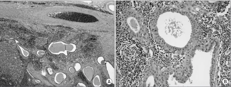
Fig. 6. Microscopic findings. The tumor consists of cystic spaces lined by bilayered oncocytic epithelium and lymphoid stroma (H&E staining, A: x40, B: x200).
Chul-Hwan Kim et al: Warthin's tumor of the parotid gland: a case report. J Korean Assoc Oral Maxillofac Surg 2012
1981;39:362-6.
4. Heller KS, Attie JN. Treatment of Warthin's tumor by enucleation.
Am J Surg 1988;156:294-6.
5. Albrecht H, Arzt L. Papillare cystadenome in lymphdrusen. Frankfurt Z Pathol 1910;4:47-69.
6. Thompson AS, Bryant HC Jr. Histogenesis of papillary cysta- denoma lymphomatosum (Warthin's tumor) of the parotid salivary gland. Am J Pathol 1950;26:807-49.
7. Teymoortash A, Krasnewicz Y, Werner JA. Clinical features of cystadenolymphoma (Warthin's tumor) of the parotid gland: a retrospective comparative study of 96 cases. Oral Oncol 2006;42:
569-73.
8. Maiorano E, Lo Muzio L, Favia G, Piattelli A. Warthin's tumour:
a study of 78 cases with emphasis on bilaterality, multifocality and association with other malignancies. Oral Oncol 2002;38:35-40.
9. Chapnik JS. The controversy of Warthin's tumor. Laryngoscope 1983;93:695-716.
10. Yih WY, Kratochvil FJ, Stewart JC. Intraoral minor salivary gland neoplasms: review of 213 cases. J Oral Maxillofac Surg 2005;63:
805-10.
11. Lamelas J, Terry JH Jr, Alfonso AE. Warthin's tumor: multi- centricity and increasing incidence in women. Am J Surg 1987;
154:347-51.
12. Monk JS Jr, Church JS. Warthin's tumor. A high incidence and no sex predominance in central Pennsylvania. Arch Otolaryngol Head Neck Surg 1992;118:477-8.
13. Párraga-Linares L, Aguirre-Urízar JM, Berini-Aytés L, Gay- Escoda C. Papillary cystoadenoma lymphomatosum (Warthin- like) of minor salivary glands. Med Oral Patol Oral Cir Bucal 2009;14:e597-600.
14. Allegra SR. Warthin's tumor: a hypersensitivity disease? Ultra- structural, light, and immunofluorescent study. Hum Pathol 1971;2:
403-20.
15. Koyuncu M, Seşen T, Akan H, Ismailoglu AA, Tanyeri Y, Tekat A, et al. Comparison of computed tomography and magnetic resonance imaging in the diagnosis of parotid tumors. Otolaryngol Head Neck Surg 2003;129:726-32.
16. Stewart CJ, MacKenzie K, McGarry GW, Mowat A. Fine-needle aspiration cytology of salivary gland: a review of 341 cases. Diagn Cytopathol 2000;22:139-46.
17. Parwani AV, Ali SZ. Diagnostic accuracy and pitfalls in fine- needle aspiration interpretation of Warthin tumor. Cancer 2003;99:166-71.
18. Batsakis JG. Carcinoma ex papillary cystadenoma lympho- matosum. Malignant Warthin's tumor. Ann Otol Rhinol Laryngol 1987;96:234-5.
19. Maddox PT, Paydarfar JA, Davies L. Parotidectomy: a 17-year institutional experience at a rural academic medical center. Ann Otol Rhinol Laryngol 2012;121:100-3.
20. O'Brien CJ. Current management of benign parotid tumors--the role of limited superficial parotidectomy. Head Neck 2003;25:946- 52.
the lesion is in the deep lobe, however, deep parotidectomy is done after performing superficial parotidectomy and confirming the branches of facial nerves. Note, however, that parotidectomy with extended range reportedly results in a high ratio of complications. More specifically, approximately 8% of the patients who undergo such surgery on the area beneath the investing layer reported Frey’s syndrome19. Yet another research showed that 27% of such patients experienced facial nerve palsy, and that 2.5% experienced permanent facial nerve palsy20. Therefore, in recent years, partial parotidectomy has been used; it is basically the same with palliative superficial parotidectomy, but the tumor is removed together with normal parotid gland tissues instead of removing all investing layers of the parotid gland depending on the location of the tumor. Since Warthin’s tumor is often found at the tail part of the parotid gland, partial paroti- dectomy may be an appropriate procedure because it removes only the bottom branches of the facial nerves and the tumor including the surrounding normal tissues. In this case, the tumor was widely seated over the investing layer of the parotid gland; thus, superficial parotidectomy was conducted to reduce the risk of recurrence instead of simply removing the tumor.
This case will be presented together with the clinical characteristics and treatment details and a consideration of references as an elderly patient aged 69 years having Warthin’s tumor that was found to be typical based on CT and yielding a successful result from the surgery with no particular complication.
References
1. Warthin AS. Papillary cyst adenoma lymphomatosum: a rare teratoid of the parotid region. J Cancer Res 1929;13:116-25.
2. Chaudhry AP, Gorlin RJ. Papillary cystadenoma lympho- matosum (adenolymphoma); a review of the literature. Am J Surg 1958;95:923-31.
3. Ibi A, Yokobayashi T, Kawasaki T, Nakajima T. Bilateral Warthin's tumor: report of case and review of Japanese literature. J Oral Surg