246
Nonsurgical treatment of stylohyoid (Eagle) syndrome: a case report
Arman Taheri, Shahram Firouzi-Marani, Masoud Khoshbin
Department of Anesthesiology and Pain Management, Amir Alam Hospital, Tehran University of Medical Science, Tehran, Iran
Abstract(J Korean Assoc Oral Maxillofac Surg 2014;40:246-249)
Eagle syndrome is a rare condition caused by elongation of the styloid process or calcification of the stylohyoid ligament. Patients with Eagle syn- drome typically present with dysphagia, dysphonia, cough, voice changes, otalgia, sore throat, facial pain, foreign body sensation, headache, vertigo, and neck pain. Here we report a case in which the patient initially presented with sore throat, left-sided facial pain, and cough. This case report provides a brief review of the diagnosis and nonsurgical management of this rare syndrome.
Key words: Eagle syndrome, Dysphonia, Heterotopic ossification, Temporal bone
[paper submitted 2014. 9. 18 / accepted 2014. 9. 19]
The prevalence of styloid process elongation or stylohyoid ligament mineralization in imaging studies has been reported to be between 19.4% to 52.1% in the general population5-7, and in up to 76% of patients with temporomandibular disor- der8. However, the incidence of Eagle syndrome in the gen- eral population is underestimated since only 6% of those with an elongated styloid process have symptoms9.
Here, we present a case of Eagle syndrome treated with medical therapy.
II. Case Report
A 53-year-old woman was referred to the pain clinic at Amir Alam Hospital with a complaint of pain in the throat and the left side of the head and face. Her pain began 16 years prior following a root canal of a mandibular tooth on the left side. She at times experienced severe pain attacks lasting approximately 10 minutes with a frequency of 10 times per day, which extended from the external auditory canal to the left side of the throat. Additionally, cough was a trigger for these pain attacks. Certain odors or nervousness were also aggravating factors of her pain, while pressure on the ear and diphenhydramine were mitigating factors. The patient reported her highest and lowest pain intensity as 4 and 2, respectively, according to the numeric rating scale (NRS).
The patient had a past medical history significant for hyper- tension and favism.
The patient’s general health as well as her head and neck
I. Introduction
Eagle syndrome is a rare clinical condition caused by elon- gation of the styloid process or by mineralization (ossification or calcification) of the stylohyoid ligament1. The length of the styloid process is normally 2 to 2.5 cm, and elongation be- yond 2.5 cm causes Eagle syndrome, which was first defined in 19372,3.
This syndrome has two types, including classic and styloid- carotid syndromes. The classic syndrome, also known as stylalgia, is usually characterized by pharyngeal pain local- ized to the tonsillar fossa, referred otalgia, and neck pain.
It also may be associated with dysphagia, hypersalivation, sensation of a foreign body in the throat, and transient voice changes that are often seen following tonsillectomy4. Styloid- carotid syndrome is characterized by nonspecific symptoms caused by compression of the carotid arteries and sympathetic fibers by the styloid process4. The most common etiology of this syndrome is ossification or calcification of the stylohyoid ligament.
CASE REPORT
Arman Taheri
Department of Anesthesiology and Pain Management, Amir Alam Hospital, North Sadi, Tehran 13185-1678, Iran
TELEFAX: +98-21-66423101 E-mail: [email protected]
This is an open-access article distributed under the terms of the Creative Commons Attribution Non-Commercial License (http://creativecommons.org/licenses/by-nc/3.0/), which permits unrestricted non-commercial use, distribution, and reproduction in any medium, provided the original work is properly cited.
CC
Copyright Ⓒ 2014 The Korean Association of Oral and Maxillofacial Surgeons. All rights reserved.
http://dx.doi.org/10.5125/jkaoms.2014.40.5.246 pISSN 2234-7550·eISSN 2234-5930
Eagle syndrome
247 treatment, her pain severity improved by about 80% and her NRS score decreased to one. The patient also experienced oc- casional pain-free intervals.(Table 1)
III. Discussion
In this study, Eagle syndrome was identified in our patient, after which medical therapy was initiated with an appropriate response.
Eagle syndrome may be unilateral or bilateral, although in most cases symptoms are unilateral. According to Moon et al.10, Eagle syndrome and elongation of the stylohyoid liga- ment is typically a bilateral process, though in our patient this condition was unilateral.
A calcified stylohyoid ligament is found in 4% to 28% of normal population11. There is no direct correlation between the severity of Eagle syndrome and level of calcification12. However, there is a clear relationship between longer lengths of the styloid process and higher pain intensity and severity of Eagle syndrome8.
Pharyngeal pain with radiation to the neck and ears poses a very difficult challenge for interpretation, including a vast number of differential diagnoses13-17.(Table 2) The dif- ferential diagnosis for Eagle syndrome includes inflamma- tory disorders, masses of the pharynx and tongue base, and cranial nerve neuralgia. On the other hand, patients with an elongated styloid process manifest some transient and non- specific symptoms as well as the severe classic symptoms, which require surgery18. Traumatic fracture of the apophysis, pharyngeal infection and inflammation, rheumatoid disorders of the hyoid bone, inflammation of the muscles attached to the styloid process, osteoarthritis of the cervical vertebrae, pressure on adjacent nerves such as cranial nerve IX and the were thoroughly examined. Her skull, cranial nerves, eyes,
ears, nose, sinuses, thyroid, and dentition were unremarkable.
No cervical, axillary, epitrochlear, or inguinal adenopathy was detected. On oral and pharyngeal examination, severe tenderness was present on a bony prominence in the tonsillar cavity. Head and neck rotation was painful at the end of ac- tive range of motion. As a result, lateral skull and neck x-rays were taken that showed elongation of the styloid process, thereby confirming the diagnosis of Eagle syndrome.(Fig. 1) Laboratory studies and histopathology were not performed.
Upon diagnosis, treatment with pregabalin (75 mg/day) and amitriptyline (10 mg/day) was initiated. These medications improved the patient’s cough and reduced the pain severity by about 50% within 2 months. Three and 6 months after
Fig. 1. A lateral skull and neck X-ray shows increased length of the styloid process.
Arman Taheri et al: Nonsurgical treatment of stylohyoid (Eagle) syndrome: a case report.
J Korean Assoc Oral Maxillofac Surg 2014
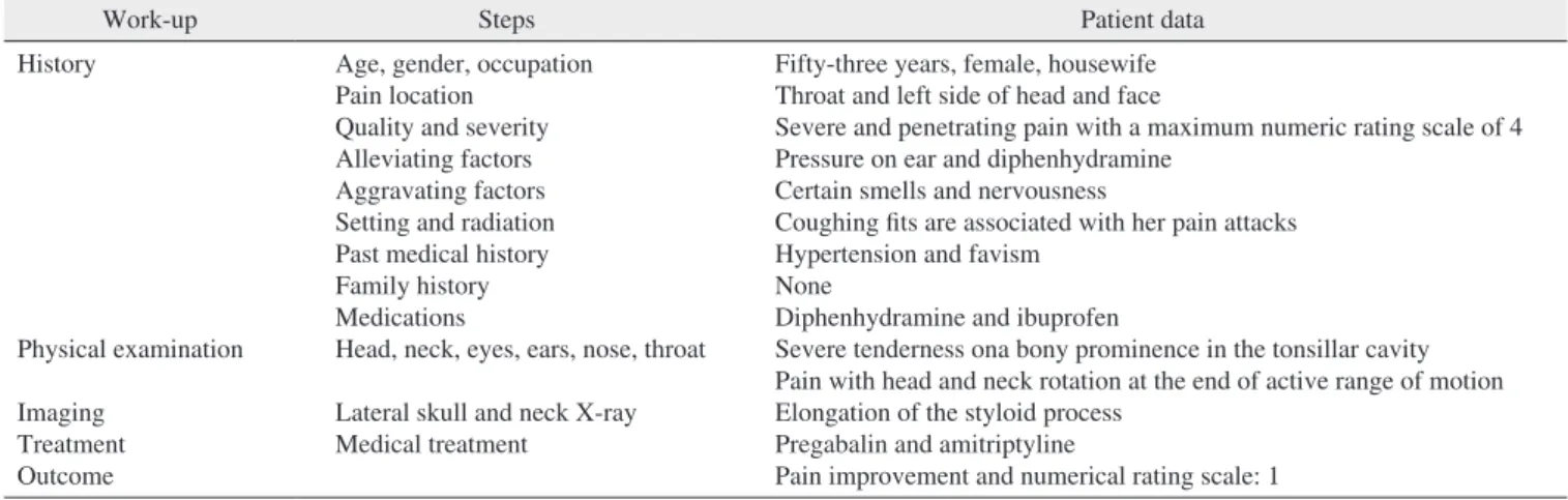
Table 1. Patient history, work-up, treatment, and outcomes
Work-up Steps Patient data
History
Physical examination Imaging
Treatment Outcome
Age, gender, occupation Pain location
Quality and severity Alleviating factors Aggravating factors Setting and radiation Past medical history Family history Medications
Head, neck, eyes, ears, nose, throat Lateral skull and neck X-ray Medical treatment
Fifty-three years, female, housewife Throat and left side of head and face
Severe and penetrating pain with a maximum numeric rating scale of 4 Pressure on ear and diphenhydramine
Certain smells and nervousness
Coughing fits are associated with her pain attacks Hypertension and favism
None
Diphenhydramine and ibuprofen
Severe tenderness ona bony prominence in the tonsillar cavity Pain with head and neck rotation at the end of active range of motion Elongation of the styloid process
Pregabalin and amitriptyline
Pain improvement and numerical rating scale: 1 Arman Taheri et al: Nonsurgical treatment of stylohyoid (Eagle) syndrome: a case report. J Korean Assoc Oral Maxillofac Surg 2014
J Korean Assoc Oral Maxillofac Surg 2014;40:246-249
248
Surgery with resection of the elongated styloid process is considered to be definitive treatment, however, surgery may be contraindicated in some cases or patients may decline operative intervention. In these cases, conservative treatment with oral medications may be considered. In cases that do not respond to medical therapies, surgery is indicated. Different medications may be used in medical management of Eagle syndrome based on the respective etiology, including anal- gesics, anticonvulsants, antidepressants, and local infiltration with steroids or long-acting local anesthetic agents20. In our case, a tricyclic antidepressant (amitriptyline) and an anticon- vulsant (pregabalin) were started after diagnosis. Our patient dramatically and persistently responded to conservative treat- ment after 3 and 6 months of therapy. Nonsurgical treatment of Eagle syndrome with gabapentin, tianeptine, tramadol, acetaminophen, local lidocaine injection and stellate ganglion block has also been reported20. However, to our knowledge, our patient is the first case of Eagle syndrome to be treated with only oral medications, while previous similar cases also utilized a stellate ganglion block2,10,14-16,20.
The exact mechanism by which medications achieve symp- tom relief in patients with Eagle syndrome is not known.
mandibular branch of the 5th cranial nerve and corda tym- pani, pathologies of the third molar, and pharyngeal scar fol- lowing tonsillectomy are other pathologies that could mimic symptoms of Eagle syndrome. Different forms of pharyngeal neuralgia may also result in similar symptoms including laryngeal neuralgia, occipital neuralgia (involving Arnold’s nerve), sphenopalatine neuralgia (secondary to sphenoeth- moid inflammation), and finally trigeminal neuralgia which may account for sporadic pains with pressure within the auditory canal19. Disorders of the temporomandibular joint constitute another possible diagnosis. In our case, most other differential diagnoses were ruled out according to the history and physical examination findings.
Clinical diagnosis rests upon previous history of trauma or tonsillectomy and palpation of the tonsillar fossa. Radiologic studies such as an orthopantomograph or lateral skull view with the head slightly extended may help to confirm this di- agnosis18. A precise history, examination, and imaging stud- ies also contribute greatly to achieving the correct diagnosis.
(Table 2) The final diagnosis in our case was confirmed by imaging.
Medical therapy is first-line treatment for Eagle syndrome.
Table 2. Differential diagnoses for pain in the head, cervicofacial, and cervicopharyngeal regions
Etiology Differential diagnosis
Vascular13
Muscle spasm13,14
Without demonstrable physical substrate Combined tension-
migraine
Meningeal inflammation Altered intracranial
pressure
Cranial neuralgias15,16 Bones and joints13,14
Ear, nose, and throat diseases
Other diseases17 Referred pain
Migraine, cluster headache, chronic tension and cervicogenic headaches, carotidynia, atypical facial pain, paroxysmal hemicranias; headaches of reactive vasodilation: fever, drug-induced, postictal, hyperthyroidism, hypoglycemia, hypoxia, hypercarbia; headaches associated with arterial hypertension: chronic severe hypertension, pheochromocytoma, coital headaches; headaches caused by cranial arteritis: temporal arteritis, etc.
Headache of posturally-induced or perilesional muscle spasm: impaired posture, cervical spondylosis and other diseases of cervical spine; myofascial pain dysfunction syndrome (headache or facial pain associated with disorders of teeth, jaws, and related structures, or TMJ syndrome); headaches associated with psychophysiologic muscular contraction: muscle contraction headaches or tension-type headaches associated with disorder of pericranial muscles Headaches of uncertain etiology: tension headaches unassociated with disorder of pericranial muscles, some forms of
posttraumatic headache; psychogenic headaches: hypochondriacal, conversional, delusional, and malingered; facial pain of uncertain etiology: atypical facial pain
Episodic migraine superimposed on chronic tension headaches, chronic daily headaches (associated with analgesic and/or ergotamine overuse, also called rebound headaches; not associated with drug overuse)
Subarachnoid hemorrhage, meningitis and meningoencephalitis, meningeal carcinomatosis
Increased intracranial pressure: intracranial mass lesions (neoplasm, hematoma, abscess, etc.), hydrocephalus, benign intracranial hypertension, venous sinus thrombosis
Decreased intracranial pressure: postlumbar puncture headaches, spontaneous hypoliquorrheic headaches Postherpetic neuralgia, glossopharyngeal, trigeminal, superior laryngeal, occipital, pterygopalatine ganglion,
intermediate nerve, geniculate neuralgia
Clicking jaw, nonerupted or distorted third molar, faulty dental prostheses, salivary gland disease, degenerative disc disease, diffuse idiopathic skeletal hyperostosis, rheumatoid arthritis, spondyloarthropathies, juvenile rheumatoid arthritis, osteomyelitis, infectious discitis, stylohyoid (Eagle) syndrome
Chronic tonsillitis, tonsillar calculi, spasm of the pharyngeal constrictor muscle, otitis, mastoiditis, fracture of the hyoid bone, pterygoidhamulus bursitis
Chronic laryngopharyngeal reflux, psychosomatic diseases, foreign bodies, inflammatory and neoplastic processes in the oropharyngeal area, pharyngeal and base of tongue tumors, nuchal cellulitis and fibrositis, neck-tongue syndrome TMJ pain, cardiac pain, diaphragmatic irritation, gastrointestinal sources (peptic ulcer disease, gallbladder, pancreas) (TMJ: temporomandibular joint)
Arman Taheri et al: Nonsurgical treatment of stylohyoid (Eagle) syndrome: a case report. J Korean Assoc Oral Maxillofac Surg 2014
Eagle syndrome
249
4. Soldati AB, Miguelote C, Quero C, Pereira R, Santos R, Soares C.
Eagle's syndrome. Arq Neuropsiquiatr 2013;71:265-6.
5. More CB, Asrani MK. Evaluation of the styloid process on digital panoramic radiographs. Indian J Radiol Imaging 2010;20:261-5.
6. Phulambrikar T, Rajeshwari A, Rao BB, Warhekar A, Reddy P. In- cidence of elongated styloid process: a radiographic study. J Indian Acad Oral Med Radiol 2011;23:S344-6.
7. Bagga MB, Kumar CA, Yeluri G. Clinicoradiologic evaluation of styloid process calcification. Imaging Sci Dent 2012;42:155-61.
8. de Andrade KM, Rodrigues CA, Watanabe PC, Mazzetto MO.
Styloid process elongation and calcification in subjects with tmd:
clinical and radiographic aspects. Braz Dent J 2012;23:443-50.
9. Ilgüy M, Ilgüy D, Güler N, Bayirli G. Incidence of the type and calcification patterns in patients with elongated styloid process. J Int Med Res 2005;33:96-102.
10. Moon CS, Lee BS, Kwon YD, Choi BJ, Lee JW, Lee HW, et al.
Eagle's syndrome: a case report. J Korean Assoc Oral Maxillofac Surg 2014;40:43-7.
11. Gorlin RJ, Cohen MM, Levin LS. Syndromes of the head and neck. New York: Oxford University Press; 1990.
12. Goaz PW, White SC. Oral radiology: principles and interpretation.
St. Louis, Missouri: Mosby; 1994.
13. Casale M, Rinaldi V, Quattrocchi C, Bressi F, Vincenzi B, Santini D, et al. Atypical chronic head and neck pain: don't forget Eagle's syndrome. Eur Rev Med Pharmacol Sci 2008;12:131-3.
14. Gelabert-González M, García-Allut A. Eagle syndrome. An un- usual cause of neck pain. Neurocirugia (Astur) 2008;19:254-6.
15. Chi J, Harkness M. Elongated stylohyoid process: a report of three cases. N Z Dent J 1999;95:11-3.
16. Nishihara K, Hanakita J, Kinuta Y, Kondo A, Yamamoto Y, Kishimoto S. Three cases of Eagle's syndrome. No Shinkei Geka 1986;14(3 Suppl):441-5.
17. Eversole LR. Clinical outline of oral pathology: diagnosis and treatment. 2nd ed. Philadelphia: Lea and Febiger; 1978.
18. Prasad KC, Kamath MP, Reddy KJ, Raju K, Agarwal S. Elongated styloid process (Eagle's syndrome): a clinical study. J Oral Maxil- lofac Surg 2002;60:171-5.
19. Mortellaro C, Biancucci P, Picciolo G, Vercellino V. Eagle's syn- drome: importance of a corrected diagnosis and adequate surgical treatment. J Craniofac Surg 2002;13:755-8.
20. Han MK, Kim DW, Yang JY. Non surgical treatment of Eagle's syndrome: a case report. Korean J Pain 2013;26:169-72.
The cause of pain in Eagle syndrome is the stimulation of adjacent nerves by the elongated styloid process and second- ary induced inflammation. It seems that medications such as anticonvulsants and antidepressants may reduce nerve stimulation and consequently pain intensity by altering the concentration of neurotransmitters, and analgesics such as nonsteroidal antiinflammatory drugs may improve pain by reducing inflammation in adjacent tissues.
In conclusion, lateral skull imaging in cases suspicious for Eagle syndrome is recommended to confirm this diagnosis, and medical therapy should be considered as first-line treat- ment for this rare condition.
Conflict of Interest
No potential conflict of interest relevant to this article was reported.
Acknowledgements
The authors would like to thank the Farzan Institute for Re- search and Technology for technical assistance.
References
1. Balbuena L Jr, Hayes D, Ramirez SG, Johnson R. Eagle's syn- drome (elongated styloid process). South Med J 1997;90:331-4.
2. Eagle WW. Elongated styloid processes: report of two cases. Arch Otolaryngol 1937;25:584-7.
3. Murtagh RD, Caracciolo JT, Fernandez G. CT findings associated with Eagle syndrome. AJNR Am J Neuroradiol 2001;22:1401-2.