대한소화기학회지 2006;47:248-253 □ REVIEW □
서 론
모든 암종의 수술 전 병기가 치료방침을 결정하는 데 중 요하다는 것은 분명하다. 특히 직장암에서는 그 역할이 더 욱 중요하여, 환자의 예후를 판단하는 주요 지표일 뿐 아니 라 수술 전 추가 치료 여부를 결정하는 결정적인 판단 기준 이 되고 있다. 이는 외측절제연 침범 여부나 림프절 전이 여 부와 같이 병기 자체가 국소 재발과 밀접한 관계가 있을 뿐 아니라,1-3 수술 전 고용량 동시항암화학방사선치료 또는 저
용량 방사선치료가 일부 진행 직장암에서 국소재발을 의미 있게 줄였다는 보고들이 발표되면서 수술 전 병기 결정에 대한 관심이 늘고 또한 이를 바탕으로 수술 전 방사선치료 의 역할에 대한 논의가 뜨겁게 이루어지고 있기 때문이 다.4-6 이로써 직장암 환자에서 향후 치료방향 결정과 환자 의 예후 판단을 위해 정확한 영상 병기 결정은 결정적인 사 항이 되었다.
수술 전 영상 검사로는 경직장 초음파(transrectal ultrason- ography, TRUS), 컴퓨터 단층촬영(computed tomography, CT), ꠏꠏꠏꠏꠏꠏꠏꠏꠏꠏꠏꠏꠏꠏꠏꠏꠏꠏꠏꠏꠏꠏꠏꠏꠏꠏꠏꠏꠏꠏꠏꠏꠏꠏ
연락처: 김주희, 135-720, 서울시 강남구 도곡동 146-92 연세대학교 의과대학 영동세브란스병원 영상의학과 Tel: (02) 2019-3510, Fax: (02) 3462-5472
E-mail: [email protected]
ꠏꠏꠏꠏꠏꠏꠏꠏꠏꠏꠏꠏꠏꠏꠏꠏꠏꠏꠏꠏꠏꠏꠏꠏꠏꠏꠏꠏꠏꠏꠏꠏꠏꠏ Correspondence to: Joo Hee Kim, M.D.
Department of Diagnostic Radiology, Yongdong Severance Hospital Yonsei University College of Medicine, 146-92 Dogok-dong, Gangnam- gu, Seoul 135-720, Korea
Tel: +82-2-2019-3510, Fax: +82-2-3462-5472 E-mail: [email protected]
직장암 수술 전 Magnetic Resonance Imaging 병기 결정의 임상 중요성
연세대학교 의과대학 세브란스병원 영상의학과
김 주 희
Clinical Significance of Preoperative Magnetic Resonance Imaging in Staging of Rectal Cancer
Joo Hee Kim, M.D.
Department of Diagnostic Radiology, Severance Hospital, Yonsei University College of Medicine, Seoul, Korea
Rectal cancer carries poor prognosis because of metastasis and local recurrence. Local recurrence has a profound effect on morbidity and quality of life. Randomized trials have proven that neoadjuvant treatment can significantly reduce local recurrence rate in some selected cases of advanced rectal cancer. Therefore, preoperative staging of rectal cancer has an important impact on treatment plan. Two main factors in predicting the local recurrence are known as the circumferential resection margin (CRM) and the nodal status. Recently, high-resolution magnetic resonance imaging (MRI) is regarded as a superior modality in the preoperative assessment of CRM with high accuracy and reproducibility. However, the results of imaging in predicting of nodal involvement are not satisfactory.
In order to increase the accuracy of preoperative nodal staging, several efforts were done to evaluate lymph node by MRI or by pelvic MRI using lymph node-specific contrast agent (ultrasmall superparamagnetic iron oxide, USPIO).
In this review, the role of MRI in preoperative evaluation of rectal cancer will be discussed. (Korean J Gastro- enterol 2006;47:248-253)
ꠏꠏꠏꠏꠏꠏꠏꠏꠏꠏꠏꠏꠏꠏꠏꠏꠏꠏꠏꠏꠏꠏꠏꠏꠏꠏꠏꠏꠏꠏꠏꠏꠏꠏꠏꠏꠏꠏꠏꠏꠏꠏꠏꠏꠏꠏꠏꠏꠏꠏꠏꠏꠏꠏꠏꠏꠏꠏꠏꠏꠏꠏꠏꠏꠏꠏꠏꠏꠏꠏꠏꠏꠏꠏꠏꠏꠏꠏꠏꠏꠏꠏꠏꠏꠏꠏꠏꠏꠏꠏꠏꠏꠏꠏꠏꠏꠏꠏꠏꠏꠏꠏꠏꠏꠏꠏꠏꠏꠏꠏꠏꠏꠏ
Key Words: Rectum, neoplasm; Magnetic resonance imaging; Staging
김주희. 직장암 수술 전 Magnetic Resonance Imaging 병기 결정의 임상 중요성 249
자기공명영상(magnetic resonance imaging, MRI) 등이 이용되 고 있다. 이 중 고해상 골반 MRI는 가장 신뢰할 수 있는 검 사이다.7-9 이는 MRI의 조직 대조도가 뛰어나서 복잡한 골반 강 내 구조들을 잘 구별하여 보여줄 수 있어서 골반강 장기 인 직장암 검사에 유용하고, 병기 결정에 중요한 지표가 되 는 직장의 고유근층(muscularis propria)과 직장간막근막(me- sorectal fascia)이 MRI에서 가장 잘 보이기 때문이다(Fig. 1).
T 병기 결정이나 N 병기 결정뿐 아니라 외측절제연의 침범 여부를 판단하는 데에 있어 MRI의 역할이 널리 알려지면서 많은 기관에서 MRI를 선호하고 있다. 이번 종설에서는 직 장암의 병기 결정에 있어서 MRI의 역할과 그 의의를 알아 보고자 한다.
고해상 골반 MRI(high resolution pelvic MRI)
MRI는 방사선 피폭 없이 높은 해상도의 연조직 대조 영 상을 원하는 여러 평면으로 얻을 수 있다. 동체코일(body coil)을 사용한 초기 MRI 결과들은 그다지 만족스럽지 못하 였고 CT에 비해서도 큰 장점이 없었다.10-12 이는 관내코일 (endoluminal coil)이 개발되면서 개선되었는데 뛰어난 공간 분해능으로 인해 초기 대장암에서의 T 병기 결정에 현저한 향상을 보였다.13,14
그러나 관내코일을 이용한 MRI에는 몇 가지 제한점이 있 는데, 코일에서 멀어지면 급격한 신호감소가 나타나서 영상
범위가 제한되고 근위 대장암에서는 사용이 어려울 뿐 아니 라 짧지 않은 촬영시간 동안 직장 내 코일을 삽입함에 따라 통증과 불편감이 크고 실패율이 높은 편이다. 특히 제한적 인 영상 범위는 직장간막 근막의 확인이나 주변 골반강 장 기의 확인이 어려워 진행된 직장암에서는 오히려 정확도를 떨어뜨린다. 따라서 최근에는 위상배열 골반다중코일 (phased array pelvic multicoil)을 이용한 MRI가 표준검사로 인정되고 있다.7,8,15
직장암을 위한 MRI를 시행할 때 장세척은 필요하지 않으 며 buscopan과 같은 연축억제제는 필요에 따라 사용할 수 있다. 직장암 환자의 MRI에서는 3 mm 두께의 고해상도로 시상면을 얻고 이를 바탕으로 주병변에 대해 수직 평면을 얻는 것이 중요하다. Gadolinium 제제의 비특이적인 조영제 의 사용은 직장암의 수술 전 평가에는 큰 도움이 되지 않으 므로 통상적으로 사용할 필요가 없고,16 따라서 비교적 간단 하게 검사를 마칠 수 있다.
직장암 진단을 위한 기본 해부학
직장암 수술에 있어 직장간막 근막을 따라 예리하게 절개 박리하여 직장과 직장간막을 한 덩어리(en bloc)로 제거하는 전직장간막절제술(total mesorectal excision, TME)이 표준치료 로 인정되고 있다. 따라서 전직장간막절제술을 위한 해부구 조의 이해와 이를 근거로 한 MRI 영상 판독이 필수이다.
근위직장의 전방과 외측은 복막에 싸여 있지만 근위 직장 의 후방과 원위 직장은 장막과 복막이 없고 직장간막 근막 에 의해 직장간막을 둘러싸이게 된다. 전방의 근막은 Deno- nvillier 근막이라 명명하고 있으며, 이는 남자의 정낭과 전 립선, 여자의 직장질중격(rectovaginal septum)의 바로 후방에 위치하게 된다. 이러한 직장간막 근막과 Denonvillier 근막은 전직장간막절제술의 수술 절개선이 되므로 이를 외측절제 연이라고 하며, 이러한 직장간막 주위 근막과 종양의 관계 를 명확히 제시하는 것이 MRI 영상진단의 중요한 역할이다 (Fig. 1). 직장간막은 원위부와 전방부에서 그 지방의 양이 적어 직장벽과의 거리가 가까워지고 따라서 판독이 어렵 다.17
T 병기와 외측절제연(circumferential resection margin, CRM) 결정
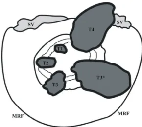
AJCC (American Joint Committee on Cancer)에서 2002년에 개편하여 제시한 TNM 병기 결정에는 대장-직장암이 총체 적으로 기술되어 있고 직장암이 따로 기술되어 있지 않다.18 이를 근거로 직장암의 T 병기를 나누면(Fig. 2),
Fig. 1. MR anatomy of the rectum, the mesorectum and mesorectal fascia. Axial T2-weighted fast spin echo MR image shows the intact hypointense rectal wall (black arrows). The rectum is surrounded by the mesorectal fat (*) and is enveloped by thin mesorectal fascia (white arrows). The mesorectal fascia and Denonvillier's fascia (arrowheads) are clearly visualized as an- atomic borders (CRM) of TME.
MR, magnetic resonance; CRM, circumferential resection margin;
TME, total mesorectal excision.
250 The Korean Journal of Gastroenterology: Vol. 47, No. 4, 2006
T1 종양이 점막하조직을 침범한 경우 T2 종양이 직장고유근을 침범한 경우
T3 종양이 직장고유근을 지나 장막하조직으로 침범한 경우(근위직장암), 또는 복막이 없는 직장 주위 조직 으로 침범한 경우(원위직장암)
T4 주변 장기로 침범하였거나, 내장측 복막이 뚫린 경 우(근위직장암)
으로 설명할 수 있다. 따라서 복막에 둘러싸이지 않은 원위 직장암의 경우는 주변 장기 침윤 외에는 다른 기준이 없으 며, 직장간막근막의 침윤 여부가 병기 결정에 영향을 주지 않는다. 즉, 국소재발과 밀접한 관계가 있다고 알려진 외측 절제연의 침윤 여부가 T 병기 결정과 상관이 없는 문제가 발생한다. 또한 수술 방법 결정에 중요한 영향을 주는 내측 괄약근의 침범 여부도 따로 기술이 없다.
따라서 현재의 T3 병기 종양을 MRI를 통해 좀더 자세히 나누어 환자의 치료방침 결정에 도움을 주는 것이 필요하다 (Fig. 3). 그 평가 방법에는 크게 두 가지가 있다. 첫째, 외측 절제연(직장간막근막)으로부터 종양(주요 종양, 종양침착물 또는 침범한 림프절을 모두 포함)까지 가장 가까운 거리를 측정하는 것이다. 이는 직장간막근막을 직접 침범하였거나 직장간막 근막에서부터의 거리가 가까운 경우 국소 재발률 이 의미있게 증가하기 때문이다. 외측절제연이 침범(CRM involvement) 또는 외측절제연이 위협 받는 경우(threatened CRM)를 명시해 주는 것이 좋다(Fig. 3C, D). 외측절제연이 Fig. 2. Schematic drawing of T stage in rectal cancer. Axial section
through the main tumor and the mesorectum shows a rectal tumor and its relation to the mesorectal fascia. Tumor that invades through the muscularis propria into the perirectal tissue is classified as T3. However, both tumors with a wide CRM and with a close or involved CRM are same T3 in present TNM staging. When the tumor is close to mesorectal fascia (T3*) on MRI, detail discrimina- tion is needed because of its higher rate of local recurrence.
CRM, circumferential resection margin; MRI, magnetic resonance imaging; SV, seminal vesicle; MRF, mesorectal fascia.
Fig. 3. Examples of the diverse T3 rectal cancers. All carcino- mas are proven as pT3. (A) Good T3 and T3A, (B) good T3 and T3B, (C) bad T3 (threatened CRM) and T3A, (D) bad T3 (CRM involvement) and T3B.
White lines indicate the shortest distance from tumor to meso- rectal fascia.
CRM, circumferential resection margin.
A B
Kim JH. Clinical Significance of Preoperative Magnetic Resonance Imaging in Staging of Rectal Cancer 251
위협 받는 경우는 보고자에 따라 차이가 있으나 종양에서부 터 직장간막근막까지의 최단 거리가 1 또는 2 mm 이내로 하는 것이 보통이다.8,19,20 또 하나의 평가 방법은 직장벽(직 장고유근)으로부터 주요 종양이 얼마나 바깥으로 자랐는지 를 측정하는 것이다. 종양이 직장벽 바깥으로 5 mm 이내까 지 나간 경우를 T3A (Fig. 3A, C)로, 5 mm 이상 나간 경우 를 T3B로 부르자는 주장도 있다(Fig. 3B, D).21 이렇게 다양 한 모습의 T3 병기 종양은 그 성격에 따라 수술 전 치료방 침을 어떻게 달리할지 더욱 연구가 필요하다.
N 병기 결정
현재 AJCC에서 제시하는 N 병기는 전이가 있는 국소 림 프절의 개수에 따라 결정된다.18
N0 국소 림프절 전이가 없는 경우
N1 1-3개의 국소 림프절에 전이가 있는 경우 N2 4개 이상의 국소 림프절에 전이가 있는 경우
국소재발의 주요 요인으로는 앞서 말한 외측절제연과 함께 림프절 전이 여부(nodal status)가 중요하다.22,23 외측절제연의 평가는 현재까지 MRI가 가장 정확한 검사방법으로 간주되 고 있으나 N 병기는 아직도 해결되지 않은 숙제로 남아 있 다. 양성 반응 림프절과 전이 림프절의 크기가 서로 중복되 어 있어 아직 그 감별에 대한 크기 기준이 확립되어 있지 않다. 또한 직장암에서는 반수 이상의 전이가 직경 5 mm
이하의 작은 림프절에서 발생하여 크기를 기준으로 판단할 때 쉽게 미세전이를 간과하게 된다.24,25 따라서 이들의 감별 은 MRI에서도 쉽지 않고 지금까지의 MRI 예측도는 만족스 럽지 않다.
최근에는 크기 기준뿐 아니라 림프절 모양으로 전이 여부 를 판단하고자 하는 노력들이 있다.26,27 림프절의 경계가 불 분명하거나(indistinct) 뾰족뾰족한 경우(speculated), 또는 반 점의 불균질(mottled heterogeneity)이 있는 경우 전이 림프절 일 가능성이 높으며, 크기와 함께 이러한 림프절 모양을 함 께 고려하면 N 병기의 예측에 있어 그 정확도를 높일 수 있 다. 또한 최근에 FDA의 공인을 받고 국내에서도 상용화 예 정인 초소형 초상자성 산화철(ultrasmall superparamagnetic iron oxide, USPIO) 제제를 이용한 MRI가 N 병기 결정에 도 움을 줄 것으로 기대한다. 이 제제는 정상 림프절의 대식세 포에 의해 포식되는 자기공명 조영제로서, 정상 림프절에 섭취되어 자화율 인공물(susceptibility artifact)에 의한 T2* 영 상에서 신호강도 감소를 초래하며, 이로 인해 정상 림프절 은 검은 신호강도를 보이는 원리를 이용한 것이다. 전립선 암이나 두경부암 환자에서의 림프절 전이 평가에 유용하게 이용되고 있다.28,29 최근에는 USPIO 제제를 이용한 MRI가 직장암 환자에서의 직장간막 림프절 감별에도 유용하다는 보고가 나오기 시작하였다.30 좀 더 연구가 필요하기는 하 나, 양성 반응 림프절과 전이 림프절의 감별이 어려운 경우, 특히 수술 방법 결정에 매우 중요한 골반벽 림프절이나 동 맥 주위 림프절 전이 여부 판단이 어려운 경우에 적용 가능 할 것으로 보인다.
결 론
직장암의 수술 전 정확한 병기 결정은 환자의 치료방침 결정에 매우 중요하다. 특히 T 병기나 N 병기뿐 아니라 외 측절제연을 평가하는 것이 직장암 평가에 있어 가장 중요한 항목이다. 이는 직장암의 수술 전 평가가 다른 암종에서와 같이 단순히 절제 가능성만을 판단하는 것이 아니라 수술 전 방사선치료와 같은 부가 치료가 필요한지를 판단하는 데 에 큰 영향을 미치기 때문에 더욱 중요하다. MRI는 직장암 의 수술 전 평가에서 가장 믿을 수 있는 검사로 여겨지고 있는데, 이는 외측절제연의 평가에서 가장 정확하고 일관된 검사로 알려져 있고, 림프절 전이의 평가에 있어서도 크기 뿐 아니라 형태 평가나 특이조영제를 사용함으로써 그 정확 도를 높일 수 있는 가능성이 있기 때문이다. 또한 항문괄약 근이나 주변 골반 장기와의 구별이 가장 용이하여, 원위 직 장암에서 특히 그 역할이 중요하게 부각되고 있다.
결론으로 직장암 환자의 경우 수술 전에 MRI로 정확한 수술 전 평가가 필요하며, 이를 근거로 여러 과간의 긴밀한 Fig. 4. A 62-year old female with T3 N1 rectal cancer. Oblique
coronal T2-weighted fast spin echo MR image revealed a small, but mottled heterogeneous node with indistinct margin (arrow). There is no other significant node in remaining area. Single metastatic regional node was proved.
MR, magnetic resonance.
252 대한소화기학회지: 제47권 제4호, 2006
협조를 통한 협동치료로 환자에게 가장 적합한 치료방법을 결정하는 것이 무엇보다 중요하다.
참고문헌
1. Quirke P, Durdey P, Dixon MF, Williams NS. Local recur- rence of rectal adenocarcinoma due to inadequate surgical resection. Histopathological study of lateral tumour spread and surgical excision. Lancet 1986;2:996-999.
2. Adam IJ, Mohamdee MO, Martin IG, et al. Role of circum- ferential margin involvement in the local recurrence of rectal cancer. Lancet 1994;344:707-711.
3. Birbeck KF, Macklin CP, Tiffin NJ, et al. Rates of circum- ferential resection margin involvement vary between surgeons and predict outcomes in rectal cancer surgery. Ann Surg 2002;
235:449-457.
4. Improved survival with preoperative radiotherapy in resectable rectal cancer. Swedish Rectal Cancer Trial. N Engl J Med 1997;336:980-987.
5. Kapiteijn E, Marijnen CA, Nagtegaal ID, et al. Preoperative radiotherapy combined with total mesorectal excision for rese- ctable rectal cancer. N Engl J Med 2001;345:638-646.
6. Sauer R, Becker H, Hohenberger W, et al. Preoperative versus postoperative chemoradiotherapy for rectal cancer. N Engl J Med 2004;351:1731-1740.
7. Brown G, Richards CJ, Newcombe RG, et al. Rectal carcin- oma: thin-section MR imaging for staging in 28 patients.
Radiology 1999;211:215-222.
8. Beets-Tan RG, Beets GL, Vliegen RF, et al. Accuracy of magnetic resonance imaging in prediction of tumour-free resec- tion margin in rectal cancer surgery. Lancet 2001;357:497-504.
9. Bissett IP, Fernando CC, Hough DM, et al. Identification of the fascia propria by magnetic resonance imaging and its relevance to preoperative assessment of rectal cancer. Dis Colon Rectum 2001;44:259-265.
10. Butch RJ, Stark DD, Wittenberg J, et al. Staging rectal cancer by MR and CT. AJR Am J Roentgenol 1986;146:1155-1160.
11. Zerhouni EA, Rutter C, Hamilton SR, et al. CT and MR imaging in the staging of colorectal carcinoma: report of the Radiology Diagnostic Oncology Group II. Radiology 1996;
200:443-451.
12. Guinet C, Buy JN, Ghossain MA, et al. Comparison of mag- netic resonance imaging and computed tomography in the preoperative staging of rectal cancer. Arch Surg 1990;125:385- 388.
13. Gualdi GF, Casciani E, Guadalaxara A, d'Orta C, Polettini E,
Pappalardo G. Local staging of rectal cancer with transrectal ultrasound and endorectal magnetic resonance imaging: com- parison with histologic findings. Dis Colon Rectum 2000;43:
338-345.
14. Zagoria RJ, Schlarb CA, Ott DJ, et al. Assessment of rectal tumor infiltration utilizing endorectal MR imaging and compa- rison with endoscopic rectal sonography. J Surg Oncol 1997;
64:312-317.
15. Blomqvist L, Machado M, Rubio C, et al. Rectal tumour staging: MR imaging using pelvic phased-array and endorectal coils vs endoscopic ultrasonography. Eur Radiol 2000;10:653- 660.
16. Vliegen RF, Beets GL, von Meyenfeldt MF, et al. Rectal cancer: MR imaging in local staging--is gadolinium-based contrast material helpful? Radiology 2005;234:179-188.
17. Peschaud F, Cuenod CA, Benoist S, et al. Accuracy of mag- netic resonance imaging in rectal cancer depends on location of the tumor. Dis Colon Rectum 2005;48:1603-1609.
18. American Joint Committee on Cancer. AJCC Cancer Staging.
New York: Springer-Verlag, 2002.
19. Brown G, Radcliffe AG, Newcombe RG, Dallimore NS, Bourne MW, Williams GT. Preoperative assessment of pro- gnostic factors in rectal cancer using high-resolution magnetic resonance imaging. Br J Surg 2003;90:355-364.
20. Nagtegaal ID, Marijnen CA, Kranenbarg EK, van de Velde CJ, van Krieken JH; Pathology Review Committee; Cooperative Clinical Investigators. Circumferential margin involvement is still an important predictor of local recurrence in rectal carcinoma: not one millimeter but two millimeters is the limit.
Am J Surg Pathol 2002;26:350-357.
21. Merkel S, Mansmann U, Siassi M, Papadopoulos T, Hohen- berger W, Hermanek P. The prognostic inhomogeneity in pT3 rectal carcinomas. Int J Colorectal Dis 2001;16:298-304.
22. Abulafi AM, Williams NS. Local recurrence of colorectal can- cer: the problem, mechanisms, management and adjuvant the- rapy. Br J Surg 1994;81:7-19.
23. Spinelli P, Schiavo M, Meroni E, et al. Results of EUS in detecting perirectal lymph node metastases of rectal cancer: the pathologist makes the difference. Gastrointest Endosc 1999;49:
754-758.
24. Kotanagi H, Fukuoka T, Shibata Y, et al. The size of regional lymph nodes does not correlate with the presence or absence of metastasis in lymph nodes in rectal cancer. J Surg Oncol 1993;54:252-254.
25. Andreola S, Leo E, Belli F, et al. Manual dissection of adenocarcinoma of the lower third of the rectum specimens for detection of lymph node metastases smaller than 5 mm. Cancer
김주희. 직장암 수술 전 Magnetic Resonance Imaging 병기 결정의 임상 중요성 253
1996;77:607-612.
26. Brown G, Richards CJ, Bourne MW, et al. Morphologic predictors of lymph node status in rectal cancer with use of high-spatial-resolution MR imaging with histopathologic com- parison. Radiology 2003;227:371-377.
27. Kim JH, Beets GL, Kim MJ, Kessels AG, Beets-Tan RG.
High-resolution MR imaging for nodal staging in rectal cancer:
are there any criteria in addition to the size? Eur J Radiol 2004;52:78-83.
28. Harisinghani MG, Barentsz J, Hahn PF, et al. Noninvasive detection of clinically occult lymph-node metastases in prostate
cancer. N Engl J Med 2003;348:2491-2499.
29. Sigal R, Vogl T, Casselman J, et al. Lymph node metastases from head and neck squamous cell carcinoma: MR imaging with ultrasmall superparamagnetic iron oxide particles (Sine- rem MR)--results of a phase-III multicenter clinical trial. Eur Radiol 2002;12:1104-1113.
30. Koh DM, Brown G, Temple L, et al. Rectal cancer: mesorectal lymph nodes at MR imaging with USPIO versus histopath- ologic findings--initial observations. Radiology 2004;231:91- 99.