13
Circulating Tumor Cells in Lung Cancer
Circulating Tumour Cells (CTCs) can be released from the primary lung tumour into the bloodstream and they may colonize distant organs and give rise to metastasis. The presence of CTCs in the blood has been documented more than a century ago, and ultrasensitive methods have been recently developed to detect circulating tumour cells (CTCs) in the peripheral blood of lung cancer patients. Most CTCs require an initial enrichment step, since CTCs are a very rare event. The different technologies and also the differences among the screened populations make the clinical significance of detecting CTCs difficult to interpret. Peripheral blood analyses are more convenient for patients than invasive BM sampling and many research groups are currently assessing the clinical utility of CTCs for assessing the prognosis and monitoring the response to systemic therapies in lung cancer patients. Here we will review the different assays that are currently available for CTC detection and analysis of lung cancer. Moreover, molecular analyses of CTCs have provided new insights into the biology of metastasis of lung cancer with important implications for the clinical management of lung cancer patients. (J Lung Cancer 2011;10(1):
13 25)
Key Words: Circulating neoplastic cells, Lung neoplasms
Sei Hoon Yang, M.D., Ph.D.1 and Yun-Sik Yang, M.D., Ph.D.2,3
1Department of Internal Medicine,
2Genome Research Center for Im- mune Disorders, and 3Department of Ophthalmology, Wonkwang University School of Medicine, Iksan, Korea Received: June 2, 2011 Revised: June 13, 2011 Accepted: June 17, 2011 Address for correspondence Sei Hoon Yang, M.D., Ph.D.
Department of Internal Medicine, Wonkwang University Hospital, 344-2, Sinyong-dong, Iksan 570-711, Korea Tel: 82-63-859-2582
Fax: 82-63-855-2025
E-mail: [email protected] This study was supported by grants from the Korea Health 21 R&D Project (Ministry of Health, Welfare and Family Affairs, Republic of Korea, A010251).
This paper was reported to Lung Cancer Symposium 2011 in the Korean Academy for Tuberculosis and Respiratory Diseases.
서 론
원발성 폐암환자의 5년 생존율은 15% 이하이고, 초기 폐 암환자의 약 50%는 재발하거나, 수술적으로 종양을 제거한 후에도 5년 내 전이가 일어난다. 이는 최근 진단법으로 찾 아낼 수 없는 잠재적 전이세포가 존재함을 시사한다. 폐암 환자의 약 40%에서는 컴퓨터 단층촬영(computed tomo- graphy, CT), 양전자방출 단층촬영(positron emission tomo- graphy, PET)과 같은 최근 진단 기법으로 원격전이를 찾아 낼 수 있으나(1), 임상적으로 찾을 수 없는 원격전이가 진단 당시, 암 치료 도중, 오랜 추적기간에도 발생한다는 점은 매 우 중요한 문제점이다. 대부분 암환자에서 종양세포는 분 명한 원격 전이와 관계없이 혈액 중에 종양세포가 순환을
하는데, 이를 circulating tumor cells (CTCs)라고 한다. 최근 에 이런 CTCs 검출에 대한 많은 연구가 이루어져 암환자의 진단 및 치료 향상을 가져올 수 있을 것으로 기대된다(2).
이에 CTCs의 최근 검출 방법들과 임상적인 의의에 대하여 알아보고자 한다.
Metastasis and Circulating Tumor Cells (CTCs)
전이는 고형암환자에서 사망에 이르게 하는 주요한 원인
이 된다. 전이에 대한 개념은 1889년 Paget이 종양세포가 원
발성 종양에서 떨어져 나와 혈액으로 들어가고 원위 장기
로 이동하여, 안착한 후에 전이가 일어난다는 “Seed and
Soil” 가설을 발표한 이후(3) 현재 종양세포의 원격전이는
세 가지 경로를 통하여 일어난다. 첫째 혈류를 통해, 둘째
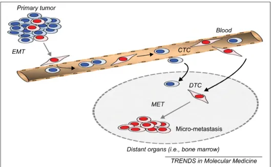
Fig. 1. Putative roles of EMT and mesenchymal-to-epithelial transition (MET) in tumour cell dissemination. In the primary tumour, a subpopulation of tumour cells (red) acquires a mesenchymal-like migratory phenotype during tumour progression. They lose their epithelial properties (ie, downregulate EpCAM and CK) through the EMT process and enter the bloodstream. These specific CTCs are thought to have stem cell properties. After extravasation into distant organs, these tumour cells (DTCs) have to re-express their epithelial properties through MET to form tumour cell clusters (micrometastases). Another subpopulation of CTCs, which is not able to undergo EMT (blue), can also disseminate through the bloodstream into distant organs but lacks cancer stem cell properties and, therefore, does not form (micro)metastases. These cells are detected by current CTC technologies, whereas the EMT-induced CTCs (red) are missed. This figure is reprinted from the article by Pantel K, et al. (42).
림프관을 통해, 셋째 늑막강, 심낭, 복강을 통해 전파된다 (4). 혈행성 전이는 원격 전이가 일어나는 가장 흔한 경로로 disseminated tumor cells (DTC)이 혈액을 타고 순환하고, 골 수에서 휴면기를 갖은 후 2차적인 전이 장소로 이동하기 위해 혈액으로 다시 들어간다(5). 최근 Bernards와 Weinberg (6)는 원발성 종양이 성장 초기에 전이능력을 갖는다는 새 로운 전이 모델을 제시하였고, DTC는 원발성 종양 성장 초 기에 이미 림프절, 말초 혈액, 골수에서 검출된다고 하였다.
또한 전이에 있어 필수적인 Epithelial-to-mesenchymal transi- tion (EMT) paradigm에 따르면 mesenchyme을 침범한 종양 세포는 cell-cell adhesion이 소실되어 혈액으로 들어가서 환 자의 혈액 샘플에서 CTCs로 검출된다(7,8) (Fig. 1). 말초 혈 액에서의 CTCs 검출은 140년 전에 오스트리아 병리학자인 Ashworth가 처음 기술하였으며(9), CTCs는 원발성 종양에 서 유래된 드문 세포로 최근에 검출방법에 대한 연구의 진 전으로 조기 검출, 진단, 예후, 암 감시, 전이과정의 생물학 이해에 커다란 희망을 주고 있다.
Circulating Tumor Cells (CTCs) Detection 많은 연구자들은 과거 수십 년 동안 CTCs를 enrichment,
identification 하기 위해 신뢰성 있는 방법의 개발에 몰두해 왔다. CTC는 10
5∼10
7mononuclear cell 당 한 개 정도로 매 우 적기 때문에 높은 성공률로 분리하기 위해서는 enri- chment 단계가 필요하다. 현재까지 대부분 enrichment 방법 은 CTCs를 찾기 위해 특이한 마커을 사용하여 백혈구와 구 별하는 방법으로 개발하였다. 가장 흔한 상피세포 마커로 는 cytokeratins (CKs), EpCAM (상피세포에 존재하는 adhesion molecule)이 있다. CTCs identification은 혈액 속의 non-tumour hematopoietic cells와 암세포를 구별할 수 있을 정도로 충분한 민감도와 특이도를 갖아야만 한다. 최근 identifcation은 immunomediated, cytometric, PCR-based strate- gies로 CTCs를 구별하려고 노력하고 있다. 그럼에도 불구하 고 아직까지 잘 디자인된 비교 연구가 없어서 이들의 결과 를 비교하기는 매우 어렵다.
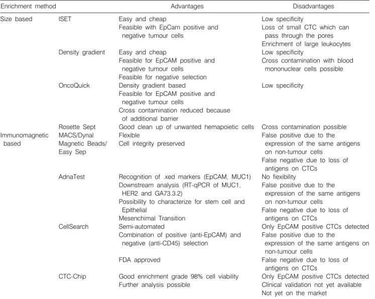
Enrichment Process
가장 흔한 방법으로 filtration, density gradient, immuno-
magnetic enrichment 방법이 있다. Table 1에서는 enrichment
방법들을 소개하고 각 방법의 장점, 단점을 요약하였다.
Table 1. Summary of Different CTC Enrichment Approaches
Enrichment method Advantages Disadvantages
Size based ISET Easy and cheap Low specificity
Feasible with EpCam positive and Loss of small CTC which can negative tumour cells pass through the pores
Enrichment of large leukocytes
Density gradient Easy and cheap Low specificity
Feasible for EpCAM positive and Cross contamination with blood negative tumour cells mononuclear cells possible Feasible for negative selection
OncoQuick Density gradient based Low specificity
Feasible for EpCAM positive and negative tumour cells
Cross contamination reduced because of additional barrier
Rosette Sept Good clean up of unwanted hemapoietic cells Cross contamination possible
Immunomagnetic MACS/Dynal Flexible False positive due to the
based Magnetic Beads/ Cell integrity preserved expression of the same antigens
Easy Sep on non-tumour cells
False negative due to loss of antigens on CTCs
AdnaTest Recognition of .xed markers (EpCAM, MUC1) No fiexibility
Downstream analysis (RT-qPCR of MUC1, False positive due to the HER2 and GA73.3.2) expression of the same antigens Possibility to characterize for stem cell and on non-tumour cells
Epithelial False negative due to loss of
Mesenchimal Transition antigens on CTCs
CellSearch Semi-automated Only EpCAM positive CTCs detected
Combination of positive (anti-EpCAM) and False positive due to the
negative (anti-CD45) selection expression of the same antigens on non-tumour cells
FDA approved False negative due to loss of
antigens on CTCs
CTC-Chip Good enrichment grade 98% cell viability Only EpCAM positive CTCs detected Further analysis possible Clinical validation not yet available
Not yet on the market This Table is reprinted from the article by Mostert B, et al. (44).
1) Filtration
ISET (Isolation by Size of Epithelial Tumour cells) 방법은 8μm pore filter를 사용하여 작은 백혈구와 큰 상피세포를 크기에 의해 직접 분류하는 방법이다(10). 여과 후에 CTCs 로 추정되는 세포를 고정하고 CKs와 같은 다른 마커들로 염색한다. 단점은 큰 백혈구는 filter에 걸리고, 작은 CTCs는 filter을 통해 빠져나가서 민감도와 특이도가 낮다.
2) Density gradient
Density gradient centrifugation 방법은 Ficoll-Hypaque을 사 용하여 CTCs, mononuclear cells (<1.077 g/mL)와 blood cells, granulocytes (>1.077 g/mL)을 분리할 수 있다. 그러나
혈장층으로 세포 이동, 세포 응집으로 CTCs를 쉽게 잃어버
리고, Ficoll-Hypaque에 오래 동안 접촉 시 세포에 독성을 나
타낸 점이 단점이다. Oncoquick (Greiner Bio-One, Fricken-
hausen, Germany)은 special membrane을 사용하여 서로 다른
층이 오염되지 않게 하는 density gradient enrichment법이다
(11,12). RosetteSep
TM(Stemcell Technologies, Vancouver,
British Columbia, Canada)은 negative selection에 의한 density
gradient 방법으로, 항체 칵테일이 cell surface markers나 혈
구세포를 인지하여 immunorosettes을 형성하고 CTCs를 혈
장과 buoyant density medium 사이에서 수집한다(13,14).
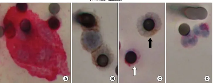
Fig. 2. Epithelial (E-cadherin) and mesenchymal (vimentin) markers in circulating tumor microemboli (CTM) and circulating tumor cells (CTCs) profiled by dual-color IHC. Blood samples from lung cancer patients were subjected to ISET filtration and CTM/CTCs were profiled for both epithelial (E-cadherin) and mesenchymal (vimentin) markers by dual-color IHC. (A) Vimentin-positive CTM (cytoplasm stained in red) with negative staining for E-cadherin (brown). (B) E-cadherin positive CTM (brown) without vimentin expression. (C) Displays CTC (black arrow), which is positively stained for E-cadherin in cytosol and negative for vimentin. White blood cells (white arrow in C and cells in D) serve as control and demonstrate positive staining for vimentin, negative staining for E-cadherin, and typical neutrophil nuclear morphology. This picture is reprinted from the article by Hou JM, et al. (17).
Immunomagnetic Cell Enrichment
Immunomagnetic cell enrichment은 magnetic bead을 이용하 여 분리하는 기술로 상피세포 마커들을 표적으로 하는 항 체(CKs, EpCAM, CEA, HER2, EGFR 등)를 magnetic bead와 결합하여 CTCs를 백혈구로부터 positive selection 하는 방법 이다. 다른 방법으로는 anti-CD45와 anti-CD61 항체(leuko- cytes, megakaryocytes)를 이용하여 negative selection 하는 방 법이다. 이 방법은 세포용해 없이 enriched CTCs를 쉽게 얻 을 수 있다. 그러나 false positive selection이 있을 수 있는데 이는 비 상피세포(non-epithelial cells)에서 상피세포 마커 발 현이 발생할 수 있기 때문이다(11). AdnaTest (AdnaGen AG, Langenhagen, Germany)는 EpCAM, Mucin 1 (MUC1) 항체를 사용하여 CTCs를 수집한 후에 용해해서 mRNA를 추출하 여 MUC1, HER2, surface glycoprotein GA73.3.-2 마커들을 이 용하여 RTqPCR을 통해 CTCs를 확인한다. Veridex (Johnson and Johnson, Raritan, NJ, USA)사의 Cell Search System
TM은 미국 FDA의 승인을 받은 semi-automated analyzer로서 EpCAM 항체를 입힌 ferrofluid nanoparticles을 사용한다.
Enriched CTCs는 four colour semi-automated fluorescent microscope인 cell spotter analyzer (Veridex)를 사용하여 계산 한다(15). 그러나 아직까지 universal marker가 없다는 점과 서로 다른 종양조직에서 서로 다른 유전적 특징을 보이기
도 하지만, 같은 조직에서도 서로 다른 유전특징을 보이기 도 하는 종양의 이질성이 이 방법의 제한점이다. 또한 CKs 는 활성화된 백혈구에서도 발현될 수 있고, 또한 EpCAM marker는 Epithelial to mesenchimal transition (EMT)을 통과 할 때 하향조절(down-regulation) 되어 CTCs가 소실될 수 있 다. CTCs의 검출은 전통적인 영상진단 시기보다 조기에 암 진행을 신뢰할 수 있게 예측할 수 있다. 그러나 확실한 원격 전이가 일어난 일부 환자에서 CTCs는 검출되지 않을 수 있 는데, 이는 최근 검출 방법으로는 CTCs with an EMT phenotype을 놓칠 수 있기 때문이다(16). Hou 등(17)은 폐암 환자의 CTC와 Circulating Tumor Microemboli (CTM)에서 epithelial markers와 mesenchymal marker가 이질적으로 발현 됨을 확인하였고, 이는 incomplete EMT가 존재함을 보고하 였다(Fig. 2).
Identification Process
수집된 CTCs의 origin, genetic profile을 확립하기 위하여
identification 과정이 요구된다. 이 과정은 nucleic acid con-
tent나 단백질을 이용하여 확인한다. Table 2에는 identi-
fication의 여러 가지 방법에 대한 장점과 단점을 기술하였
다.
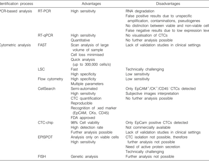
Table 2. Summary of Different CTC Identification Approaches
Identification process Advantages Disadvantages
PCR-based analysis RT-PCR High sensitivity RNA degradation
False positive results due to unspecific amplification, contaminations, pseudogenes No distinction between viable and non-viable cells False negative results due to low expression level
RT-qPCR High sensitivity No visualisation of CTCs
Quantitative No further analysis possible
Cytometric analysis FAST Scan analysis of large Lack of validation studies in clinical settings volume of sample
Cell loss minimised Quick analysis
(up to 300,000 cells/s)
LSC Fast Technically challenging
High specificity Low sensitivity Flow cytometry High specificity Low sensitivity
Multiple parameters
CellSearch Semi-automated Only EpCAM+/CK+/CD45−CTCs detected High sensitivity Subjective images interpretation
CTC quantification No further analysis possible Reproducible
Recognition of .xed marker (EpCAM, CKs, CD45) FDA approved
CTC-chip 98% Cell viability Only EpCam positive CTCs detected High detection rate Not commercially available
Further analysis possible Lack of validation studies in clinical settings EPISPOT Analysis only on viable cells CTC isolation not possible, therefore
High sensitivity further analysis not possible Need of active protein secretion Technically challenging
FISH Genetic analysis Further analysis not possible
This Table is reprinted from the article by Alunni-Fabbroni M, et al. (43).
1) PCR-based analysis
CTC genetic screening을 위한 가장 강력한 기술로서 immunocytochemistry보다 민감도가 높다(18,19). Reverse transcription PCR (RT-PCR)은 CTCs의 표적 mRNAs를 증폭 하는 방법으로, 최근에 민감도와 특이도를 높이기 위해 multiplex RT-PCR approach가 개발되었고, 이는 동시에 하나 의 마커보다는 많은 마커들을 선별 검사할 수 있는 기회를 제공한다.
2) Cytometric methods
단일항체를 이용하여 암세포를 분류하고 셈하는 방법으 로 가장 많이 사용하는 항체는 CKs, EpCAM이다. 이 방법 은 세포용해 과정이 없이 세포형태를 온전히 유지하여 scan 할 수 있다. 그러나 CK 같은 것은 백혈구를 검출할 수도
있고, EpCAM 경우는 CTC 세포가 하향조절 되어 있어 특이 도가 낮다는 단점이 있다. FAST (Fiber-optic Array Scanning Technology)는 정제단계와 세포손실 없이 매우 빨리 scanning 할 수 있고, 많은 양의 샘플을 처리할 수 있다. 그 러나 clinical validation은 아직 시도되지 않았다(20). Flow cytometry는 CTCs 검출에 높은 특이도를 보이고, size, viability, DNA content, 다른 markers 발현을 동시에 알아낼 수 있다. 그러나 민감도가 낮아 매번 많은 양의 샘플이 필요 하다는 단점도 있다(21,22). 그 외에도 ACIS (Automated Cellular Imaging System, DAKO, Denmark), ARIOL (Applied Imaging Corp. San Jose, CA)과 같은 scanning system이 있다.
최근에 자동화 기술이 발달하여 enrichment, staining, scann-
ing을 동시에 할 수 있는 기술이 개발되었다. CellSearch
System
TM(Veridex LLC, Warren, NJ, USA)은 이 중 하나로서
enriched anti-EpCAM(+) CTC을 CK8, 18, 19, CD45, DAPI로
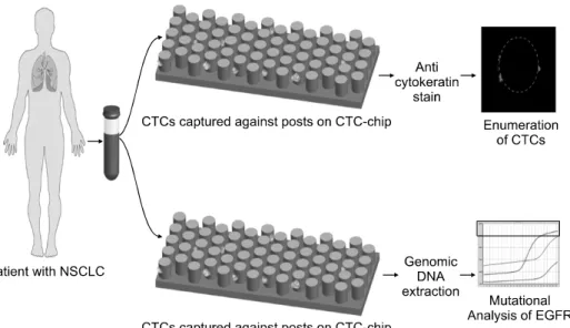
Fig. 3. Illustrative cartoon demonstrating a patient with non-small cell lung cancer (NSCLC) donating a tube of peripheral blood which is then processed in the circulating tumor cell (CTC)-chip immediately, without any required preprocessing. CTCs are captured against the sides of the anti-Ep-CAM-coated posts (epithelial cell adhesion molecule), and then can be stained with fluore- scently-labeled markers for enumeration or undergo genomic DNA extraction for epidermal growth factor receptor mutation or other molecular analysis. This picture is reprinted from the article by Sequist LV, et al. (28).
Fig. 4. Deposition of a single CTC on the AmpliGrid chip. (A) Ampli- Grid chip with 48 reaction sites for single cell deposition and PCR analysis. (B) A single CTC is deposited with a capillary on the reaction site of the Ampligrid chip.
CTCs are normally resuspended in physiological buffer and the volume of deposition is approxi- mately 20 nl. (C) A single CTC can be easily visualised on the reac- tion site when the DNA is stained with Hoechst dye. This pricture is reprinted from the article by Alunni-Fabbroni M, et al. (43).
염색하여 Cell Spotter analyzer (Veridex)를 사용하여 CK+/
DAPI+/CD45− 세포를 CTC 로 검출한다(15). Cell Search System
TM는 유일하게 FDA에서 승인된 방법으로 진행성 유 방암(23,24), 전립선암(25), 대장암(26)에서 사용한다. “CTC- chip”은 새로운 기술로서 anti-EpCAM coated antibodies을 78.000 microposts으로 배열하여 혈액이 지나가게 하여 EpCAM 양성 세포를 검출하는 방법이다. 단점은 EpCAM 항체만 가능하다는 것이다(27). Sequist 등(28)은 CTC-chip을
이용하여 폐암으로 진단 받은 68명의 환자의 116개 혈액 샘 플에서 CTCs를 평균 155 cells/mL±236을 분리하였다고 보 고 하였다(Fig. 3). 그러나 아직까지 대규모의 clinical vali- dation이 이루어지지 않았다.
그 외 antibody-based approach 방법으로 EPISPOT (Epi-
thelial Immunospot), ELISPOT (Enzyme- Linked Immunosor-
bent assay) technology 가 있다(29,30). EPISPOT는 CD45 양
성세포를 제거한 후 세포고사 세포는 검출하지 않고 살아
Table 3. Clinical Relevance of CTC Detection in Breast Cancer Patients
Study Tumour stage Method Number
of patients
CTC detection
rate (%) Clinical results
Cristofanilli Metastatic breast CellSearch 177 49 CTCs P 5/7.5 ml associated with reduced
et al. (23) cancer PFS (p<0.001) and OS (p<0.001)
Nolé Metastatic breast CellSearch 80 61 CTCs P 5/7.5 ml associated with reduced
et al. (45) cancer PFS (p=0.002)
Budd Metastatic breast CellSearch 138 25.4 CTC evaluation after 4 weeks from the
et al. (46) cancer start of chemotherapy are better predictor
of OS compared to radiologic response evaluation performed at 10 weeks De Giorgi Metastatic breast CellSearch 115 21 Univariate analysis: CTCs levels at
et al. (47) cancer 9.12 weeks (p<0.0001) and FDG-PET/CT
(p≤0.001) associated with OS Multivariate analysis: only CTCs levels at 9.12 weeks (p<0.001) associated with OS
Pierga Locally advanced CellSearch 118 27 CTCs P 1/22.5 ml associated with reduced
et al. (48) breast cancer DFS (0.017)
Stathopoulou Early breast cancer RT-PCR (CK19) 148 29.7 Reduced DFS (p=0.0007) and OS
et al. (49) (stage I-II) (p=0.01)
Xenidis Early breast cancer RT-PCR (CK19) 167 21.6 Reduced DFS (p<0.00001) and OS
et al. (50) (stage I-II) (p=0.008)
Ignatiadis Early breast cancer RT-PCR (CK19) 444 40.8 Reduced DFS (p<0.001) and OS
et al. (51) (stage I-III) (p=0.001)
Apostolaki Early breast cancer RT-PCR (HER2) 214 21 Reduced DFI (p=0.006), no association
et al. (52) (stage I-II) with OS (p=0.2)
Xenidis Early breast cancer RT-PCR (CK19) 437 32.7 Reduced DFS (p<0.001) and OS
et al. (53) (stage I-III) (p=0.001)
Rack Early breast cancer CellSearch 1,500 9 CTCs>1/22.5 mL associated with reduced
et al. (54) (stage I-III) DFS (p=0.04) and OS (p=0.03)
This Table is reprinted from the article by Alunni-Fabbroni M, et al. (43).
있는 세포만을 검출하는 방법이다. 이 방법은 표적세포와 의 직접적인 접촉을 피하기 위하여 48시간이라는 짧은 기 간에 배양하여 MUC1, CK19와 같은 단백질을 분비하는 세 포만 검출한다. 그러나 임상적 시도는 이루어지지 않았다.
Genetic Characterization
CTCs를 분리한 후에는 genetic characterization이 필요하 다. 유전적인 분석은 분리한 CTCs가 아직도 악성 세포의 특징을 가지고 있는지, 원발성 종양과 유전학적으로 같은 지를 확인하는 작업이다. 이런 정보를 통하여 암의 진행을 monitoring 할 수 있고, 치료적인 접근을 할 수 있다.
AmpliGrid (Beckman Coulter Genomics, Munich, Germany)는 PCR based chip으로 분리된 CTCs 각각의 세포들이 유전적 정보가 다른지를 확인 할 수 있는 방법이다(31,32) (Fig. 4).
상업화 되어 있지만 아직까지 대규모 임상 시도는 없는 실 정이다.
FISH (Fluorescent In Situ Hybridization)는 CTC genotyping
에 유용한 검사로서 gene translocation이나 amplification 변 화를 염색체상에서 특이 DNA 염기서열의 유, 무로 확인하 는 방법이다(33,34). CGH (Comparative Genomic Hybridiz- ation)는 세포유전학적 검사방법으로 DNA에서 유전자 copy number의 변화를 찾아내는 방법이다(35).
Clinical Application of CTCs in Lung Cancer Patients
CTCs에 대한 연구는 많은 악성종양에서 진행되고 있으 나, 폐암에서는 아직까지 CTCs를 이용한 임상결과는 적은 편이고, 많은 임상 결과는 주로 유방암에서 연구결과들이 발표되었다(Table 3).
1) CTC as a diagnostic maker in primary lung cancer
Tanaka 등(36)은 폐암환자의 말초 혈액에서 CTCs가
30.6% 검출됨을 보고하였고, CTC 수의 증가는 종양의 진
행, 특히 원격 전이와 연관되어 있고, 전이가 있는 환자에서
1개 이상 CTC가 검출될 경우 CTC 검사의 민감도와 특이도
Fig. 5. (A) Distribution of CTC count in primary lung cancer patients without distant metastasis and in patients with distant metastasis. (B) ROC curves for CTC count and serum CEA level to predict the absence or presence of distant metastasis among primary lung cancer patients. CTC: Circulating tumor cell, CEA: Carcinoembryonic Antigen. This picture is reprinted from the article by Tanaka F, et al. (36).
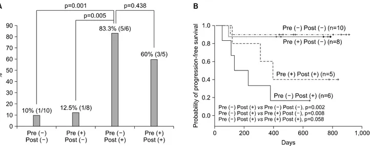
Fig. 6. Disease progression and progression-free survival in surgically resected NSCLC patients according to paired presurgery and postsurgery TTF-1 (+) CTC status. When pairing presurgery and postsurgery TTF-1 (+) CTC status and allocating patients to four groups, the Pre (−) Post (+) group (TTF-1 (+) CTCs were not present at presurgery, but detected at postsurgery time) most frequently developed disease progression (A), and showed shortest progression-free survival (B) as compared to other three groups. This picture is reprinted from the article by Yoon SO, et al. (37).
는 71.0%, 83.3%이고 이는 원발성 폐암환자에서 원격전이 의 surrogate marker로 사용할 수 있다고 보고하였다(Fig. 5).
2) Prognosis of lung cancer patients
여러 가지 기술적인 제한에도 불구하고, 임상연구는 짧 은 기간에 이루어졌다. 그러나 결론을 내리는 어려움이 있 다. 서로 다른 분석, 서로 다른 CTC 정의, 서로 다른 임상 집단, 제한된 환자 수, 서로 다른 병기 등에 의해 서로 다른
결과를 보인다. 대부분의 임상연구는 CTCs와 Disease Free
Survival (DFS), Progression Free Survival (PFS), Overall
Survival (OS)과 관련된 연구다. 말초 혈액을 통한 CTC의 연
속적인 분석은 쉽게 접근할 수 있어 많은 연구자들이 최근
에 CTC의 임상적인 유용성을 평가하고 있다. 유방암, 전립
선암, 대장암에서 Cell Search System을 이용하여 진행된 암
환자에서 CTC 검출과 전이 진행이 상관관계가 있음을 보
고 하였다(23). 이는 초기 병기에 CTC 검출이 가능하고 전
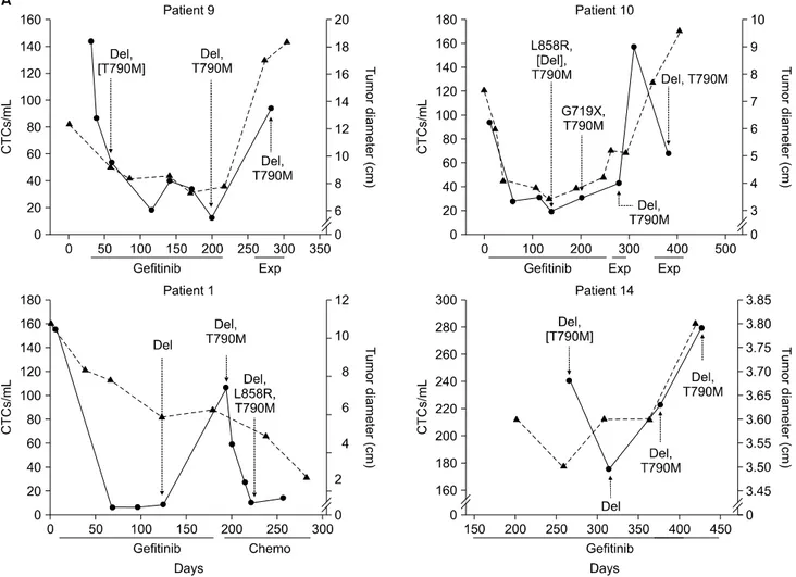
Fig. 7. Serial Analyses of Circulating Tumor Cells during Therapy. (A) Shows serial analyses of the numbers of circulating tumor cells (CTCs) per milliliter and the radiographic tumor burden in centimeters in four patients with non-mall-cell lung cancer with EGFR mutations, as measured at multiple time points during the course of treatment with gefitinib, another chemotherapy agent (chemo), or an experimental agent (exp). The duration of each therapy is indicated by the gray bars. The genotypes of circulating tumor cells are shown for various time points. Mutations in brackets are those that were present at low allele frequencies. (B) SARMS analysis of EGFR genotypes in Patient 9 shows an increased abundance of the T790M drug-resistance allele during disease progression. Arrows denote the cycle of threshold for amplification cycles (Ct) required for detection of the primary mutation (Del or Deletion, referring to the grouped exon 19 deletions) and the T790M mutation, as compared with the exon 2 control. ΔCt reflects the difference in allele frequency between the primary mutation and T790M in the tumor-biopsy specimen, the circulating tumor cells that were isolated at the time of response to gefitinib therapy, and the circulating tumor cells that were isolated at the time of disease progression. This picture is reprinted from the article by Maheswaran S, et al. (38).
이성 재발의 위험을 예측할 수 있지만, 너무 적은 CTC가 (<5 cell/10 mL blood) 존재하여 검출 방법에 따라 양성율이 10∼100%로 큰 변화가 있다.
Yoon 등(37)은 79명 resected non-small cell lung cancer 환 자의 수술 전, 후 말초 혈액에서 thyroid transcription factor-1 (TTF-1)와 cytokeratin19 (CK19) mRNA markers로 검출된 CTC는 수술 전 TTF-1 (+) CTCs 36.1% (22/61), 수술 후 37.5% (18/48)이고, CK19 (+) CTCs는 수술 전 42.6%
(26/61), 수술 후 25.0% (12/48)로 보고 하였다. 또한 수술 후
TTF-1 (+) and/or CK19 (+) CTCs는 종양의 진행(p=0.004)
과 의미 있는 연관성을 보여주었고, 짧은 disease progression-
free survival (p=0.006)을 보여 주었다. 그리고 수술 후 TTF-1
(+) CTCs-positive status는 TTF-1 (+) CTCs-negative status
보다 의미 있는 종양의 진행(p=0.004)과 짧은 disease
progression-free survival (p=0.004)을 보여 주었다. 특히 수술
후 TTF-1 (+) CTCs를 가진 환자는 현저히 짧은 disease
progression-free survival을 보였다(p<0.001). TTF-1 mRNA-
expressing CTCs는 임상적으로 발현되기 전에 disease
Fig. 7. Continued.
progression의 surrogate predictor로 사용할 수 있다고 보고 하였다. 수술 후에 TTF-1 (+) CTCs status의 monitoring은 수술적으로 절제 가능한 비소세포폐암 환자 중 고위험 환 자를 찾아내는데 도움이 된다고 하였다(Fig. 6).
3) Monitoring chemotherapy in lung cancer patients
CTCs 분석은 항암치료에 대한 반응을 monitoring 하는데 임상적인 의미를 갖는다. Randomised trial SWOG S0500는 진행성 유방암에서 1차 항암치료 3주 후에 OS, PFS를 향상 시키는지에 대해 CTC level을 측정하여 임상적으로 pro- gression 되기 전에 다음 항암치료 regimen을 바꿔야 하는지 를 알아보고자 하는 임상 시험이 진행 중이다.
Maheswaran 등(38)은 2008년 NEJM에서 27명의 전이성 비소세포폐암에서 평균 74 CTCs/mL이 분리되었고, 12명 중 11명의 CTCs (92%)에서 EGFR mutation을 찾아 낼 수 있 었고, tyrosine kinase inhibitors 치료를 받고 있는 EGFR mutation 환자의 CTCs에서 T790M mutation을 관찰하였다고
보고하였다. CTCs의 연속적인 분석에서 CTCs의 감소는 영 상학적인 종양 반응과 관련되어 있고 CTCs 수의 증가는 종 양의 악화를 예측할 수 있으며, 일부에서는 추가적인 EGFR mutations의 발현과 관련되어 있다고 보고 하였다(Fig. 7).
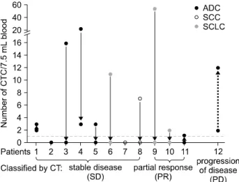
Wu 등(39)은 새로 진단된 41명의 폐암환자에서 CTC 양 성률(2/7.5 mL blood)은 8% (ADC stage III), 75% (squamous cell carcinoma stage III), 60% (SCLC stage III)인 반면, 46%
(ADC IV), 71% (SCLC IV)로 보고하였다. 또한 12명의 환자 에서 2회 1차 항암치료 후 CTC level 과 방사선학적 반응 사이에 높은 상관관계를 보여 항암치료 효과를 빨리 평가 할 수 있는 임상적 도구로 사용할 수 있다고 보고하였다 (Fig. 8).
4) Identification of therapeutic targets
CTCs는 유방암, 대장암, 폐암에서는 세포분열이 일어나
지 않기 때문에 항암치료는 한계가 있다(40,41). 휴지기나
세포분열이 일어나지 않는 CTCs를 죽이기 위해서는 전통
Fig. 8. Correlation of circulating tumor cell (CTC) counting and computed tomography (CT) scanning with a follow-up study.
Correlation of CT scanning and CTC counting was evaluated on a small scale of follow-up study. CTC was enumerated from each patient before chemotherapy started. After two courses of first line chemotherapy, patient CTC was measured, followed by CT examination. Eight of 12 patients (patient 1-) including 5 adenocarcinoma (ADC), 2 squamous cell carcinoma (SCC) and 1 small cell lung cancer (SCLC) were classified by CT as stable disease. Among those 8 patients, 5 patients dropped their CTC to 0, 1 patient dropped from 21 to 3, 2 patients remained 0, and in 1 patient the count increased from 2 to 3.
Three patients had a partial radiologic response: For partial response (PR) patients (patients 9-1), one was noted to have a CTC drop from 52 to 0, while the CTC in the other 2 patients remained below 2. Only one patient was noted to have an increasing CTC, from 2 to 12 after treatment, and this patient was confirmed to have progressive disease (PD). Clinical responses are classified by CT scanning according to the Response Evaluation Criteria in Solid Tumors (RECIST). This pricture is reprinted from the article by Wu C, et al. (39).
적인 치료에 CTCs를 표적으로 하는 치료를 추가하는 것이 임상 종양학에서 새로운 치료시대를 예고하는 것이다.
향후 전망
현재 CTC 검출방법은 상피 종양세포와 정상 간엽 혈액 세포를 구별할 수 있는 EMT phenotype을 갖은 CTCs를 검출 하기 위한 마커를 찾는 것이 향후 과제이다. 더욱이 CTCs의 세부적 분자생물학적 분석을 위한 robust multiplex tech- nology를 개발하고 대규모 임상시험이 향후 필요하다. 또한 CTCs 연구방향은 CTC 분석을 통해 전신적 항암치료의 효 능을 real-time monitoring 하고, 약물 민감도 또는 내성과 관 련된 분자생물학적 표적을 찾는데 임상적 의미가 있다고 하겠다. 특히 폐암환자에서는 CTCs가 metastatic foci를 형성
할 수 있는 능력이 있는지, EGFR mutation과 같은 생물학적 특성이 있는지, CTC test가 임상적으로 치료의 surrogate marker로 의미가 있는지에 대해 향후 지속적인 연구가 필요 하다.
REFERENCES
1. Socinski MA, Crowell R, Hensing TE, et al. Treatment of non-small cell lung cancer, stage IV: ACCP evidence-based clinical practice guidelines (2nd edition). Chest 2007;132(3 Suppl):277S-289S.
2. Alix-Panabières C, Riethdorf S, Pantel K. Circulating tumor cells and bone marrow micrometastasis. Clin Cancer Res 2008;14:5013-5021.
3. Paget S. The distribution of secondary growths in cancer of the breast. 1889. Cancer Metastasis Rev 1989;8:98-101.
4. Fidler IJ. Tumor heterogeneity and the biology of cancer invasion and metastasis. Cancer Res 1978;38:2651-2660.
5. Riethdorf S, Wikman H, Pantel K. Review: biological relevance of disseminated tumor cells in cancer patients. Int J Cancer 2008;123:1991-2006.
6. Bernards R, Weinberg RA. A progression puzzle. Nature 2002;418:823.
7. Kalluri R, Weinberg RA. The basics of epithelial-mesen- chymal transition. J Clin Invest 2009;119:1420-1428.
8. Thiery JP. Epithelial-mesenchymal transitions in tumour progression. Nat Rev Cancer 2002;2:442-454.
9. Ashworth TR. A case of cancer in which cells similar to those in the tumors were seen in the blood after death. Aust Med J 1869;14:146-147.
10. Vona G, Sabile A, Louha M, et al. Isolation by size of epithelial tumor cells: a new method for the immunomor- phological and molecular characterization of circulatingtumor cells. Am J Pathol 2000;156:57-63.
11. Gertler R, Rosenberg R, Fuehrer K, Dahm M, Nekarda H, Siewert JR. Detection of circulating tumor cells in blood using an optimized density gradient centrifugation. Recent Results Cancer Res 2003;162:149-155.
12. Müller V, Stahmann N, Riethdorf S, et al. Circulating tumor cells in breast cancer: correlation to bone marrow micro- metastases, heterogeneous response to systemic therapy and low proliferative activity. Clin Cancer Res 2005;11:3678-3685.
13. Naume B, Borgen E, Tøssvik S, Pavlak N, Oates D, Nesland JM. Detection of isolated tumor cells in peripheral blood and in BM: evaluation of a new enrichment method. Cytotherapy 2004;6:244-252.
14. Hayes GM, Busch R, Voogt J, et al. Isolation of malignant B cells from patients with chronic lymphocytic leukemia (CLL) for analysis of cell proliferation: validation of a simplified method suitable for multi-center clinical studies.
Leuk Res 2010;34:809-815.
15. Allard WJ, Matera J, Miller MC, et al. Tumor cells circulate in the peripheral blood of all major carcinomas but not in
healthy subjects or patients with nonmalignant diseases. Clin Cancer Res 2004;10:6897-6904.
16. Müller V, Alix-Panabières C, Pantel K. Insights into minimal residual disease in cancer patients: implications for anti-cancer therapies. Eur J Cancer 2010;46:1189-1197.
17. Hou JM, Krebs M, Ward T, et al. Circulating tumor cells as a window on metastasis biology in lung cancer. Am J Pathol 2011;178:989-996.
18. Smith BM, Slade MJ, English J, et al. Response of circulating tumor cells to systemic therapy in patients with metastatic breast cancer: comparison of quantitative polymerase chain reaction and immunocytochemical techniques. J Clin Oncol 2000;18:1432-1439.
19. Lambrechts AC, Bosma AJ, Klaver SG, et al. Comparison of immunocytochemistry, reverse transcriptase polymerase chain reaction, and nucleic acid sequence-based amplification for the detection of circulating breast cancer cells. Breast Cancer Res Treat 1999;56:219-231.
20. Krivacic RT, Ladanyi A, Curry DN, et al. A rare-cell detector for cancer. Proc Natl Acad Sci USA 2004;101:10501-10504.
21. Cruz I, Ciudad J, Cruz JJ, et al. Evaluation of multiparameter flow cytometry for the detection of breast cancer tumor cells in blood samples. Am J Clin Pathol 2005;123:66-64.
22. Simpson SJ, Vachula M, Kennedy MJ, et al. Detection of tumor cells in the bone marrow, peripheral blood, and apheresis products of breast cancer patients using flow cytometry. Exp Hematol 1995;23:1062-1068.
23. Cristofanilli M, Budd GT, Ellis MJ, et al. Circulating tumor cells, disease progression, and survival in metastatic breast cancer. N Engl J Med 2004;351:781-791.
24. Cristofanilli M, Hayes DF, Budd GT, et al. Circulating tumor cells: a novel prognostic factor for newly diagnosed metastatic breast cancer. J Clin Oncol 2005;23:1420-1430.
25. Danila DC, Heller G, Gignac GA, et al. Circulating tumor cell number and prognosis in progressive castration-resistant prostate cancer. Clin Cancer Res 2007;13:7053-7058.
26. Cohen SJ, Punt CJ, Iannotti N, et al. Relationship of circulating tumor cells to tumor response, progression-free survival, and overall survival in patients with metastatic colorectal cancer. J Clin Oncol 2008;26:3213-3221.
27. Nagrath S, Sequist LV, Maheswaran S, et al. Isolation of rare circulating tumour cells in cancer patients by microchip technology. Nature 2007;450:1235-1239.
28. Sequist LV, Nagrath S, Toner M, Haber DA, Lynch TJ. The CTC-chip: an exciting new tool to detect circulating tumor cells in lung cancer patients. J Thorac Oncol 2009;4:281-283.
29. Alix-Panabières C, Vendrell JP, Pellé O, et al. Detection and characterization of putative metastatic precursor cells in cancer patients. Clin Chem 2007;53:537-539.
30. Alix-Panabières C, Brouillet JP, Fabbro M, et al. Charac- terization and enumeration of cells secreting tumor markers in the peripheral blood of breast cancer patients. J Immunol Methods 2005;299:177-188.
31. Holodick NE, Repetny K, Zhong X, Rothstein TL. Adult BM generates CD5+ B1 cells containing abundant N-region
additions. Eur J Immunol 2009;39:2383-2394.
32. Waak J, Weber SS, Waldenmaier A, et al. Regulation of astrocyte inflammatory responses by the Parkinson's disease- associated gene DJ-1. FASEB J 2009;23:2478-2489.
33. Meng S, Tripathy D, Frenkel EP, et al. Circulating tumor cells in patients with breast cancer dormancy. Clin Cancer Res 2004;10:8152-8162.
34. Meng S, Tripathy D, Shete S, et al. HER-2 gene amplification can be acquired as breast cancer progresses. Proc Natl Acad Sci USA 2004;101:9393-9398.
35. Schmidt-Kittler O, Ragg T, Daskalakis A, et al. From latent disseminated cells to overt metastasis: genetic analysis of systemic breast cancer progression. Proc Natl Acad Sci USA 2003;100:7737-7742.
36. Tanaka F, Yoneda K, Kondo N, et al. Circulating tumor cell as a diagnostic marker in primary lung cancer. Clin Cancer Res 2009;15:6980-6986.
37. Yoon SO, Kim YT, Jung KC, Jeon YK, Kim BH, Kim CW.
TTF-1 mRNA-positive circulating tumor cells in the peripheral blood predict poor prognosis in surgically resected non-small cell lung cancer patients. Lung Cancer 2011;71:209-216.
38. Maheswaran S, Sequist LV, Nagrath S, et al. Detection of mutations in EGFR in circulating lung-cancer cells. N Engl J Med 2008;359:366-377.
39. Wu C, Hao H, Li L, et al. Preliminary investigation of the clinical significance of detecting circulating tumor cells enriched from lung cancer patients. J Thorac Oncol 2009;4:
30-36.
40. Pantel K, Schlimok G, Braun S, et al. Differential expression of proliferation-associated molecules in individual micrometa- static carcinoma cells. J Natl Cancer Inst 1993;85:1419-1424.
41. Pantel K, Izbicki JR, Angstwurm M, et al. Immunocytological detection of bone marrow micrometastasis in operable non-small cell lung cancer. Cancer Res 1993;53:1027-1031.
42. Pantel K, Alix-Panabieres C. Circulating tumour cells in cancer patients: challenges and perspectives. Trends Mol Med 2010;16:389-406.
43. Alunni-Fabbroni M, Sandri MT. Circulating tumour cells in clinical practice: Methods of detection and possible charac- terization. Methods 2010;50:289-297.
44. Mostert B, Sleijfer S, Foekens JA, et al. Circulating tumor cells (CTCs): detection methods and their clinical relevance in breast cancer. Cancer Treat Rev 2009;35:463-474.
45. Nolé F, Munzone E, Zorzino L, et al. Variation of circulating tumor cell levels during treatment of metastatic breast cancer:
prognostic and therapeutic implications. Ann Oncol 2008;19:
891-897.
46. Budd GT, Cristofanilli M, Ellis MJ, et al. Circulating tumor cells versus imaging--predicting overall survival in metastatic breast cancer. Clin Cancer Res 2006;12:6403-6409.
47. De Giorgi U, Valero V, Rohren E, et al. Circulating tumor cells and [18F]fluorodeoxyglucose positron emission tomo- graphy/computed tomography for outcome prediction in metastatic breast cancer. J Clin Oncol 2009;10:3303-3311.
48. Pierga JY, Bidard FC, Mathiot C, et al. Circulating tumor cell
detection predicts early metastatic relapse after neoadjuvant chemotherapy in large operable and locally advanced breast cancer in a phase II randomized trial. Clin Cancer Res 2008;14:7004-7010.
49. Stathopoulou A, Vlachonikolis I, Mavroudis D, et al.
Molecular detection of cytokeratin 19-positive cells in the peripheral blood of patients with operable breast cancer:
evaluation of their prognostic significance. J Clin Oncol 2002;20:3404-3412.
50. Xenidis N, Perraki M, Kafousi M, et al. Predictive and prognostic value of peripheral blood cytokeratin-19 mRNA- positive cells detected by real-time polymerase chain reaction in node-negative breast cancer patients. J Clin Oncol 2006;24:3756-3762.
51. Ignatiadis M, Xenidis N, Perraki M, et al. Different prognostic
value of cytokeratin-19 mRNA positive circulating tumor cells according to estrogen receptor and HER2 status in early-stage breast cancer. J Clin Oncol 2007;25:5194-5202.
52. Apostolaki S, Perraki M, Pallis A, et al. Circulating HER2 mRNA-positive cells in the peripheral blood of patients with stage I and II breast cancer after the administration of adjuvant chemotherapy: evaluation of their clinical relevance. Ann Oncol 2007;18:851-858.
53. Xenidis N, Ignatiadis M, Apostolaki S, et al. Cytokeratin-19 mRNA-positive circulating tumor cells after adjuvant chemo- therapy in patients with early breast cancer. J Clin Oncol 2009;
27:2177-2184.
54. Rack B, Schindlbeck C, Jückstock J, et al. Prevalence of CA 27.29 in primary breast cancer patients before the start of systemic treatment. Anticancer Res 2010;30:1837-1841.