I. 서론
임프란트는 여러 학자들의 장기적인 노력과 연구 로 부분 또는 완전 무치악 환자에 유용한 보철적 술 식임이 입증되어 왔고, 높은 임상적 성공률과 예측 가능성을 보이며 저작력의 회복뿐만 아니라 심미적, 발음적 능력의 증진에 큰 역할을 담당하고 있다1-12). 이러한 임프란트 시술이 성공하기 위해서는 임프란 트와 치조골 사이에 연조직의 개재없이 골유착 (osseointegration)이 일어나야 하며6,8,13), 골유착이 일어난다고 하더라도 적지만 합병증이 발생하고 이 런 가능성은 항상 존재한다. 대표적인 것으로 임프 란트 주위염이 있으며 그 원인은 생역학적 그리고 교합적 과부하와 세균적 원인으로 발생한다14-21). 특 히, 치과용 임프란트는 구강내에 계속 노출되어 있어 세균오염의 문제가 있으며 세균의 조성상 자연치와
유사하나10,22) 임프란트 주위 조직은 자연치 치주조
직보다 세균에 더 민감하다23). 이것은 자연치에서 섬 유다발이 백악질에 대해 수직적으로 주행하는데 반 해 임프란트에서는 표면에 대해 평행하게 주행하기 때문이며 이로 인해 치은 연하 치태 축적 시 치근단 쪽으로 세균의 파급이 더 빠르게 진행된다24,25). 따라 서 임프란트 치료를 받은 환자에서 초기 치태 제거 는 더 중요하며26)치태나 치석 제거를 위한 여러 구 강 위생 술식 및 기구조작은 임프란트 산화막에 변
화를 야기시키고 표면의 거칠기를 변화시켜 세균성 치태나 치석 형성을 촉진시킬 수 있으므로 세심한 주의가 요구된다27-33).
임프란트 주위염은 임상적, 방사선학적 진단을 통 해 이루어질 수 있으며, 진단을 위해 치은 색조, 치주 낭 깊이, 부착 수준, 탐침시 출혈, 화농, 열구액 유출 량, 치태 지수, 치은 지수, 동요도, 산소압 수준, 조직 교원질 분해 효소 활성도, 치은 연하 세균총, 백혈구 와 같은 국소적인 숙주 요인 검사 그리고 확진적인 조직학적 소견들이 보고되어졌다. 골 소실을 동반한 임프란트 주위염의 결손부는 스스로 치유되지 않으 므로 임프란트와 재 골유착을 위해 외과적 시술이 필요하며 여기에는 조직 유도 재생술이 주로 이용된 다. 또한 임프란트 표면은 치태와 치석으로 오염되 어 있으므로 이 시술은 세척 및 해독과정이 포함된 다. 이 과정에 기계적, 화학적 방법이 이용되었으며, 사용되는 재료로 air-powder abrasive, citric acid(pH 1), 1% chloramine T solution, tetracycline HCl, chlorohexidine, plastic instrument, distilled water, laser 등이 있다. 이 중 구연산과 공기-분말 연마는 현 재 가장 많이 사용되는 재료이다.
공기-분말 연마는 1980년 이후 많이 사용되는 기 구로 치아의 치태나 착색을 제거하는데 효과적이며 주로 55에서 60psi 압력에서 사용된다34-36). Barnes 등37)은 공기-분말 연마는 임프란트의 표면 구조에 유
공기-분말 연마와 구연산의 적용시간에 따른 임프란트 표면 거칠기의 변화에 관한
주사탐침현미경적 연구
박민서ㆍ정진형ㆍ임성빈 단국대학교 치과대학 치주과학교실
대한치주과학회지 : Vol. 30, No. 4, 2000
해하지 않고 착색 또는 치태를 제거할 수 있다고 하 였으며, Parham 등38)은 임프란트 표면의 세균제거에 공기-분말 연마의 사용이 효과적이라고 하였다. 또 Dennison 등39)은 공기-분말 연마가 내독소를 제거하 는 데 가장 효과적인 방법이며, 수종의 임프란트 중 선반 가공된 타이타늄 임프란트에 더 유용하다고 하 였다.
구연산은 과거 치근면 처치 시에 신생 백악질 형성 을 촉진하기 위하여 주로 사용되었다. Zablotsky 등
40,41)은 pH 1 구연산을 HA 임프란트 표면에 처리할
경우 효과적으로 세균독소를 제거할 수 있으나 임프 란트 표면에 손상을 일으킨다고 하였다.
서술한 바와 같이 임프란트는 관리의 어려움과 염 증에 대한 취약성으로 자연치에 비해 치주염 발생 가능성이 높으며, 질환 발생 시 치료의 어려움이 많 다. 또한 임프란트의 표면은 오염되고 이 부분의 치 유는 지연된다. 따라서 임프란트의 골유착을 위한 골재생술 과정에 있어 표면 처치가 필요하고 이것은 임프란트 표면의 변화를 야기 시킬 수 있다. 본 연구 의 목적은 최근까지 임프란트의 표면 처리에 가장 효과적인 것으로 알려진 공기-분말 연마와 구연산의 적용시간을 변화시켜 선반 가공된 타이타늄에 적용 한 후 주사탐침 현미경을 이용하여 표면의 변화를 관찰하고 이 두 제재가 매식체의 표면 거칠기에 어 떤 영향을 미치는지 거칠기 분석 프로그램을 통해 수치화해서 알아보고자 하였다.
II. 연구재료 및 방법
1. 연구재료 1) 실험 모형
주사탐침현미경으로 표면 거칠기를 수치화 할 목 적으로 직경 10mm, 높이 2mm의 machined pure titanium 원통형 모델을 매식체와 동일한 표면 거칠 기를 갖도록 주문 제작(AVANA, Soomin co., KOREA)하였다(Figure 1).
2) 공기-분말 연마기
MicroprophyTM(Danville Engineering, Inc., USA)
를 55psi 압력 하에서 증류수와 분말(sodium bicar- bonate, sodium laurel, etc)을 2:1로 혼합한 후 10mm 거리에서 분사하여 사용하였다(Figure 2).
3) 구연산 용액(pH 1)
실온 상태에서 anhydrous citric acid를 포화 상태 가 될 때까지 용해시킨 후 pH-meter로 측정한 후 No. 1 Whatman filter에 통과시켜 pH 1 용액을 만들 었다.
2. 연구방법
1) 대조군 및 실험군
10개의 모형 중 아무 처리도 하지 않은 군을 대조 군으로 하였으며, 여기에 Microprophy를 이용한 공 기-분말 연마를 1분간 처리한 군을 실험 1군으로 하 였다. 다시 실험 1군에 구연산을 cotton pellet으로 30초, 1분, 3분, 5분을 연속적으로 처리하여 각각을 실험 2, 3, 4, 5군으로 분류하였다. 즉 30초 처리 후 1 분간 증류수로 세척한 후 실험 2군을 관찰하고 다시 여기에 30초를 더 처리하여 관찰한 것이 실험 3군이 다. 실험 4, 5군도 위와 동일한 방법으로 관찰하였다.
2) 관찰 및 측정방법
주사탐침현미경(Scanning Probe Microscope, Nanoscope IIIa, Veeco Inc., USA)을 이용하여 실험 모형의 중심 직경 3mm이내의 범위에 임의적으로 100㎛×100㎛ 크기의 정사각형 표면 양상을 관찰하 였다(Figure 3). 관찰된 2차원적, 3차원적 소견은 거 칠기의 높낮이를 등고선과 같이 높이에 따라 다른 색으로 표현되었고 이 색으로 표현된 것을 수치로 환산하는 거칠기 분석 프로그램을 통해 평균 표면 거칠기인 Ra(Mean Surface Roughness, nm)값을 구 하였다.
3) 통계 처리
Windows용 SPSS ver. 8.0(SPSS Inc., USA)을 사용 하여 각 군의 표면 거칠기의 평균과 표준편차를 구 하고 공기-분말 연마와 구연산의 표면 거칠기에 미
치는 영향을 알아보기 위하여 paired-t test를 이용하 여 통계 처리하였다.
III. 연구결과
1. 표면 거칠기 분석주사탐침현미경을 통한 평균 표면 거칠기(Ra)값은 아무 처치도 하지 않은 노출된 매식체인 대조군에서 가장 크고 두 번째는 공기-분말 연마를 1분간 시행한 실험 1군이었으며 다음으로는 실험 5군, 실험 2군, 실험 3군, 실험 4군의 순이었다. 표면 거칠기의 감소 수준은 대조군과 실험 1군 사이에서 가장 크고 실험
2군과 실험 3군에서 가장 작았으며, 실험 4군과 실험 5군은 반대로 표면 거칠기가 증가하였다(Table 1, Figure 4).
공기-분말 연마 1분 후 표면 거칠기는 대조군과 비 교했을 때 감소량은 평균 12.6nm였으며 이것은 통 계적으로 유의성이 있는 것으로 나타났다(Table 2).
또한 실험 1군과 다른 실험 군간의 표면 거칠기 감 소량은 실험 2군, 3군, 4군, 5군 각각에 대해 평균 2.6nm, 4.8nm, 7.6nm, 0.6nm였으며, 실험 5군을 제 외하고는 통계적으로 유의성이 있었다. 그리고 실험 2군과 실험 4군 사이에는 표면 거칠기의 감소가, 실 험 4군과 실험 5군 사이에는 표면 거칠기의 증가가 통계적으로 유의성이 있었다(Table 3).
Figure 4. Mean and Standard deviation of the Surface roughness(nm)
평균 표면거칠기 표준편차
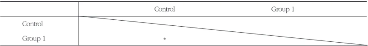
Table 1. Mean and standard deviation of the surface roughness(nm)
Control Group 1 Group 2 Group 3 Group 4 Group 5
Mean 133.4 120.8 118.2 116.0 113.2 120.2
St. D. 9.83 6.82 5.98 11.21 6.13 3.59
Table 2. Statistical difference of the surface roughness after air-powder abrasive treatment(*: p<0.05)
Control Group 1
Control
Group 1 *
2. 주사탐침현미경적 소견
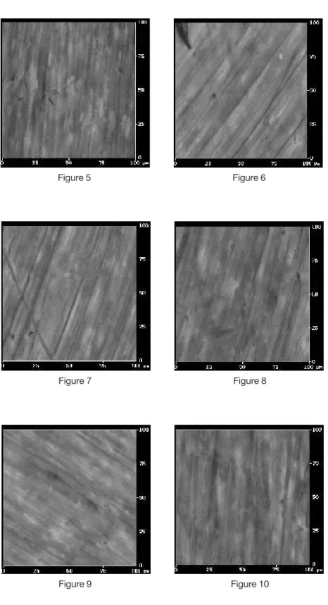
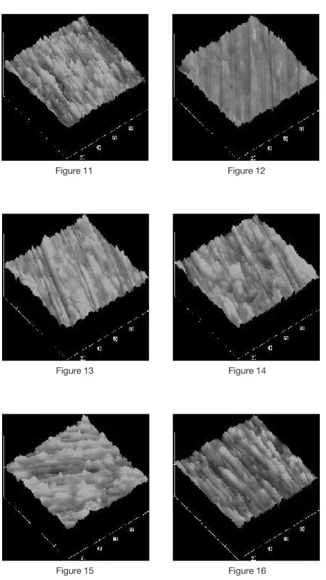
모든 군(대조군 및 실험군)의 2차원적 3차원적 소 견에서 생물학적 오염은 관찰되지 않았다(Figure 5- 16). 대조군 표면은 기계적으로 가공한(milling) 홈이 불균일하게 관찰되었으며, 같은 milling line에서도 높낮이의 차가 심하였다(Figure 5, 11). 그러나 실험 1군의 표면은 균일하였으며, 같은 milling line에 유 사한 높낮이를 보였고 이로 인해 표면 자체가 가장 매끄러워 보였다(Figure 6, 12). 실험 1군에 구연산을 적용할수록 적용시간에 따라서 균일한 홈이 점점 더 불규칙적인 양상을 보였으며, 실험 5군으로 갈수록 표면의 형태가 실험 1군보다 대조군에 더 유사하게 관찰되었다(Figure 7-10, 13-16).
V. 총괄 및 고찰
최근 사용되어지고 있는 임프란트는 1952년 Bränemark에 의해 도입되었고 광범위한 실험적, 임 상적 연구를 거쳐 오늘날 많은 부분에 사용되어지고 있다. 특히 치과영역에서의 임프란트는 개개 치아의 수복 및 계속 가공의치의 지대치 그리고 무치악 환 자에서 가철성 보철물의 지지에 널리 이용되고 있으 며, 저작 효율의 증가와 더불어 기능적, 심미적인 개 선을 가져왔다.
이런 임프란트 재료로는 titanium, hydroxyapatite, alumina oxide 등이 있으며,4)주로 titanium이 널리 사용되고 있다. titanium은 생체 적합성과 낮은 부식
33,42,43)을 보이는데 이러한 성질은 titanium 금속의 산
화막이 가지는 안정성에 의한 것으로 알려졌다. tita- nium은 공기 중에 노출되면 TiO, TiO2, Ti2O3등의 산화막을 형성하는데 이중 TiO2가 가장 안정한 상태 인 것으로 밝혀져 있다. 이렇게 불활성화된 titanium 은 생리적 환경에서는 이론적으로 안정하게 되어 생 체에 사용시에 부식을 방지하는 역할을 하게 된다.
그러나 임프란트는 구강내로 노출되어 하중 및 치 태와 치석의 침착이 가능하고 특히 titanium 임프란 트는 높은 자유-표면 에너지(free-surface energy) 때 문에 자연치보다 더 치태 형성이 잘 되며44-46), 거친 임프란트 표면은 평활한 임프란트 표면에 비해 25배 정도 더 많은 세균 부착을 가진다44). 또한 임프란트 주위 조직은 자연치의 치주조직과 다소 유사하나 치 주인대가 없고 교원질 섬유가 임프란트 표면에 평행 하게 배열되며, 교원질 섬유에 혈관공급이 적어 병원 성에 대한 방어기전이 더 약하다. 그러므로 자연치 에 비해 염증에 더 민감하고 비가역적 골 소실이 발 생한다.
임프란트 주위염의 골소실의 특징은 골 소실이 치 관부에서 시작되나, 근단부는 골과 임프란트 유착이 유지되므로 임프란트 표면전체에 골 소실이 발생할 때까지 임상적으로 동요도가 없다는 것이다15,18,47,48). 또한 임프란트 주위염 시 임프란트 표면은 유약한 조직 세포, 세균 그리고 이러한 세균 부산물에 둘러 싸여 미세한 불규칙성이 증가되고 노출된 임프란트 표면은 오염되어 치유되는 데 어려움이 있다.
많은 치과의사들이 임프란트 주위염의 치료 방법 Table 3. Statistical difference of the surface roughness between citric acid application time(*: p<0.05)
Group 1 Group 2 Group 3 Group 4 Group 5
Group 1
Group 2 *
Group 3 *
Group 4 * *
Group 5 *
에 대하여 연구했으며, 임프란트 골 결손부에 조직 유도 재생술이 성공적인 치료방법으로 제시되었다
49-55). 조직 유도 재생술은 자연치에서와 유사한 방법
으로 시행되며 재생술시 재 골유착(re-osseointegra- tion)을 얻기 위하여 이전의 오염된 임프란트에 대한 표면 처치(surface preparation)가 요구되었다. 최근 의 연구들에서 이런 목적을 달성하는데 공기-분말 연마와 과포화 구연산 처리가 가장 효과적인 것으로 입증되어 왔고38,40) 차폐막과 표면 처리를 사용하여 임프란트 결손부를 치료한 연구들이 보고되어지고 있다.
그러나 표면 처리는 임프란트의 표면의 변화를 야 기시킬 수 있다. Rapley 등56)은 다양한 구강 위생 기 구를 사용하여 titanium implant abutment에 적용시 켜 표면 거칠기를 비교하였을 때 공기-분말 연마가 거칠기를 약간 감소시킨다고 보고하였고, Chairay 등
57)도 machined implant와 plasma- sprayed implant에 공기-분말 연마를 시행한 결과 implant의 body부분에 서는 machined 군(Branemark , 3i )과 plasma- sprayed 군(3i , ITI ) 모두에서 표면 거칠기가 감소 하고 neck 부위에서는 machined 군 중 Branemark implant에서만 표면 거칠기가 감소했다고 보고했다.
Zablotsky 등40)은 여러 가지 화학적 제재가 burnish- ing 효과에 의해 HA implant 표면의 Lipopolysaccha- ride를 제거할 수 있다고 하였다. 본 실험에서 machined implant 표면 거칠기의 변화에 대해 관찰한 결과 공기-분말 연마와 구연산 모두 공기 중에 노출 된 임프란트 표면의 거칠기를 감소시킨다는 결과를 얻었다. 또한 Bollen 등58)은 임프란트의 표면 거칠기 에 대해 특정한“threshold Ra”(0.2㎛)이하의 표면 거 칠기는 치은 연상과 연하의 세균 조성에 별다른 영 향을 끼치지 않는다고 하였으며, 본 실험 결과 나온 표면 거칠기도 모두 0.15㎛이하로 측정되었다(Fig- ure 4, Table 1).
Mouhyi 등59)은 6가지 화학적, 기계적 제재를 사용 하여 임프란트의 표면 구성 성분에 대해 관찰한 결 과 구연산 30초 처치 후 증류수로 5분간 세척했을 때 초기 임프란트 표면과 가장 유사한 구성 성분 비율 이 형성됨을 보고하고 공기-분말 연마 시에는 임프
란트 표면에 나트륨 성분이 많이 관찰된다고 하였 다. Rapley 등56)도 공기-분말 연마시 기계적으로 산 화막이 제거되며, 화학적 성분들이 염기화되어 부식 증가를 야기한다고 하였다.
본 실험에서도 주사탐침현미경의 2차원, 3차원적 소견을 관찰하여 보았을 때 대조군에서 산화막의 형 성으로 인한 불규칙한 모습을 볼 수 있었다. 하지만 공기-분말 연마 후 이런 형태의 소견은 사라지고 비 교적 매끄러운 표면으로 규칙적인 milling line을 보 여 산화막이 제거되었음을 볼 수 있었고 이러한 현 상과 공기-분말 연마시 볼록한 표면이 균일하게 깍 여서 비교적 평탄해 짐으로서 표면 거칠기가 감소한 것으로 생각되었다. 그러나, 이러한 소견은 구연산 을 적용함에 따라 다시 불규칙한 소견이 관찰되는 데 이것은 염기화된 임프란트 표면이 점차 산성화되 고 산화막이 형성되어지는 것으로 보이며 구연산 처 치 후 산화막의 형성, 부식 등은 표면 물성 검사를 통 하여 검사되어야 할 것으로 사료된다. 산 적용 시간 이 길어질수록 이러한 양상은 심화되고 3분까지는 burnishing 효과로 표면 거칠기가 감소했을 것으로 생각되며, 5분과 같이 오랜 시간의 적용은 더 많은 산성화에 의해 거칠기를 다시 증가시킨다고 사료된 다.
V. 결론
주사탐침현미경을 이용한 표면 거칠기를 조사하 기 위해 특별히 10개의 machined pure titanium 원통 형 모델을 제작하고 공기-분말 연마와 구연산(pH 1) 의 적용에 따른 표면 거칠기의 변화를 알아보기 위 해 실험 전과 공기-분말 연마 1분 후 그리고 구연산 적용 30초, 1분, 3분, 5분 시에 표면의 거칠기를 연속 적으로 각 과정마다 측정하였다. 그 후 측정된 값의 평균과 표준 편차를 구하고, 변화량을 통계 처리하여 다음과 같은 결과를 얻었다.
1. Titanium의 표면 거칠기는 공기-분말 연마 1분 간 처치 후 감소하였고, 통계적으로 유의성이 있었다.(p<0.05)
2. 공기-분말 연마가 된 Titanium에 구연산을 처리 했을 때, 적용시간에 따른 표면 거칠기는 3분 적용시에 가장 낮았으며, 다음으로 1분, 30초, 5 분의 순이었다.
3. 구연산 적용 후, 3분까지 표면 거칠기는 모든 군이 적용 전보다 통계적으로 유의성있는 감소 를 보였으나, 30초와 1분 그리고 1분과 3분 사 이에는 통계적 유의성이 없었으며 30초와 3분 사 이 에 는 통 계 적 으 로 유 의 성 이 있 었 다.(p<0.05)
4. Titanium의 표면은 공기 중에 노출 시 산화막이 형성되었고 공기-분말 연마 후에는 산화막이 제거되었으나, 구연산의 적용으로 다시 형성되 었다.
노출된 machined pure titanium에 공기-분말 연마 와 구연산의 적용은 표면 거칠기를 감소시킬 수 있 었으나, 구연산의 오랜 시간의 적용은 이와는 반대로 거칠기를 증가시켰다. 구연산이 machined pure tita- nium 표면 거칠기에 가장 효과적인 시간은 3분이었 으나, 이것을 임상에 적용 시 너무 길 것이라 생각되 며, 30초 적용하는 것은 3분 적용시와 통계적으로 유 의성이 있다. 따라서 임상에 사용시 시간적 질적으 로 구연산을 1분 정도 적용하는 것이 표면 거칠기 측 면에서 본다면 가장 유용하리라 사료된다.
VI. 참고문헌
1. Adell R, Lekholm U, Rockler B, Bränemark PI: A 15-year study of osseointegrated implants in the treatment of edentulous jaw: Int J Oral Surg:
1981; 10: 387-416
2. Zarb GA, Schmitt A: The longitudinal clinical effectiveness of osseointegrated dental implants.
The Toronto study: part I, surgical results: J Pros- thet Dent: 1990; 63: 451-457
3.Ericsson I, Lekholm U, Bränemark PI, Lindhe J, Glantz PO, Nyman S: A clinical evaluation of fixed bridge restorations supported by the
combination of tooth and osseointegrated titanium implants: J Clin Periodontol: 1986;
13: 307-312
4. Meffert RM, Langer B, Fritz ME: Dental implant : A review: J Periodontol: 1992; 63: 859-870 5. Babbush CA: Surgical atlas of implant tech-
niques. Philadelphia. WB Saunders Co., 1980 6. Hobo S, Ichida E, Garcia LT: Osseointegration
and occlusal rehabilitation. Quintessence Pub- lishing Co., 1989
7. Mickinney RV Jr: Endosteal dental implant, Mosby-Year book 1991
8. Albreksson T, Zarb G, Worthinton P, Eriksson RA: The long-term efficacy of currently used dental implants: A review and prognosis criteria for success: Int J Oral Maxillofac Implants: 1986;
1: 11-25
9. Albreksson T, Dahl E, Enbom L: Osseointegrated oral implants. A Swedish multicenter study of 8,139 consecutively inserted Nobelpharma implants: J Periodontol: 1988; 59: 287-296 10. Leckholm U, Ericsson I, Adell R, Slots J: The con-
ditions of the soft tissues at tooth and fixture abutments supporting fixed bridge. A microbio- logical and histological study: J Clin Periodontol:
1986; 13: 558-562
11. d'Hoedt B, Schulte W: A comparative study of results with various endosseous implant system:
Int J Oral Maxillofac Implants: 1989; 4: 95-105 12. Finger IM, Guerra IR: Prosthetic considerations
in reconstructive implantology: Dent North Am:
1986; 30: 69-83
13. Albrektsson T, Bränemark PI, Hansson HA, Lindstorm J: Osseointegrated titanium implants:
Acta Othop Scand: 1981; 52: 155-170
14. Meffert RM, Block MS, Kent JN: What is osseoin- tegration?: Int J Periodont Rest Dent: 1987; 7: 9- 21
15. Newman MG, Flemmig TF: Periodontal consid-
erations of implants and implant associated microbiota: J Dent Educ: 1988; 52: 737-744 16. Lekholm U, Adell R, Lindhe J, Bränemark PI,
Ericsson B, Rocker B: Marginal tissue reactions at osseointegrated fixtures(II). A cross-sectional ret- rospective study: Int J Oral Maxilofac Surg: 1986;
15: 53-61
17. Hickey JS, O'Neal RB, Scheidt MJ, Strong SL, Tur- geon D, Dyke TEV: Microbiologic characteriza- tion of ligature-induced peri-implant- itis in the microswine model: J Periodontol: 1991; 548-553 18. Lindhe J, Berglundh T, Ericsson I, Liljenberg B,
Marinello C: Experimental breakdown of peri- implant and periodontal tissues: Clin Oral Impl Res: 1992; 3: 9-16
19. Lang NP, Bragger U, Walther D, Beamer B, Komman KS: Ligature-induced peri-implant infection in cynomolgus monkey: Clin Oral Impl Res: 1993; 4: 2-11
20. Shou S, Holmstrup P, Keiding NE: Microbiology of ligature-induced marginal inflammation around osseointegrated implants and ankylosed teeth in cynomolgus monkeys(Macaca fascicu- laris): Clin Oral Impl Res: 1996; 7: 190-200 21. Schou S, Holmstrup P, Stolze K, Hjorting-Hansen
E, Kornman KS: Ligature-induced marginal inflammation around osseointegrated implants and ankylosed teeth. Clinical and radiographic observations in cynomolgus monkey(Macaca fascicularis): Clin Oral Impl Res: 1993; 4: 12-22 22. Holt R, Newman M, Kratochvil F: The clinical
and microbial characterization of peri-implant environment: J Dent Res: 1986; 65(Spec Issue):
257
23. Ericsson I, Berglundh T, Marinello CP, Liljenberg B, Lindhe J: Long standing plaque and gingivitis at implants and teeth in dogs: Clin Oral Impl Res: 1992; 3: 99-103
24. Berglundh T, Lindhe J, Ericsson I, Marinello CP,
Liljenberg B, Thomsen P: The soft tissue barrier at implants and teeth: Clin Oral Impl Res: 1991;
2: 81-90
25. Berglundh T, Lindhe J, Ericsson I, Marinello CP, Liljenberg B: Soft tissue reactions to de novo plaque formation on implants and teeth. Anex- perimental study in the dogs: Clin Oral Impl Res:
1992; 3: 1-8
26. Quirynen M, van Steenberghe D: Bacterial adhe- sion to oral implants and assessment of attach- ment and marginal bone level: Dtsch Zahn ztl Z: 1993; 48:158-160
27. Swartz MI, Phillips RW: Comparison of bacterial accumulation on rough and smooth enamel sur- faces: J Periodontol: 1957; 28:304-307
28. Clayton JA, Green E: Roughness of pontic mate- rials and dental plaque: J Prosth Dent Res: 1970;
23: 407-411
29. Baier RE, Meenaghan MA, Hartman LC: Implant surface characteristics and tissue reaction: J Oral Implantol: 1988; 13:594
30. Meffert RM: The soft tissue interface in dental implantology: J Dent Ed: 1988; 52:810
31. Balshi TJ: Hygiene maintenance procedures for patients treated with the tissue integrated pros- thesis(osseointegration): Quintenssence Intern:
1986; 17: 95-102
32. Ducheyne P, Willems G, Martens M, Helsen J:
Invivo metal-ion release from porous titanium- fiber material: J Biomed Mater Res: 1984; 18:
293-308
33. Klauber C, Lenz LJ, Henry PJ: Oxide thickness and surface contamination of six endosseous dental implants determined by electron spec- troscopy for chemical analysis: A preliminary report: Int J Oral Maxillofac Implants: 1990; 5:
264-271
34. Weaks LM, Lescher NB, Barnes CM, Holroyd SV:
Clinical evaluation of The Prophy-jet(R) as an
instrument for routine removal of tooth stain and plaque: J Periodontol: 1984; 55: 486-488
35. Horning G: Clinical use of an sir-powder abra- sive: Compend Contin Educ Dent: 1987; 8: 652- 662
36. Cooley RL, Brown FH, Lubow RM: Evaluation of air-powder abrasive prophylaxis unit: Gen Dent:
1990; 38: 24-27
37. Barnes CM, Fleming LS, Muenninghoff LA: An SEM evaluation of the in-vivo effects of an air- abrasive system on various implant surfaces: Int J Oral Maxillofac Implants: 1991; 6: 463-469 38. Parham PL, Cobb CM, French AA, Love JW,
Drisko CL, Killoy WJ: Effects of an air-powder abrasive system on plasma-sprayed titanium implant surfaces: An in vitro evaluation: J Oral Implantol: 1989; 15: 78-86
39. Dennison DK, Huerzeler MB, Quinones C, Caffesse RG: Contaminated implant surfaces: An in vitro comparison of implant surface coating and treatment madalities for decontamination: J Periodontol: 1994; 65: 942-948
40. Zablotsky MH, Diedrich DL, Meffert RM: Detoxi- fication of endotoxin-contaminated titanium and hydroxyapatite-coated surfaces utilizing various Chemotherapeutic and mechanical modalities:
Implant Dent: 1992; 1: 154-158
41. Zablotsky MH, Diedrich DL, Meffert RM, Wittrig E: The abiliry of various chemotherapeutic agents to detoxify the endotoxin infected HA- coated implant surface: Int J Oral Implant: 1991;
8: 45-51
42. Kasemo B: Biocompatibility of titanium implants: Surface science aspects: J Prosthet Dent: 1983; 49:832-837
43. Kasemo B, Lausmaa J: Biomaterial and implant surfaces: On the role of cleanliness contamina- tion and preparation procedures: J Biomed Mater Res: Applied materials: 1988; 22:145-158
44. Quirynen M, Bollen CML: The influence of sur- face roughness and surface-free energy on supra- and subgingival plaque formation in man:
A review literature: J Clin Periodontol: 1995; 22:
1-14
45. Quirynen M, Van Der Mei C, Bollen CML, Schotte A, Marechal M, Doornbrusch GI: An in vivo study of the influence of the surface rough- ness of implants on the microbiology of supra- and subgingival plaque: J Dent Res: 1993; 72:
1304-1309
46. Quirynen M, Van Der Mei C, Bollen CML, Van Den Bossche LH, Doornbrush GI, van Steen- berghe D: The influence of surface-free energy on supragingival plaque microbiology: An in vivo study on implants: J Periodontol: 1994; 65:
162-167
47. Newman MG, Flemmig TF: Periodontal consid- erations of implants and implant associated microbiota: Int J Oral Implantol: 1988; 5: 65-70 48. Rosenberg ES, Torosian JP, Slots J: Microbial dif-
ferences in two clinically distinct types of failures of osseointegrated implants: Clin Oral Impl Res:
1991; 2: 134-144
49. Choi P, Oyen O, Bissada N: Guided tissue regeneration and bone formation around endosseous dental implants: J Dent Res: 1989;
68: abstr no. 264
50. Zablotsky MH, Meffert RM, Caudill R, Evans G:
Histological and clinical comparisons of guided tissue regeneration on dehisced hydroxyapatite- coated and titanium implant surfaces: A pilot study: Int J Oral Maxillofac Implants: 1991; 6:
294-303
51. Becker W, Becker BE, Handelsman M, Celletti R, Ochsenbein C, Hardwick R, Langer B: Bone for- mation at dehisced dental implant sites treated with implant augmentation material: A pilot study in dogs: Int J Periodont Rest Dent: 1990;
10: 93-101
52. Dahlin C, Sennerby L, Lekholm U, Lindhe A, Nyman S: Generation of new bone around tita- nium implants using a membrane technique: An experimental study in rabbits: Int J Oral Maxillo- fac Implants: 1989; 4: 19-25
53. Nyman S, Lang N, Buser D, Bragger U: Bone regeneration adjacent to titanium dental implants using guided tissue regeneration: A report of two cases: Int J Oral Maxillofac Implants: 1990;
5: 9-14
54. Jovanovic SA, Kenney EB, Carranza FA, Donath K: The regenerative potential of plaque-induced peri-implant bone defects treated by a sub- merged membrane technique: A experimental study: Int J Oral Maxillofac Implants: 1993; 8: 13- 18
55. Jovanovic SA, Spiekermann H, Richter EJ: Int J Oral Maxillofac Implants: 1992; 7: 233-245 56. Rapley JW, Swan RH, Hallmon WW, Mills MP:
The surface characteristics produced by various oral hygiene instruments and materials on titani- um implant abutments: Int J Oral Maxillofac Implants: 1990; 5: 47-52
57. Chairay JP, Boulekbache H, Jean A, Soyer A, Bouchard P: Scanning electron microscopic eval- uation of effects of an air-abrasive system on dental implants: A comparative in vitro study between machined and plasma-sprayed titanium surfaces: J Periodontol: 1997; 68: 1215-1222 58. Bollen CML, Papioannou W, Van Elder J, Schep-
ers E, Quirynen M, Van Steen berghe D: The influence of abutment surface roughness on plaque accumulation and peri-implant mucositis:
Clin Oral Impl Res: 1996; 7: 201-211
59. Mouhyi J, Sennerby L, Pireaux JJ, Dourov N, Nammour S, Van Reck J: An XPS and SEM eval- uation of six chemical and physical techniques for cleaning of contaminated titanium implants:
Clin Oral Impl Res: 1998; 9: 185-194
사진부도 설명
Figure 1. A view of experimental machined titanium model Figure 2. A view of microprophy for air-powder abrasive Figure 3. A view of SPM
Figure 5. Two dimensional view of air contamination surface in the control group by SPM
Figure 6. Two dimensional view of air-powder abrasive treated surface for 1 minute in the experimental group 1 by SPM
Figure 7. Two dimensional view of citric acid treated surface for 30 seconds in the experimental group 2 by SPM Figure 8. Two dimensional view of citric acid treated surface for 1 minute in the experimental group 3 by SPM Figure 9. Two dimensional view of citric acid treated surface for 3 minutes in the experimental group 4 by SPM Figure 10. Two dimensional view of citric acid treated surface for 5 minutes in the experimental group 5 by SPM Figure 11. Three dimensional view of air contamination surface in the control group by SPM
Figure 12. Three dimensional view of air-powder abrasive treated surface for 1 minute in the experimental group 1 by SPM
Figure 13. Three dimensional view of citric acid treated surface for 30 seconds in the experimental group 2 by SPM
Figure 14. Three dimensional view of citric acid treated surface for 1 minute in the experimental group 3 by SPM Figure 15. Three dimensional view of citric acid treated surface for 3 minutes in the experimental group 4 by SPM Figure 16. Three dimensional view of citric acid treated surface for 5 minutes in the experimental group 5 by SPM
Figure 1. Experimental machined titanium model Figure 2. Microprophy
Figure 3. Scanning Probe Microscope
Figure 4. Mean and Standard deviation of the surface roughness Figure 5. Two dimensional SPM view(control group)
Figure 6. Two dimensional SPM view(experimental I group) Figure 7. Two dimensional SPM view(experimental II group) Figure 8. Two dimensional SPM view(experimental III group) Figure 9. Two dimensional SPM view(experimental IV group) Figure 10. Two dimensional SPM view(experimental V group) Figure 11. Three dimensional SPM view(control group) Figure 12. Three dimensional SPM view(experimental I group) Figure 13. Three dimensional SPM view(experimental II group) Figure 14. Three dimensional SPM view(experimental III group) Figure 15. Three dimensional SPM view(experimental IV group) Figure 16. Three dimensional SPM view(experimental V group)
사진부도 ( I )
Figure 1
Figure 2
Figure 3
사진부도 ( II )
Figure 5 Figure 6
Figure 7 Figure 8
Figure 9 Figure 10
사진부도 ( III )
Figure 11 Figure 12
Figure 13 Figure 14
Figure 15 Figure 16
-Abstracts-
The SPM Study on the Change of Titanium Surface Roughness following Air-powder Abrasive
and Application Time of Citric Acid
Min-Seo Park, Chin-Hyung Chung, Sung Bin Lim
Department of Periodontology, College of Dentisry, Dan-kook University
The Peri-implantitis causes inflammation of periodontal tissue and bone loss. It contaminates surface of implants. Therefore, guided bone regeneration has been used for the treatment of this disease. For the re- osseointegration of the exposed surface, various mechanical and chemical methods have been used for clean- ing and detoxication of implant surface. Among these methods, air-powder abrasive and oversaturated citrate are known to be most effective. However, these treatments may deform implant surface. In this research, changes of surface roughness they were examined.
10 experimental machined titanium cylinder models were fabricated to be used for control groups. Each of them was air-powder abraded for 1 minute and they were named group 1. And then, group 1 were burnished with cotton pellets soaked with citrate for 30 seconds(Group 2), 1 minute(Group 3), 3 minutes(Group 4), and 5 minutes(Group 5) burnishing were applied for grouping respectively. Each group were examined with SPM, and their surface roughness were measured and analyzed.
1. Surface roughness of titanium decreased when it was air-powder abraded for 1 minute. It was statistically significant.
2. When Air-powder abraded titanium were treated with citrate for 3 minutes, Their surface roughness was the lowest. Titanium treated for 1 minute was the second lowest and 30 seconds was the third and titani- um burnished for 5 minutes was the highest.
3. Surface roughness of titanium which was treated with citrate was decreased till 3 minutes, which was sta- tistically significant. There was no statistical significance from 30 seconds to 1 minute and from 1 minute to 3 minutes, and there was statistical significance from 30 seconds to 3 minutes.
4. Oxide layer was formed when titanium is exposed to air, and it was removed when air-powder abraded.
It was made when treated with citrate.
It is thought that citrate treatment is necessary after the air-powder abrasion, and 1 minute is clinically and qualitatively adequate for burnishing time of citrate.