REVIEW ARTICLE
위선종의 내시경 치료
허철웅, 김병욱
가톨릭대학교 의과대학 인천성모병원 소화기내과
Endoscopic Treatment of Gastric Adenoma
Cheal Wung Huh and Byung-Wook Kim
Division of Gastroenterology, Department of Internal Medicine, Incheon St. Mary's Hospital, College of Medicine, The Catholic University of Korea, Incheon, Korea
Gastric adenoma (dysplasia) is a precancerous lesion. Therefore, managements of gastric adenomas are important for preventing the development of gastric cancers and for detecting gastric cancers at earlier stages. The Vienna classification divides gastric ad- enomas into two categories: high-grade dysplasia and low-grade dysplasia. Generally, endoscopic resection is performed for ad- enoma with high-grade dysplasia due to the coexistence of carcinoma and the potential of progression to carcinomas. However, the treatments of adenoma with low-grade dysplasia remain controversial. Currently two treatment strategies for the low-grade type have been suggested; First is the ‘wait and see’ strategy; Second is endoscopic treatment (e.g., endoscopic mucosal resection, endoscopic submucosal dissection, or argon plasma coagulation). In this review, we discuss the current optimal strategies for endoscopic man- agement of gastric adenoma. (Korean J Gastroenterol 2017;70:115-120)
Key Words: Gastric adenoma; Dysplasia; Endoscopic treatment
Received August 9, 2017. Revised September 1, 2017. Accepted September 5, 2017.
CC This is an open access article distributed under the terms of the Creative Commons Attribution Non-Commercial License (http://creativecommons.org/licenses/
by-nc/4.0) which permits unrestricted non-commercial use, distribution, and reproduction in any medium, provided the original work is properly cited.
Copyright © 2017. Korean Society of Gastroenterology.
교신저자: 김병욱, 21431, 인천시 부평구 동수로 56, 가톨릭대학교 의과대학 인천성모병원 소화기내과
Correspondence to: Byung-Wook Kim, Division of Gastroenterology, Department of Internal Medicine, Incheon St. Mary's Hospital, College of Medicine, The Catholic University of Korea, 56 Dongsu-ro, Bupyeong-gu, Incheon 21431, Korea. Tel: +82-32-280-5052, Fax: +82-32-280-5987, E-mail: [email protected]
Financial support: None. Conflict of interest: None.
서 론
위암은 전 세계에서 네 번째로 흔히 발생하는 암으로, 한국 을 포함한 동아시아 국가에서 특히 그 발생 빈도가 높다.1위 선종(gastric adenoma) 또는 위이형성(gastric dysplasia)은 위암의 전구 병변으로, 이에 대한 진단 및 치료는 위암의 조기 발견 및 예방에 매우 중요하다.2
현재 대부분의 임상 의사들은 위선종과 위이형성을 비슷한 의미로 혼용하여 사용하고 있다. 하지만 엄밀하게 위선종은 주변 점막의 위축성 위염과 같은 염증과 관련 없이 발생한 양성 신생물로 정의할 수 있는 반면, 위이형성은 주변 점막의 염증과 관련이 있는 양성 신생물이라는 차이가 있다.3 병리학 적으로는 위선종의 경우 주변 점막에 비해 융기되어 있는 병
변을 일컬으며, 위이형성은 주변 점막과 동일한 높이의 병변을 일컫는다.4이에 본고에서는 저도 이형성 선종(gastric adenoma with low grade dysplasia), 고도 이형성 선종(gastric ad- enoma with high grade dysplasia)으로 통일하여 사용하고 자 한다.
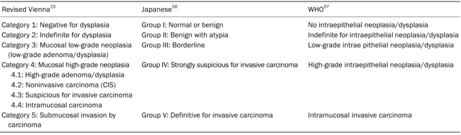
위암과 고도 이형성 선종을 감별할 때 일본 병리학자들과 서양 병리학자들 사이에 이견이 있다. 일본 병리학자들은 점 막 고유층의 침범 여부에 관련 없이 세포학적 구조의 변화에 따라 암을 진단하는 반면, 서양 병리학자들은 점막 고유층의 침범 여부에 따라서 암을 진단하였다.5이에 대한 혼돈을 줄이 고자 1998년에 Vienna 분류를 발표하였고, 2000년에 수정된 Vienna 분류법을 발표하였다(Table 1).6,7
현재 고도 이형성 선종의 치료는 이견이 없는 반면에 저도
Table 1. Classification Systems of Gastric Epithelial Neoplasia
Revised Vienna15 Japanese56 WHO57
Category 1: Negative for dysplasia Group I: Normal or benign No intraepithelial neoplasia/dysplasia
Category 2: Indefinite for dysplasia Group II: Benign with atypia Indefinite for intraepithelial neoplasia/dysplasia Category 3: Mucosal low-grade neoplasia
(low-grade adenoma/dysplasia)
Group III: Borderline Low-grade intrae pithelial neoplasia/dysplasia
Category 4: Mucosal high-grade neoplasia Group IV: Strongly suspicious for invasive carcinoma High-grade intraepithelial neoplasia/dysplasia 4.1: High-grade adenoma/dysplasia
4.2: Noninvasive carcinoma (CIS) 4.3: Suspicious for invasive carcinoma 4.4: Intramucosal carcinoma Category 5: Submucosal invasion by
carcinoma
Group V: Definitive for invasive carcinoma Intramucosal invasive carcinoma
WHO, World Health Organization; CIS, carcinoma in situ.
이형성 선종의 치료는 논란이 있다. 본고에서는 고도 및 저도 이형성 선종의 치료, 특히 내시경적 치료에 초점을 맞추어 살 펴보고자 한다.
본 론
고도 이형성 선종과 저도 이형성 선종은 모두 암으로 진행 될 가능성이 있다. 여러 보고에 따르면 고도 이형성 선종은 약 60-85% 이상에서 악성 변화를 보였다.8-14 따라서, 고도 이 형성 선종은 반드시 내시경 치료를 통하여 제거할 것을 권유 하고 있다.15 하지만 저도 이형성 선종은 현재까지 치료에 대 해 이견이 있다. 저도 이형성 선종은 암으로 진행될 가능성이 10% 미만으로 낮고,8,16 일부에서는 추적 관찰 중 자연적으로 소실되기도 한다.11,17-19
고도 및 저도 이형성 선종의 치료에 대해 여러 가이드라인 이 제시되었는데, 개정된 Vienna 분류에서는 고도 이형성 선 종은 내시경 치료를 권유하고, 저도 이형성 선종은 내시경 치료 혹은 추적 관찰을 권유하고 있다.15최근 American Society for Gastrointestinal Endoscopy 가이드라인과 British Society of Gastroenterology 가이드라인은 위선종의 크기 및 분화도와 상관없이 가능하면 내시경 절제를 권고하고 있다.20,21 유럽 가 이드라인 역시 고도 이형성 선종은 반드시 내시경 절제가 필 요하며, 저도 이형성 선종이라고 하더라도 정확한 병리학적 진단을 위해 내시경 절제를 시행할 것을 권고하고 있다.22
1. 내시경 점막 절제술(endoscopic mucosal resection, EMR) 내시경 점막 절제술은 선종의 치료뿐만 아니라 진단 목적 으로도 사용된다. 선종의 진단에 있어서 생검 겸자를 이용한 조직 검사와 내시경 절제 후 조직 검사 결과의 불일치가 비교 적 높은 것으로 알려져 있다. 최근 한 메타 분석은 생검 겸자 를 이용한 조직 검사에서 저도 이형성 선종으로 진단되어 내
시경 절제를 받은 환자들을 분석하였을 때, 약 25%의 환자에 서 고도 이형성 선종이나 조기 위암이 관찰되었다.23 이러한 불일치는 고도 이형성 선종의 경우에는 더욱 심하게 나타나, 약 80%의 환자에서 내시경 절제 후 조기 위암이 관찰되었 다.24 따라서 내시경 점막 절제술은 선종에 대한 치료 목적뿐 만 아니라 생검 겸자의 조직학적 불일치를 극복할 수 있는 진단적 목적, 즉 ‘total biopsy’ 측면에서도 의미가 있을 수 있다.
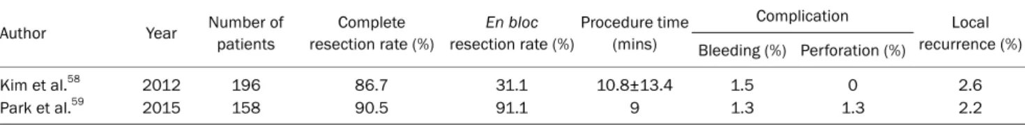
내시경 점막 절제술은 고식적 점막 절제술(conventional EMR, snaring technique)뿐만 아니라 점막층 절개 후 점막 절제술(EMR with circumferential precutting, EMR-P), 이 중 겸자공 내시경을 이용한 점막 절제술(EMR using a du- al-channel endoscope, EMR-D), 내시경 선단에 캡을 씌운 점막 절제술(cap-assisted EMR, EMR-C), 밴드로 묶은 후 점 막 절제술(ligation-assisted EMR, EMR-L) 등 여러 변형된 점막 절제술 방법이 사용되고 있다.25-28 내시경 점막 절제술은 내시경 점막하 박리술(endoscopic submucosal dissection) 에 비해 시술 시간이 짧고, 출혈이나 천공과 같은 합병증이 적다는 장점이 있다.29 하지만 내시경 점막 절제술은 내시경 점막하 박리술에 비해서 일괄 절제율, 완전 절제율이 낮다는 단점이 있다(Table 2). 특히, 병변의 크기가 2 cm 이상으로 큰 경우나 병변의 위치에 따라서 일괄 절제율과 완전 절제율 은 크게 감소하는 것으로 알려져 있다.30-33 이로 인해 불완전 절제와 분할 절제가 많아져 국소 재발률이 높아질 수 있는 단 점이 있다.34 따라서, 내시경 점막 절제술은 위암의 가능성이 높은 고도 이형성 선종의 치료에는 효과적이지 못하다. 또한 생검 겸자를 이용한 조직 검사에서 저도 이형성 선종으로 진 단된 경우라도 병변의 크기가 2 cm 이상인 경우, 함몰이 동반 된 경우, 표면이 거칠거나 붉은색으로 보이는 경우, 자발적 출 혈 등 내시경 절제 후 고도 이형성 선종이나 암으로 진단될 가능성이 높은 병변의 치료에는 내시경 점막 절제술이 효과적
Table 2. Treatment Outcomes and Complications of Endoscopic Mucosal Resection for Gastric Adenoma
Author Year Number of
patients
Complete resection rate (%)
En bloc resection rate (%)
Procedure time (mins)
Complication Local
recurrence (%) Bleeding (%) Perforation (%)
Kim et al.58 2012 196 86.7 31.1 10.8±13.4 1.5 0 2.6
Park et al.59 2015 158 90.5 91.1 9 1.3 1.3 2.2
Table 3. Treatment Outcomes and Complications of Endoscopic Submucosal Dissection for Gastric Adenoma
Author Year Number of
patients
Complete resection rate (%)
En bloc resection rate (%)
Procedure time (mins)
Complication Local
recurrence (%) Bleeding (%) Perforation (%)
Kato et al.24 2011 468 97.0 97.0 59.0 5.4 4.3 N/A
Choi et al.60 2012 282 96.1 N/A 26.4 1.4 0 1.4
Kim et al.58 2012 56 98.2 75.0 43.1±23.7 10.7 0 1.8
Jung et al.46 2012 204 95.4 91.7 53.1±38.1 2 1 0.5
Lee et al.48 2017 113 89.4 100 N/A 5.3 0.9 3.5
N/A, not available.
Table 4. Treatment Outcomes and Complications of Argon Plasma Coagulation for Gastric Adenoma
Author Year Number of
patients Hospital days Need to admission (%)
Procedure time (mins)
Complication Local
recurrence (%) Bleeding (%) Perforation (%)
Lee et al.48 2009 57 N/A N/A 15.0±5.0 1.7 1.7 7.0
Jung et al.46 2013 116 1.2±2.3 31 7.8±5.1 1.7 0 3.8
Ahn et al.47 2013 71 N/A N/A N/A 1.4 0 21.1
Lee et al.45 2017 97 1.6±2.0 42 N/A 0 0 15.3
N/A, not available.
이지 못할 수 있다.35-38
2. 내시경 점막하 박리술(endoscopic submucosal dissection) 내시경 점막하 박리술은 내시경 점막 절제술과는 달리 병 변의 크기와 위치에 관계 없이 일괄 절제, 완전 절제를 할 수 있다는 장점이 있다. 따라서, 내시경 점막하 박리술은 조기 위암과 동일하게 치료를 권고하는 고도 이형성 선종의 치료에 있어 가장 효과적인 치료라고 할 수 있다. 하지만 저도 이형성 선종의 내시경 치료에 있어서는 아직 명확히 정립된 바 없다.
현재까지의 보고를 종합해보면 고도 및 저도 이형성 선종에 대한 내시경 점막하 박리술의 치료 성적은 매우 우수하다 (Table 3). 또한, 내시경 점막하 박리술은 내시경 점막 절제술 에 비해 일괄 절제율, 완전 절제율이 높고 국소 재발률이 낮다 고 알려져 있다. 하지만 내시경 점막하 박리술은 내시경 점막 절제술에 비해서 출혈, 천공 등과 같은 합병증의 발생 빈도가 높고 시술 시간이 많이 소요된다는 단점이 있다.39,40 또한 내 시경 점막하 박리술은 시술자의 숙련도에 따라 시술의 성공이 크게 좌우된다는 제한점이 있다.41,42출혈, 천공과 같은 대부 분의 내시경 점막하 박리술의 합병증은 내시경적으로 치료가
가능하고, 내시경 점막하 박리술로 인한 심각한 합병증은 매 우 적다고 알려져 있다. 고도 및 저도 이형성 선종의 생검 겸 자를 이용한 조직 검사와 내시경 절제 후 조직 검사 결과의 불일치가 높다는 점을 고려하면 내시경 점막하 박리술은 고도 이형성 선종과 저도 이형성 선종 중에 내시경 절제 후 고도 이형성 선종이나 암으로 불일치될 가능성이 높은 병변에 대해 서 효과적인 치료 방법이라고 할 수 있다.
3. 아르곤 플라즈마 응고술(argon plasma coagulation) 아르곤 플라즈마 응고술은 이온화된 아르곤 가스를 통하여 조직으로 전해지는 에너지를 이용하여 조직과 직접 닿지 않고 전기 소작을 하는 방법이다.43,44 아르곤 플라즈마 응고술은 이 론적으로는 점막층뿐 아니라 점막하층의 표층까지도 근절할 수 있다고 알려져 있다.45 아르곤 플라즈마 응고술은 내시경 절제에 비해 시술 시간이 짧고, 출혈이나 천공의 위험이 적으 며, 입원이 필요 없고, 시술 비용이 적게 들며, 시술자의 숙련 도가 크게 중요하지 않다는 장점이 있다(Table 4).45-48 여러 보고에 따르면, 아르곤 플라즈마 응고술은 1-2 cm 미만의 크 기가 작은 육안적으로 편평한 고도 및 저도 이형성 선종의
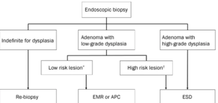
Fig. 1. Proposed treatment strategy algorithm for gastric adenoma diagnosed by endoscopic biopsy; *low risk lesion: size<2 cm, grossly flat type, whitish color with smooth surface; †high risk lesion: size≥2 cm, grossly depressed type, surface erythema or unevenness, presence of spontaneous bleeding. EMR, endoscopic mucosal resection; APC, argon plasma coagulation; ESD, endoscopic submucosal dissection.
치료에 효과적이라고 알려져 있다. 하지만 크기가 2 cm 이상 일 경우, 육안적으로 융기형이나 점막하 식염수 주입 시 병변 이 떠오르지 않는 경우, 40 W 등 낮은 전압으로 아르곤 플라 즈마 응고술을 시행하였을 경우에는 그 효과가 떨어져 재발률 이 상승하는 것으로 알려져 있다.45-48 또한 아르곤 플라즈마 응고술은 내시경 절제와 다르게 선종 전체의 조직 검사 결과 를 확인할 수 없기 때문에 고도 이형성 선종이나 고위험 저도 이형성 선종의 치료에는 제한적이다. 따라서 육안적으로 평면 형인 1 cm 크기 미만의 작은 저도 이형성 선종의 치료에는 아르곤 플라즈마 응고술로 치료를 시도해 볼 수 있겠지만 충 분한 60-80 W의 높은 전압으로 충분한 시간 동안 치료하여야 한다. 또한 국소 재발률이 높은 점을 감안하여 정기적인 내시 경 추적 관찰이 반드시 필요하다.
4. 단순 추적 관찰(wait and see)
고도 이형성 선종의 경우에는 반드시 내시경 절제술이 필 요하다. 하지만 일부 연구에서 저도 이형성 선종의 경우에는 추적 기간 동안 자연적으로 소실되었고,11,17,19
Helicobacter pylori
(H. pylori
)균이 있는 경우 제균 치료 후 저도 이형성 선종이 소실되었음을 보고한 바 있다.17,49이를 근거로 일부에 서는 저도 이형성 선종에서는 치료 없이 1년마다 조직 검사를 동반한 내시경 추적 관찰을 추천하였다.50,51 하지만 이러한 반 복적인 위 내시경 검사는 환자 입장에서 육체적, 정신적 부담 및 경제적 부담이 커질 수 있다. 또한 무엇보다 저도 이형성 선종의 생검 겸자 조직과 내시경적 절제술 이후 조직 간의 심한 불일치와 저도 이형성 선종의 암 발전 가능성 등을 고려 하면 단순 추적 위 내시경 검사만은 위험 부담이 클 수밖에 없다. 그리고 내시경 절제술을 이용한 치료가 보급되고 기술 이 발전한 국내에서는 단순 추적 관찰은 좋은 치료 전략이라 고 보기는 어렵다.5. 내시경 절제 후 추적 관찰(follow up after endoscopic resection)
생검 겸자로 위선종이 발견된 경우, 동시성 병변의 선종이 나 암이 존재할 위험이 있기 때문에 반드시 전체 위를 다시 한 번 자세히 관찰하여야 한다. 또한 고도 및 저도 이형성 선종 에 대한 내시경 치료 후에는 이시성 병변의 발생 위험이 높으 므로 주기적인 내시경 추적 관찰이 필요하다.36,52 American Society for Gastrointestinal Endoscopy 가이드라인은 내시 경 절제 후 1년 뒤 추적 내시경 검사를 권고하고 있으며, British Society of Gastroenterology 가이드라인은 고도 이 형성 선종이나 불완전 절제 후에는 6개월, 그 외의 경우에는 1년 뒤 추적 내시경 검사를 권고하고 있다. 또한 이러한 환자 군에서
H. pylori
가 있을 경우 제균 치료하는 것이 이시성 병변의 발생을 줄일 수 있다고 알려져 있기 때문에 가능한
H.
pylori
제균 치료를 하는 것이 좋다.53-55결 론
고도 이형성 선종의 경우에는 조기 위암에 준한 내시경 절 제 등 적극적인 치료가 반드시 필요하다. 반면에 저도 이형성 선종의 치료에는 이견이 있는 실정이다. 하지만 생검 겸자를 이용한 조직 검사와 내시경 절제 후 조직 검사 결과의 불일치 가능성이 있으므로 진단 목적 및 치료 목적으로 내시경 절제 를 시행하는 것이 좋겠다. 현재까지의 연구를 근거로 생검 겸 자를 이용한 조직 검사 결과에 따른 치료 전략을 Fig. 1에 정리하였다. 고도 및 저도 이형성 선종에 대한 내시경 절제 후
H. pylori
가 검출되는 경우, 이시성 병변의 발생 위험을 낮 추기 위해 제균 치료를 시행하는 것이 좋을 것으로 생각한다.REFERENCES
1. Kamangar F, Dores GM, Anderson WF. Patterns of cancer in- cidence, mortality, and prevalence across five continents: defin- ing priorities to reduce cancer disparities in different geographic regions of the world. J Clin Oncol 2006;24:2137-2150.
2. Lauwers GY, Riddell RH. Gastric epithelial dysplasia. Gut 1999;
45:784-790.
3. Lewin KJ. Nomenclature problems of gastrointestinal epithelial neoplasia. Am J Surg Pathol 1998;22:1043-1047.
4. Iacobuzio-Donahue CA, Montgomery EA. Gastrointestinal and liver pathology. 2nd ed. Philadelphia: Saunders, 2012.
5. Schlemper RJ, Kato Y, Stolte M. Review of histological classi- fications of gastrointestinal epithelial neoplasia: differences in diagnosis of early carcinomas between Japanese and Western pathologists. J Gastroenterol 2001;36:445-456.
6. Stolte M. The new Vienna classification of epithelial neoplasia of
the gastrointestinal tract: advantages and disadvantages. Virchows Arch 2003;442:99-106.
7. Schlemper RJ, Riddell RH, Kato Y, et al. The Vienna classification of gastrointestinal epithelial neoplasia. Gut 2000;47:251-255.
8. Yamada H, Ikegami M, Shimoda T, Takagi N, Maruyama M. Long-term follow-up study of gastric adenoma/dysplasia. Endoscopy 2004;36:
390-396.
9. Kokkola A, Haapiainen R, Laxén F, et al. Risk of gastric carcinoma in patients with mucosal dysplasia associated with atrophic gas- tritis: a follow up study. J Clin Pathol 1996;49:979-984.
10. Fertitta AM, Comin U, Terruzzi V, et al. Clinical significance of gastric dysplasia: a multicenter follow-up study. Gastrointestinal Endoscopic Pathology Study Group. Endoscopy 1993;25:265-268.
11. Di Gregorio C, Morandi P, Fante R, De Gaetani C. Gastric dysplasia.
A follow-up study. Am J Gastroenterol 1993;88:1714-1719.
12. Rugge M, Farinati F, Di Mario F, Baffa R, Valiante F, Cardin F.
Gastric epithelial dysplasia: a prospective multicenter follow-up study from the Interdisciplinary Group on Gastric Epithelial Dysplasia.
Hum Pathol 1991;22:1002-1008.
13. Lansdown M, Quirke P, Dixon MF, Axon AT, Johnston D. High grade dysplasia of the gastric mucosa: a marker for gastric carcinoma.
Gut 1990;31:977-983.
14. Saraga EP, Gardiol D, Costa J. Gastric dysplasia. A histological fol- low-up study. Am J Surg Pathol 1987;11:788-796.
15. Dixon MF. Gastrointestinal epithelial neoplasia: Vienna revisited.
Gut 2002;51:130-131.
16. Rugge M, Cassaro M, Di Mario F, et al. The long term outcome of gastric non-invasive neoplasia. Gut 2003;52:1111-1116.
17. Suzuki S, Gotoda T, Suzuki H, et al. Morphologic and histologic changes in gastric adenomas after helicobacter pylori eradication:
a long-term prospective analysis. Helicobacter 2015;20:431-437.
18. Srivastava A, Lauwers GY. Gastric epithelial dysplasia: the Western perspective. Dig Liver Dis 2008;40:641-649.
19. Bearzi I, Brancorsini D, Santinelli A, Rezai B, Mannello B, Ranaldi R. Gastric dysplasia: a ten-year follow-up study. Pathol Res Pract 1994;190:61-68.
20. ASGE Standards of Practice Committee, Evans JA, Chandrasekhara V, et al. The role of endoscopy in the management of premalignant and malignant conditions of the stomach. Gastrointest Endosc 2015;82:1-8.
21. Goddard AF, Badreldin R, Pritchard DM, Walker MM, Warren B;
British Society of Gastroenterology. The management of gastric polyps. Gut 2010;59:1270-1276.
22. Dinis-Ribeiro M, Areia M, de Vries AC, et al. Management of pre- cancerous conditions and lesions in the stomach (MAPS): guide- line from the European Society of Gastrointestinal Endoscopy (ESGE), European Helicobacter Study Group (EHSG), European Society of Pathology (ESP), and the Sociedade Portuguesa de Endoscopia Digestiva (SPED). Endoscopy 2012;44:74-94.
23. Zhao G, Xue M, Hu Y, Lai S, Chen S, Wang L. How commonly is the diagnosis of gastric low grade dysplasia upgraded following endo- scopic resection? A meta-analysis. PLoS One 2015;10:e0132699.
24. Kato M, Nishida T, Tsutsui S, et al. Endoscopic submucosal dis- section as a treatment for gastric noninvasive neoplasia: a multi- center study by Osaka University ESD Study Group. J Gastroenterol 2011;46:325-331.
25. de Melo SW Jr, Cleveland P, Raimondo M, Wallace MB, Woodward T. Endoscopic mucosal resection with the grasp-and-snare techni- que through a double-channel endoscope in humans. Gastrointest Endosc 2011;73:349-352.
26. Yamamoto H, Kawata H, Sunada K, et al. Success rate of curative endoscopic mucosal resection with circumferential mucosal in- cision assisted by submucosal injection of sodium hyaluronate.
Gastrointest Endosc 2002;56:507-512.
27. Suzuki Y, Hiraishi H, Kanke K, et al. Treatment of gastric tumors by endoscopic mucosal resection with a ligating device. Gastrointest Endosc 1999;49:192-199.
28. Inoue H, Endo M, Takeshita K, Yoshino K, Muraoka Y, Yoneshima H. A new simplified technique of endoscopic esophageal mu- cosal resection using a cap-fitted panendoscope (EMRC). Surg Endosc 1992;6:264-265.
29. Kim JW, Jang JY. Optimal management of biopsy-proven low-grade gastric dysplasia. World J Gastrointest Endosc 2015;7:396-402.
30. Oka S, Tanaka S, Kaneko I, et al. Advantage of endoscopic sub- mucosal dissection compared with EMR for early gastric cancer.
Gastrointest Endosc 2006;64:877-883.
31. Tanabe S, Koizumi W, Kokutou M, et al. Usefulness of endoscopic aspiration mucosectomy as compared with strip biopsy for the treatment of gastric mucosal cancer. Gastrointest Endosc 1999;
50:819-822.
32. Kojima T, Parra-Blanco A, Takahashi H, Fujita R. Outcome of en- doscopic mucosal resection for early gastric cancer: review of the Japanese literature. Gastrointest Endosc 1998;48:550-554;
discussion 554-555.
33. Inoue H, Takeshita K, Hori H, Muraoka Y, Yoneshima H, Endo M.
Endoscopic mucosal resection with a cap-fitted panendoscope for esophagus, stomach, and colon mucosal lesions. Gastrointest Endosc 1993;39:58-62.
34. Szalóki T, Tóth V, Tiszlavicz L, Czakó L. Flat gastric polyps: results of forceps biopsy, endoscopic mucosal resection, and long-term follow-up. Scand J Gastroenterol 2006;41:1105-1109.
35. Lim H, Jung HY, Park YS, et al. Discrepancy between endoscopic forceps biopsy and endoscopic resection in gastric epithelial neoplasia. Surg Endosc 2014;28:1256-1262.
36. Won CS, Cho MY, Kim HS, et al. Upgrade of lesions initially diag- nosed as low-grade gastric dysplasia upon forceps biopsy follow- ing endoscopic resection. Gut Liver 2011;5:187-193.
37. Cho SJ, Choi IJ, Kim CG, et al. Risk of high-grade dysplasia or carci- noma in gastric biopsy-proven low-grade dysplasia: an analysis using the Vienna classification. Endoscopy 2011;43:465-471.
38. Park DI, Rhee PL, Kim JE, et al. Risk factors suggesting malignant transformation of gastric adenoma: univariate and multivariate analysis. Endoscopy 2001;33:501-506.
39. Park YM, Cho E, Kang HY, Kim JM. The effectiveness and safety of endoscopic submucosal dissection compared with endo- scopic mucosal resection for early gastric cancer: a systematic review and metaanalysis. Surg Endosc 2011;25:2666-2677.
40. Cao Y, Liao C, Tan A, Gao Y, Mo Z, Gao F. Meta-analysis of endo- scopic submucosal dissection versus endoscopic mucosal re- section for tumors of the gastrointestinal tract. Endoscopy 2009;
41:751-757.
41. Oda I, Odagaki T, Suzuki H, Nonaka S, Yoshinaga S. Learning
curve for endoscopic submucosal dissection of early gastric can- cer based on trainee experience. Dig Endosc 2012;24 Suppl 1:129-132.
42. Sakamoto T, Saito Y, Fukunaga S, Nakajima T, Matsuda T. Learning curve associated with colorectal endoscopic submucosal dis- section for endoscopists experienced in gastric endoscopic sub- mucosal dissection. Dis Colon Rectum 2011;54:1307-1312.
43. Grund KE. Argon plasma coagulation (APC): ballyhoo or break- through? Endoscopy 1997;29:196-198.
44. Grund KE, Storek D, Farin G. Endoscopic argon plasma coagu- lation (APC) first clinical experiences in flexible endoscopy.
Endosc Surg Allied Technol 1994;2:42-46.
45. Lee DH, Bae WK, Kim JW, et al. The usefulness of argon plasma coagulation compared with endoscopic submucosal dissection to treat gastric adenoma. Korean J Gastroenterol 2017;69:283-290.
46. Jung SJ, Cho SJ, Choi IJ, et al. Argon plasma coagulation is safe and effective for treating smaller gastric lesions with low-grade dysplasia: a comparison with endoscopic submucosal dissection.
Surg Endosc 2013;27:1211-1218.
47. Ahn JY, Choi KD, Na HK, et al. Clinical outcomes of argon plasma coagulation for the treatment of gastric neoplasm. Surg Endosc 2013;27:3146-3152.
48. Lee KM, Kim YB, Sin SJ, et al. Argon plasma coagulation with sub- mucosal saline injection for gastric adenoma on outpatient basis. Dig Dis Sci 2009;54:2623-2628.
49. Gotoda T, Saito D, Kondo H, et al. Endoscopic and histological re- versibility of gastric adenoma after eradication of helicobacter pylori. J Gastroenterol 1999;34 Suppl 11:91-96.
50. Rugge M, Nitti D, Farinati F, di Mario F, Genta RM. Non-invasive neoplasia of the stomach. Eur J Gastroenterol Hepatol 2005;17:
1191-1196.
51. Weinstein WM, Goldstein NS. Gastric dysplasia and its management.
Gastroenterology 1994;107:1543-1545.
52. Kim MK, Jang JY, Kim JW, et al. Is lesion size an independent in- dication for endoscopic resection of biopsy-proven low-grade gastric dysplasia? Dig Dis Sci 2014;59:428-435.
53. Shin SH, Jung DH, Kim JH, et al. Helicobacter pylori eradication prevents metachronous gastric neoplasms after endoscopic re- section of gastric dysplasia. PLoS One 2015;10:e0143257.
54. Bae SE, Jung HY, Kang J, et al. Effect of helicobacter pylori erad- ication on metachronous recurrence after endoscopic resection of gastric neoplasm. Am J Gastroenterol 2014;109:60-67.
55. Chon I, Choi C, Shin CM, Park YS, Kim N, Lee DH. Effect of heli- cobacter pylori eradication on subsequent dysplasia develop- ment after endoscopic resection of gastric dysplasia. Korean J Gastroenterol 2013;61:307-312.
56. Japanese Gastric Cancer Association. Japanese classification of gastric carcinoma: 3rd English edition. Gastric Cancer 2011;14:
101-112.
57. Yakirevich E, Resnick MB. Pathology of gastric cancer and its pre- cursor lesions. Gastroenterol Clin North Am 2013;42:261-284.
58. Kim SY, Sung JK, Moon HS, et al. Is endoscopic mucosal resection a sufficient treatment for low-grade gastric epithelial dysplasia?
Gut Liver 2012;6:446-451.
59. Park SM, Kim JS, Ji JS, Choi H, Lee BI, Kim BW. Efficacy of endo- scopic mucosal resections for the management of small gastric adenomas with low-grade dysplasia. Scand J Gastroenterol 2015;50:1175-1182.
60. Choi CW, Kang DH, Kim HW, Park SB, Kim S, Cho M. Endoscopic submucosal dissection as a treatment for gastric adenomatous polyps: predictive factors for early gastric cancer. Scand J Gastroenterol 2012;47:1218-1225.