알츠하이머병 환자에서 콜린에스터라제 억제제의 투여에 의한 대뇌피질 국소혈류의 변화
영남대의료원 신경과 생리검사실․대구보건대학 임상병리과1․김천대학 임상병리과2
문성식․김영활1․김병원2
Change of Regional Cerebral Blood Flow in Alzheimer's Disease Patients treated with Cholinesterase Inhibitors
Moon, Seung Sik., Kim, Young Hwal1., Kim, Byung Weon2
College of Medicine, Yeungnam University, Daegu, Korea Electrophysiolgy Laboratory Yeungnam University Hospital
Department of Clinical Pathology, Daegu Health College Daegu, Korea1 Department of Biomedical Laboratory Science, Gimcheon College, Gimcheon, Korea2
Alzheimer`s Disease(AD) is a progressive, degenerative disease that attacks the brain resulting in impaired memory, thought processing, and behavior. This is associated with a loss in presynaptic cholinergic function.
It has been suggested that cholinergic inhibitors could restore this function and improve some symptoms of AD. Previous studies have shown that cholinesterase inhibitors(ChEI) improve cognitive and global functions in patients with mild to moderate AD. This study aims to evaluate regional cerebral blood flow(rCBF) changes and contemporary clinical responses such as cognitive and psychiatric symptoms after ChEI treatment. The subjects were eight at risk AD patients(four males and four females, mean age 69.63 years) recruited from the department of Neurology at Yeungnam University Medical Center between August 2000 and April 2002. The clinical diagnosis of AD was based on DSM-Ⅳ and NINCDS-ADRDA criteria.
Huchinski ischemic scores of all the patients were below 1. The mean treatment duration was 30.38 weeks, ranging between 24 to 44 weeks. Four patients received Rivastigmine(ExelonⓇ) 12mg after titration, three patients received Donepezil(AriceptⓇ) 10mg during the whole period, and one patient received Donepezil 10mg after the initial 5mg for three weeks. The base line and follow up Ethylene Cysteine Diethylester Single Photon Emission Computed Tomography(99mTC-ECD SPECT) studies were done within one week prior to ChEI treatment and within one week following the study. Regions of interest(ROIs) were drawn over the left and right frontal lobe, temporal lobe, parietal lobe, and cerebellum. Region to cerebellar ratio(RCR)(each count in the ROIs divided by mean cerebellar ROI count) were calculated as a estimation
임상병리검사과학회지 : 35권 제1호, 68-73, 2003
1)
교신저자 : 김병원,(우)740-704 경상북도 김천시 삼락동 754번지 김천대학 임상병리과
Tel : 054-420-4048
E-mail : [email protected]
of rCBF, and base line RCRs were compared with those of the follow up. The results display an overall increase in global cerebral blood flow through K-MMSE(Korean-Mini Mental State Examination), CDR(Clinical Dementia Rating), CDR-SB(Clinical Dementia Rating-Sum of Box), GDS(Global Deterioration Scale), and NPI(Neuropsychiatric Inventory, Cummings). We found that the most significant increase in blood flow occurred in the bilateral parietal lobes(p<0.05). Reduction in the rCBF is more profound on the left hemisphere in the base line(p<0.05) where there is a significant increase of rCBF after ChEI treatment compared with to right hemisphere(p<0.05).
Key Words: Alzheimer`s disease, Cholinesterase inhibitors, Regional cerebral blood flow
I. 서 론
알츠하이머병(alzheimer`s disease : AD)은 광범위한 대 뇌피질의 퇴행성 변화에 의해서 초래되는 질환으로서 주 증상이 치매이며 그 외에 정신이상 및 이상행동도 함께 수반된다. 이러한 치매증상은 대뇌의 아세틸콜린(acetyl- choline, Ach)의 감소와 관련이 있다(Davies와 Maloney, 1976). 아세틸콜린은 acethylcholin transferase(CAT)라는 효소에 의해 생산되고 acethylcholine-esterase(acethy:
AchE)라는 효소에 의해 분해되어 그 양이 조절되는 물질 인데 콜린에스테라제 억제제(cholinesterase inhibitor, ChEI)를 이용하여 아세틸콜린의 분해과정을 억제해 줄 경우 아세틸콜린의 양이 증가되어 알츠하이머 환자의 치 매증상이 호전될 수 있다(Giacobini와 Becker, 1989).
알츠하이머 환자의 경우 뇌자기 공명영상(brain magne- tic resonance imaging: brain MRI) 촬영을 할 경우 대뇌피 질의 위축현상이 나타난다. 또한 뇌 단일광전자 단층촬영 (brain single photon emission computed tomography: brain SPECT)을 실시할 경우 역시 전반적인 대뇌피질의 뇌혈 류 감소 소견을 보이며 특히 측두엽과 두정엽의 뇌혈류 가 초기부터 감소되어 있을 뿐만 아니라 시간이 지날수 록 감소가 더 심해져서 나중에는 전두엽까지 뇌혈류가 감소된다(Brown 등, 1996). 이러한 소견은 뇌자기 공명영 상과 함께 알츠하이머를 진단하는데 있어서 매우 중요한 진단방법 중의 한 방법으로 알려져 있다(Jagust 등, 2001).
콜린에스테라제 억제제가 알츠하이머 환자의 치매증상 과 정신증상에 효과가 있다는 것은 여러 연구(Birks 등, 2000(1); Birks 등, 2000(2); Cummings, 2000; Qizilbash 등, 2000)를 통해 이미 잘 알려져 있으나 어떠한 생리적 변화
에 의해서 콜린성 시스템에 작용하여 증상을 호전시키고 질환의 경과를 완화시키는지에 대해서는 명확하지 않다.
그러나 몇 건의 알츠하이머 환자를 대상으로 한 연구에 서 콜린에스테라제 억제제 투여를 실시할 경우 저하된 대뇌피질의 전반적인 뇌혈류를 보존(Nakano 등, 2001) 해 주거나 혹은 개선(Vennerica 등, 2002) 시켜줄 뿐만 아니 라 일부분의 뇌혈류 즉, 양측 전두엽과 측두엽의 국소 뇌 혈류량(rCBF: regional Cerebral Blood Flow)을 증가시켜 주는 것으로 보고되어 있다(Staff 등, 2000).
또한 특정 정신증상과 관련되어 저하된 뇌혈류량이 정 신증상의 호전과 더불어 향상시켰다는 보고(Warren 등, 1998; Mega 등, 2000)가 있으며, Warren 등(1998)은 환각 증상을 나타내는 알츠하이머 환자에서 콜린에스테라제 억제제인 Donepezil 투여 후 99mTC-HMPAO(99mTC-Hexa- methylpropylene amine oxime)를 이용한 뇌 단일광전자 단 층촬영 검사 소견상 전두엽과 측두엽의 뇌혈류가 현저히 증가한다고 하였으며, Mega 등(2000)은 안와(orbital)부와 전두엽 후방측면(dorso-lateral frontal)부의 뇌혈류량이 투 여 전부터 심하게 저하된 알츠하이머 환자의 경우 과민 반응을 보이고 충동을 억제할 수 없을 뿐만 아니라 다행 증이 심한 정신증상 소견을 보이는데 이런 환자에게 콜 린에스테라제 억제제를 투여 할 경우 반응이 좋다고 보 고한 바 있다.
따라서 본 연구는 현재까지 연구가 이루어지지 않은 한국인 알츠하이머 환자를 대상으로 하여 콜린에스테라 제 억제제 투여 전과 투여 후의 뇌 단일광전자 단층촬영 소견을 비교하여 콜린에스테라제 억제제가 한국인 알츠 하이머 환자의 대뇌피질의 혈류량에 미치는 효과를 조사 하였다.
II. 재료 및 방법
1. 연구대상
2000년 8월부터 2002년 4월 사이에 영남대학병원 신경 과를 내원한 치매환자를 대상으로 신경학적 검사를 시행 하여 국소신경 증상이 없으면서 DSM Ⅳ 치매기준과 NINCDS/ADRDA(National Institute for Neurological and Communicative Disorders and Stroke/Alzheimer`s Disease and Related Disorders Association) criteria를 만족하고 brain MRI 소견상 다른 치매의 원인이 없는 8명(M:F=4:4) 을 잠정 알츠하이머(probable AD)로 진단하고 연구대상으 로 하였다. 모든 환자에서 Huchinski 허혈계수는 1 이하였 다. 연령은 61세에서 78세까지로 하였으며 평균연령은 69.63±5.60세였다. 콜린에스터라제 억제제로 이용되는 Rivastigmine(ExelonⓇ)과 Donepezil(AriceptⓇ)을 각각 4명 에게 투여하였다. Rivastigmine은 3mg, 6mg, 9mg을 각각 4주간씩 투여한 후 증량하여 12주간 투여하였으며 나머 지 기간은 12mg을 투여하였다. 그리고 Donepezil은 1명에 게 3주간 5mg을 투여한 후 4주 때부터 10mg을 투여하였 으며 3명은 10mg을 연구시작부터 종료시점까지 투여하 였다. 콜린에스테라제 억제제의 총 투여기간은 24주에서 44주까지로 평균기간은 30.38±7.82주였다.
2. 뇌 단일광전자 단층촬영 검사
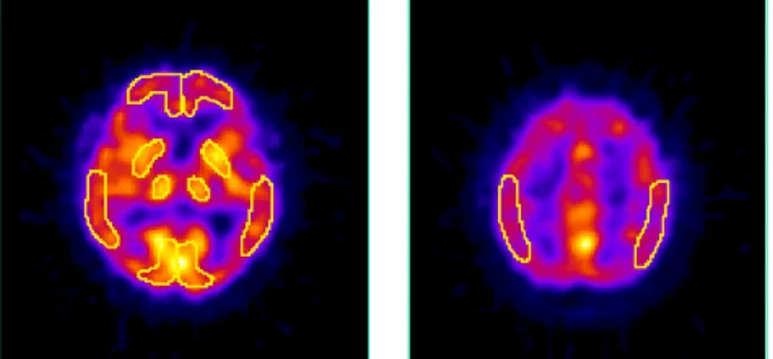
99mTC-ECD(ethylene cysteine diethylester)를 이용한 뇌 단일광전자 단층촬영 검사는 콜린에스테라제 억제제 투 여 전과 후 각 일주일이내 시행하였으며 뇌혈류량의 변 화를 비교분석 하였다. 뇌 단일광전자 단층촬영 영상은 Fanbeam Collimator를 장착한 이중 헤드 감마카메라를 이 용하여 얻었다. 환자를 희미한 불빛 조명의 조용한 방에 서 앙와위 자세로 10분간 안정시킨 후 740MBeq의 99mTC- ECD를 정맥주사하였으며 1시간 후에 자료 수집을 시작 하였다. 관심영역(ROIs : regions of interests)은 양측 대뇌 의 전두엽, 측두엽, 두정엽 및 소뇌의 8개 부위로 정하고 각각의 관심 영역에서 구한 계수율(count)을 대뇌의 계수 율로 나누어 상대적 국소 뇌혈류량(RCR : region to cerebellar ratio)을 구하였다(Fig. 1).
T
C F
P
Fig. 1. Regions of interest.
F: Frontal lobe, T: Temporal lobe, P: Parieta lobel, C: Cerebellum
대뇌반구의 혈류량은 대뇌피질 전반에 걸쳐 뇌혈류량 이 감소하는 알츠하이머에서도 대체로 혈류량의 변화가 적어 일정하게 유지되기 때문에 뇌혈류량의 정량적 지표 로 삼았다. 그리고 국소 뇌혈류량은 다음과 같은 방법으 로 산출하였다.
RCR =
Count of ROI
Mean count of cerebellar hemispheres ROIs
각 군에서 콜린에스테라제 억제제 투여 전․후 관심 영역의 국소 뇌혈류량 변화의 유의성을 비교하기 위하여 표본의 값이 정규분포를 따르는 P value test를 이용하여 통계학적 분석을 실시하였다.
III. 결 과
콜린에스테라제 억제제 투여 후 뇌혈류량은 전반적으 로 호전되는 경향을 보였다. 특히 양측 두정엽에서는 좌․우측 대뇌반구의 국소 뇌혈류량이 각각 0.06씩 증가 하여 콜린에스테라제 억제제 투여 전에 비해 뇌혈류량이 의미 있게 증가하였다(p<0.05). 양측 전두엽과 좌측 측두 엽의 국소 뇌혈류량도 콜린에스테라제 억제제 투여 후에 모두 동일하게 0.03씩 증가하였으나 통계적 유의성은 없 었고, 우측 측두엽에서는 변화가 없는 것으로 나타났다 (Table 1, Fig. 2).
좌․우측 대뇌피질의 뇌혈류량은 투여 전 평균이 좌측 에서는 0.84±0.09, 우측에서는 0.87±0.09로 나타나 좌측 에서 유의하게 감소되었고(p<0.05), 투여 후의 평균은
Table 1. Change of RCR in the right and left hemisphere of each lobe after treated with ChEI(mean ± SD)
ROIs AchEI
RCR
Frontal lobe Temporal lobe Parietal lobe
Rt Lt Rt Lt Rt Lt
Pre 0.86±0.10 0.85±0.11 0.87±0.11 0.82±0.10 0.87±0.08 0.85±0.06
Post 0.89±0.11 0.88±0.10 0.87±0.09 0.85±0.09 0.93±0.07 0.91±0.04
P value 0.52 0.35 1 0.50 0.02* 0.02*
* : P < 0.05, ROIs : Regions of interest, RCR : Region to cerebellar ratio Rt : Right, Lt : Left
0.75 0.8 0.85 0.9 0.95
F (R t) F (L t) T(R t) T(L t) P(R t) P (L t)
ROIs
R C R
P o st-A chE I P re-A chE I
*
*
*p<0.05
Fig. 2. Change of the over time in the rCBF of AD patients(n=8).
ROIs : regions of interest, RCR : region to cerebellar ratio
Table 2. Change of RCR in the right and left hemisphere after treated with ChEI(mean ± SD)
ROIs AchEI
RCR Rt hemisphere
(F+T+P)
Lt hemisphere
(F+T+P) P value
Pre 0.87±0.09 0.84±0.09 0.02*
Post 0.89±0.09 0.88±0.08 0.19
P value 0.13 0.04*
* : P<0.05, F : Frontal lobe, T : Temporal lobe, P : Parietal lobe, ROIs : Regions of interest, RCR : Region to cerebellar ratio, Rt : Right, Lt : Left
좌․우측이 0.88±0.08과 0.89±0.09로 역시 좌측의 뇌혈 류량이 콜린에스테라제 억제제 투여 후 유의하게 증가되 었다(p<0.05)(Table 2).
IV. 고 찰
잠정 알츠하이머 환자를 대상으로 하여 약 30주(7.5개
월)간 콜린에스테라제 억제제 투여를 한 결과, 양측 두정 엽의 뇌혈류량이 의미있게 증가되었다. 그리고 통계적 의 미는 없었지만 나머지 양측 전두엽과 좌측 측두엽에서도 동일한 정도의 혈류 개선효과를 보였다. 이러한 결과는 시간이 경과할수록 점점 더 진행되는 알츠하이머의 뇌혈 류량의 경우 초기에는 주로 측두엽과 두정엽의 뇌혈류가 감소되지만(Johnson 등, 1987; Hunter 등, 1989; Burns 등, 1989) 시간이 지날수록 더 심해져서 나중에는 전두엽까지 뇌혈류가 감소된다(Brown 등, 1996; Neary 등, 1987)는 보 고와 같이 약 30주간 동안의 콜린에스테라제 억제제 투 여가 두정엽의 대사를 증가시켰을 뿐만 아니라 전두엽과 측두엽에서 병의 진행을 최소한으로 완화시켰거나 혹은 뇌혈류량을 일정수준으로 유지하는데 기여했을 것으로 생각된다.
한편 두정엽의 뇌혈류량만이 유의하게 증가한 것은 신 경세포가 완전히 소실되지는 않으면서 콜린성 자극의 결 핍으로 인한 세포기능 감소증상이 있는 경증 혹은 중등 증의 알츠하이머 환자의 두정엽에 콜린에스테라제 억제 제를 투여하였기 때문에 뇌혈류량이 의미있게 증가될 수 있었던 것으로 보인다. 그리고 측두엽의 국소혈류는 해마 를 포함한 내측 일부가 초기부터 이환되어 있어서 질병 이 진행됨에 따라 더 넓게 이환되는 경향이 있는데 본 연 구에서 사용한 관심영역(ROIs)의 일부분만이 해마를 포 함한 내측 일부에 해당되기 때문에 현저한 뇌혈류량의 변화를 보기에는 한계가 있었다.
좌․우측 대뇌피질의 뇌혈류량을 비교할 경우 콜린에 스테라제 억제제 투여 전에는 우측에 비해 좌측이 유의 성있게 감소해 있는 것을 알 수 있는데(p<0.05), 이는 알 츠하이머가 주로 우세 반구인 좌측에 더 이환됨을 시사
한다. 또한 콜린에스테라제 억제제 투여 후에도 뇌혈류량 이 감소된 좌측에서 투여 전에 비해 현저하게 뇌혈류가 증가된 것을 알 수 있었다(p<0.05).
본 연구의 결과는 알츠하이머 환자를 대상으로 약 35 주간 Donepezil을 투여하였을 때 전반적인 뇌혈류량의 개 선현상을 볼 수 있었는데 이는 특히 양측 전두엽과 측두 엽의 뇌혈류량이 의미 있게 증가하였다는 연구결과(Staff 등, 2000)와는 다소 차이가 있었다. 위의 연구는 특히 전 두엽에서 뇌혈류량이 의미 있게 증가된 소견을 보이는 것으로 보고한 바 있으나 본 연구에서는 콜린에스테라제 억제제 투여 후 두정엽의 혈류가 의미 있게 개선되었는 데 이는 연구과정이 너무 길어서 자연적으로 진행하는 병의 경과로 인해 이미 신경세포가 심하게 소실된 두정 엽이나 측두엽보다 좀 더 늦게 이환된 전두엽의 신경세 포에 더 효과적으로 콜린에스테라제 억제제가 작용한 것 으로 보인다.
이와 같이 콜린에스테라제 억제제의 투여효과가 다양 할 뿐만 아니라 뇌혈류량의 변화가 일정하지 않은 이유 는 콜린에스테라제 억제제가 뇌혈관에 직접 작용하여 뇌 혈류량을 증가시킬 뿐만 아니라(Kasa 등, 2000) 시납스 후 신경활동을 자극함으로써 뇌혈류량을 증가(Furey 등, 2000)시키기 때문에 각기 다른 대뇌부위가 콜린에스테라 제 억제제에 반응하는 정도가 다르거나, 앞서 언급한 바 와 같이 투여시점 즉, 질병의 정도(경증, 중증도)에 따라 콜린에스테라제 억제제에 반응하는 정도의 차이가 있거 나, 근본적인 알츠하이머 병리가 다르거나, 또한 다른 유 전적 다형성증 등의 몇 가지 이유로 요약할 수 있다. 즉 콜린성 자극 결핍으로 인해 비효율적으로 세포기능이 저 하된 신경세포는 콜린에스테라제 억제제가 자극을 하여 도 콜린성 자극을 전달할 수 없다.
본 연구에서 알츠하이머가 심하게 진행된 경우에는 콜 린에스테라제 억제제에 의한 뇌혈류량 변화를 기대하기 어려운 것으로 판단되는 데, 이러한 결과는 콜린에스테라 제 억제제가 콜린성 자극 결핍이 매우 심한 중증 알츠하 이머 환자에서보다도 경증 및 중등증 알츠하이머에 더 효과적이며, 뇌허혈성 실험모델 쥐에서 콜린에스테라제 억제제가 전반적인 뇌혈류량을 증가시켜 주었지만 허혈 핵심부의 혈류는 개선시키지 못하였다는 연구(Scremin 등, 1997)와도 일치하는 소견이다. 즉, 알츠하이머 환자가
초기 콜린성 자극에 효과적으로 반응하는 시기가 지나 서 시간이 더욱 경과되면 더 이상 유효반응을 하지 않는 시기가 뒤따르는 것으로 생각되는데 이러한 결과는 질환 초기에 콜린에스테라제 억제제 투여를 받는 것이 질환의 예후를 어느 정도 결정짓는 매우 중요한 인자가 될 수 있 음을 시사하는 것이다.
V. 결 론
알츠하이머 환자를 대상으로 콜린에스테라제 억제제를 투여한 결과, 전반적으로 뇌혈류량이 호전되는 경향을 보 였으며 특히 양측 두정엽의 뇌혈류량이 유의하게 증가하 였다(p<0.05). 또한 콜린에스테라제 억제제 투여 전에는 우측에 비해 좌측 대뇌피질의 뇌혈류량이 유의하게 감소 해 있었으나(p<0.05) 콜린에스테라제 억제제 투여 후에는 뇌혈류량이 감소되어 있던 좌측 대뇌피질에서 투여 전에 비해 현저하게 뇌혈류량이 증가되었다(p<0.05).
이상의 결과를 통하여 콜린에스테라제 억제제가 알츠 하이머 환자의 뇌혈류량을 호전시킴을 알 수 있었다
참 고 문 헌
1. Birks JS, Meltzer D, Beppu H. Donepezil for mild and moderate Alzheimer's disease. Cochrane Database System Review 4, 2000
2. Birks J, Grimley EJ, Iakovidou V, Tsolaki M.
Rivastigmine for Alzheimer's disease. Cochrane Data- base System Review 4, 2000
3. Brown DRP, Hunter R, Wyper DJ, Patterson J, Kelly RC, Montaldi D, McCulloch J. Longitudinal changes in cognitive function and regional cerebral function in Alzheimer`s disease : A SPECT blood flow study.
Journal of Psychiatric Research 30:109-126, 1996 4. Burns A, Philpot MP, Costa DC, Ell PJ, Levy R. The
investigation of Alzheimer`s disease with single photon emission tomography. Journal of Neurology, Neuro- surgery and Psychiatry 52:248-253, 1989
5. Cummings JL. The role of cholinergic agents in the management of behavioural disturbances in Alzheimer's disease. Internation Journal of Neuropsychopharmaco- logy 3:21-29, 2000
6. Davies P, Maloney AJF. Selective loss of central cholinergic neurons in Alzheimer`s disease. Lancet 2:1403, 1976
7. Furey ML, Pietrini P, Haxby JV. Cholinergic enhance- ment and increased selectivity of perceptual processing during working memory. Science 290:2315-2319, 2000 8. Giacobini E, Becker R. Present progress and future
development in the therapy for Alzheimer`s disease.
Progress in Clinical and Biological Research 317:
1121-1154, 1989
9. Hunter R, McLuskie R, Wyper D, Patterson J, Christie JE, Brooks DN, McCulloch J, Fink G, Googwin GM.
The pattern of function-related regional cerebral blood flow investigated by single photon emission tomography with 99mTC-HMPAO in patients with presenile Alzheimer`s disease and Korsakoff`s psychosis.
Psychological Medicine 19:847-855, 1989
10. Jagust W, Thisted R, Devous MS, Van Heertum R, Mayberg H, Jobst K, Smith AD, Borys N. SEPCT perfusion imaging in the diagnosis of Alzheimer`s disease: a clinical-pathologic study. Neurology 56:950- 956, 2001
11. Johnson KA, Mueller ST, Walshe TM, English RJ, Holman BL. Cerebral perfusion imaging in Alzheimer`s disease. Use of single photon emission computed tomography and iofetamine hydrochloride I 123.
Archives of neurology 44:165-168, 1987
12. Kasa P, Papp H, KasaⅡ P, Torok I. Donepezil dose- dependently inhibits acetylcholinesterase activity in various areas and in the presynaptic cholinergic and the postsynaptic cholinoceptive enzyme-positive structures in the human and rat brain. Neuroscience 101:89-100, 2000
13. Mega MS, Dinov ID, Lee L, O'Connor SM, Masterman DM, Wilen B, Mishkin F, Toga AW, Cummings JL.
Orbital and dorsolateral frontal perfusion defect asso- ciated with behavioral response to cholinesterase inhi- bitor therapy in Alzheimer's disease. Journal of Neuro- psychiatry Clinical Neurosciences 12:209-218, 2000 14. Nakano S, Asada T, Matsuda H, Uno M, Takasaki M.
Donepezil hydrochloride preserves regional cerebral blood flow in patients with Alzheimer`s disease.
Journal of Nuclear Medicine 42:1441-1445, 2001 15. Neary D, Snowden JS, Shields RA, Burjan AW,
Northen B, MacDermott N, Prescott MC, Testa HT.
Single photon emission tomography using 99mTC- HMPAO in the investigation of dementia. Journal of Neurology, Neurosurgery and Psychiatry 50:1101-1109, 1987
16. Qizilbash N, Birks J, Lopez AJ, Lewington S, Szeto S.
Tacrine for Alzheimer's disease. Cochrance Database System Review 2, 2000
17. Scremin OU, Li MG, Scremin AM, Jenden DJ.
Cholinesterase inhibition improves blood flow in ischemic cerebral cortex. Brain Research Bullentin 42:59-70, 1997
18. Staff RT, Gemmell HG, Shanks MF, Murray AD, Venneri A. Changes in the rCBF images of patients with Alzheimer`s disease receving donepezil therapy.
Nuclear Medicine Communications 21:37-41, 2000 19. Vennerica A, Shanks MF, Staff RT, Pestell SJ, Forbes
KE, Gemmell HG, Murray AD. Cerebral blood flow and cognitive responses to rivastigmine treatment in Alzheimer`s disease. Cognitive Neuroscience and Neuropsychology 13:83-87, 2002
20. Warren S, Hier DB, Pavel D. Visual form of Alzheimer`s disease and its response to anticholine- sterase therapy. Journal of Neuroimaging 8:249-252, 1998