⋅Received:26 November 2010 ⋅Revised:28 December 2010 ⋅Accepted:29 December 2010
⋅Correspondence to:이장훈(Jang-Hoon Lee)
서울시 동대문구 회기동 1번지 경희대학교 한의과대학 간계내과학교실
Tel:+82-2-958-9118, Fax:+82-2-958-9120, E-mail:[email protected]
Palmitate로 유발된 비알코올성 지방간 모델에 대한 KH淸肝湯의 효과 연구
한창우1, 이장훈2
1부산대학교 한의학전문대학원 내과학교실, 2경희대학교 한의과대학 간계내과학교실 Original Article
Effects of KHchunggan-tang on the Nonalcoholic Fatty Liver Disease in Palmitate-induced Cellular Model
Chang-Woo Han1, Jang-Hoon Lee2
1Department of Internal Medicine, Pusan National University School of Korean Medicine
2Department of Internal Medicine, College of Oriental Medicine, Kyung Hee University
Objectives: The aim of this investigation was to evaluate the efficacy of KHchunggan-tang aqueous extract on the experimental nonalcoholic fatty liver disease(NAFLD) induced by palmitate.
Materials and Methods: To generate a cellular model of NAFLD, we used HepG2 cells, a human hepatoma cell line, treated with 0.5 mM palmitate. By this cellular model, effects of KHchunggan-tang aqueous extract were evaluated.
Intracellular lipid accumulation, free radical formation, and apoptosis were detected by Nile red staining, 2’,7’-dichloroflourescin diacetate(H2DCF-DA), and 4’,6-diamidino-2-phenylindole(DAPI)/propidium iodide(PI) staining, respectively. Some proteins related with NAFLD were determined by western blot.
Results: Typical pathological features of NAFLD occurred in the cellular model. Palmitate increased the levels of intracellular lipid vacuoles, decreased cell viability, and increased apoptosis. Palmitate increased free radical formation and lipid peroxidation, too. However, KHchunggan-tang aqueous extract reduced palmitate-induced pathologic features, i.e. steatosis, free radical formation, and apoptosis. In addition, KHchunggan-tang aqueous extract suppressed palmitate-activated c-Jun N-terminal kinase(JNK) signaling, and SP600125, a JNK inhibitor, significantly reversed the palmitate-induced pathologic changes as KHchunggan-tang aqueous extract. It means that the signaling pathway other than JNK can be involved in the KHchunggan-tang mediated cellular protection of palmitate-treated Hep G2 cells.
Conclusions: These results suggest that KHchunggan-tang aqueous extract has hepatoprotective effects on NAFLD with combined properties in cellular steatosis, ROS production, and cytoprotection, and thus may have valuable clinical applications for treatment of this chronic liver disease.
Key Words : KHchunggan-tang, NAFLD, HepG2, ROS, JNK
서 론
비알코올성 지방간 질환(Non-alcoholic fatty liver disease, NAFLD)이란 과도한 음주력이 없으면서도
알코올성 간질환과 유사한 조직학적 손상을 보이는 질환으로, 간세포 내에 중성지방의 미만성 침윤이 나타나며, 비만, 당뇨, 고지혈증 등의 대사성 질환의 일부분으로 발현되기도 한다1). 가벼운 경우 단순 지
Herb Scientific name Weight (g) 인진
택사 백출 단삼 울금 산사 구기자
지실 감초
Artemisia capillaris Alisma orientale Atractylodes japonica
Salvia miltiorrhiza Curcuma wenyujin Crataegus pinnatifida
Lycium chinense Poncirus trifoliata Glycyrrhiza uralensis
30 15 12 8 8 8 8 6 6
Total 101
Table 1. Prescription of KHchunggan-tang
방간 소견만이 나타나기도 하지만, 지방간염, 섬유 화 및 간경변에 이르기까지 단계적으로 진행할 수 있으며2), 최근 대사성 질환의 유병율 증가와 함께, 만성 간질환에서 그 비중이 점차 증가하는 것으로 보고되고 있다3).
이러한 중요성으로 인해 근래에는 비알코올성 지 방간 질환의 병리 기전과 치료 약물에 대한 연구가 활발히 이루어지고 있으나, 여전히 유용한 표준 치 료법은 제시되지 못하고 있으며, 이에 따라 비알코 올성 지방간질환에 대한 한약물의 효능 및 약리 기 전 연구도 다양하게 진행되고 있다4-7).
KH청간탕은 한의학적 이론을 바탕으로 다양한 임상적 경험과 선행 실험연구 결과7)를 반영하여 구 성하였는데, 급만성 간질환에 다용되어온 茵蔯四苓 散을 근간으로8-10), 고지혈증 및 지방간에 효능이 있 는 것으로 알려진 鬱金, 山楂, 丹參, 枸杞子, 枳實 등 을 가미한 처방이다11-14).
이에 저자는 KH청간탕이 비알코올성 지방간질환 에 미치는 영향을 관찰하기 위해, HepG2 세포에 palmitate를 처리하여 지방간질환 세포 모델을 설정 하고, KH청간탕을 투여하며 Nile red staining, 2’,7’-dichloroflourescin diacetate(H2DCF-DA), 4’,6 diamidi no- 2- phenylindole( DAPI ) /pr opidium iodide(PI) staining, western blot 등의 방법으로, 세 포 내 지질의 축적, reactive oxygen species(ROS) 생성, apoptosis 및 이러한 변화과 관련된 것으로 알려
져 있는 c-Jun N-terminal kinase(JNK) signaling의 활성 정도를 관찰하여 유의한 결과를 얻었기에 보고 하는 바이다.
재료와 방법
1. 실험에 사용된 약재
본 실험에 사용된 약재는 대한약전 및 대한약전 외 한약규격집에 근거하여 경희대 부속 한방병원에 서 구입하여 엄선한 것을 사용하였으며 내용 및 용 량은 Table 1과 같다.
2. KH청간탕 물 추출물의 조제
KH청간탕 2첩(202 g)을 환류추출기에 넣고, 3차 증류수 1000 ml를 가하여 2시간 동안 가열 추출한 후 여과하였다. 여과한 잔사에 다시 1000 ml를 가하 여 2시간 동안 환류 추출하는 재탕의 과정을 거쳐 여과한 다음, 처음 얻은 여과액과 합하여 감압농축 기 상에서 감압 농축한 후, 동결건조기로 건조하여 46 g의 KH청간탕 물 추출물을 얻었다(수율 22.8%).
3. 재료
Palmitate, 3(4,5-dimethylthiazol-2-yl)-2,5 dip- henyltetrazolium bromide(MTT), 4’,6-diamidino 2-phenylindole(DAPI), propidium iodide(PI), Nile
red 및 polyclonal rabbit anti-β-actin antibody는 Sigma-Aldrich Co.(St. Louis, MO, USA)에서 구입 하였다. 2’,7’-dichloroflourescin diacetate(H2DCF- DA)와 SP600125 [anthra(1,9-cd)pyrazol-6(2H) one]
는 Calbiochem(La Jolla, CA, USA)에서 구입하였 다. Monoclonal mouse anti-Bax, polyclonal rabbit anti-Bcl-2, horseradish peroxidase (HRP)-conjugated goat anti-rabbit IgG, goat anti-mouse IgG는 Santa Cruz Biotechnology(Santa Cruz, CA, USA)에서 구입 하였고, c-Jun N-terminal kinase (JNK)와 phospho- JNK에 특이적으로 결합하는 polyclonal antibody는 Cell Signaling(Beverly, MA, USA)에서 구입하였다.
4. 세포 배양
Human hepatoblastoma 세포주인 HepG2 세포를 Korean Cell Line Bank(Seoul, Korea)로부터 구입하 였다. 100 U/ml penicillin, 100 μg/ml streptomycin 및 10% heat-inactivated fetal bovine serum을 포함 한 Dulbecco’s modified Eagle’s medium(DMEM)을 사용하여, 37℃, 5% CO2 환경에서 배양하였다.
5. 지방산 처리
Palmitate를 isopropanol에 50 mM 농도가 되도록 녹였다. 최종 투약 농도는 0.5mM, 용매에 대하여 1% 농도였다. 배양액은 1% bovine serum albumin 을 함유한 DMEM을 사용하였다.
6. 세포 활성도의 측정
MTT assay를 통해 세포 활성도를 측정하였다.
24-well plate에 5 × 10⁴ cells/well의 밀도로 HepG2 세포를 seeding하였다. 하룻밤 경과 후, 준비된 KH 청간탕 물 추출물(0.1, 1, 10, 50, 100 μg/ml)을 투약 하고 1시간 뒤, palmitate를 처리하여 21시간 배양하 였다. KH청간탕 물 추출물과 함께 SP600125를 투약 하기도 하였다. 각 well에 MTT(final concentration, 0.1 mg/ml)를 처리하고 4시간 뒤 medium을 제거하 였다. 활성이 있는 세포들이 MTT를 환원하여 생성
한 formazan crystal을 Acidified isopropanol을 사용 하여 용해하였고, background subtraction을 650 nm 로 하고 570 nm에서의 흡광도를 측정하였다.
7. Fluorescence-activated cell sorting(FACS)을 통한 apoptosis 측정 HepG2 세포를 60mm dish에 배양하여 80%
confluence 상태가 되면, 1시간 전 KH청간탕 물 추 출물을 투약하거나, 또는 0.5 mM palmitate만으로 21시간 처리하였다. Ice-cold phosphate-buffered saline(PBS)로 세척 후, 70%(v/v) ethanol에서 4℃
냉장 상태로 하룻밤 처리하여 세포를 고정하였다.
Ice-cold PBS로 세척 후, propidium iodide(PI) staining solution(10 mM Tris-Cl, 1 mM NaCl, 0.1%
NP-40, 0.7 μg/ml RNase A, and 50 μg/ml Propidium iodide, pH 8.0)으로 혼합한 다음 실온에서 차광 상 태로 30분간 두었다. FACSCalibur flow cytometer (Becton Dickinson Immunocytometry Systems, San Jose, CA, USA)로 세포 주기 상태를 분석하였다.
각 측정 조건마다 최소 20,000개 이상의 세포를 분 석하였다.
8. 4’,6-diamidino-2-phenylindole(DAPI) staining을 통한 apoptosis 확인
6-well plates에서 80% confluence가 되도록 배양 된 HepG2 세포에 1시간 전 KH청간탕 물 추출물을 투약하거나, 또는 0.5 mM palmitate만으로 21시간 처리하였다. SP600125를 함께 투약하기도 하였다.
PBS로 세척 후, 실온에서 5분간 4% paraformaldehyde 로 고정하였고, 세포막의 투과성을 증가시키기 위해 곧 이어 0.1% Triton X-100 in PBS(v/v)로 5분간 세 척하였다. 고정된 세포들을 DAPI(2 μg/ml)로 30분 간 염색 후 세척하고 digital camera output가 장착된 Olympus JP/1X71 fluorescence microscope로 촬영 하였다. Olympus DP70-BSW software를 사용하여 고도로 응축되거나 분절된 핵을 지닌 apoptosis 세 포들을 계산하였다. 한편, 다른 실험자로 하여 저해
상도에서 얻은 4개의 영역에서 apoptosis를 일으킨 세포들을 측정하도록 하였다.
9. ROS 측정
세포 내의 ROS 생성 정도를 측정하기 위해, 형광 probe인 H2DCF-DA를 사용하였다. HepG2 세포들 을 35-mm coverglass bottom dishes(SPL life sciences, Seoul, Korea)에 seeding하고 1시간 전 KH 청간탕 물 추출물을 투약하거나, 또는 0.5 mM palmitate만으로 21시간 처리하였다. PBS로 세척 후, 5 μM H2DCF-DA을 가한 후 37℃에 30분간 두었 다가, 520 nm long-pass filter를 사용하여 Olympus FV-1000 confocal laser scanning microscope(Tokyo, Japan)로 형광을 확인하고, Olympus FV10-ASW 2.0 software로 정량화하였다.
10. 지질 과산화도 측정
지질 과산화물을 측정하기 위해 thiobarbituric acid reactive substances(TBARS)를 측정하였다. HepG2 세포들을 1시간 전 KH청간탕 물 추출물을 투약하 거나, 또는 0.5 mM palmitate만으로 21시간 처리하 였다. Ice-cold PBS로 세척 후, 세포를 모아 50 mM Tris buffer(pH 7.4, 180 mM KCl, 10 mM EDTA, 20 mM HCl)을 용매로 한 thiobarbituric acid(0.4%) 1 ml에 넣고 suspension하였다. 샘플을 sonication한 후 95℃에서 20분간 두었다. butan-1-ol 1 ml를 첨가 후 4℃, 2,000 × g으로 10분간 centrifuge하였다. 상 층액을 분리하여 spectrophotometer로 548 nm에서 의 흡광도를 측정하였다.
11. Nile red 염색으로 지방량 측정
KH청간탕 물 추출물이나 SP600125와 함께 또는 0.5 mM palmitate만으로 21시간 처리된 HepG2 세 포들을 상온에서 15분간 4% paraformaldehyde로 고 정하였다. PBS로 세척 후, 상온에서 5분간 Nile red(100 ng/ml)를 처리하여 세포 내 중성 지방을 염 색하였다. Olympus FV-1000 confocal laser scanning
microscope(Tokyo, Japan)를 이용하여 촬영하고, Olympus FV10-ASW 2.0 software를 이용하여 형광 을 정량화하였다.
12. Cytoplasm, mitochondria 및 전체 세포 추출물의 준비
HepG2 세포를 100 mm dish에서 80% confluence 가 되도록 배양 후, 1시간 전 KH청간탕 물 추출물을 투약하거나, 또는 0.5 mM palmitate만으로 21시간 처 리하였다. Ice-cold PBS로 세척 후, 세포를 모아 protease inhibitor를 포함한 S-100 buffer(20 mM HEPES, 10 mM KCl, 1.9 mM MgCl2, 1 mM EGTA, 1 mM EDTA, pH 7.5)로 suspension하였다. Ice 속에 서 20분간 보관하였다가, glass Dounce homogenizer (Wheaton, Millville, NJ, USA)로 70회 파쇄하였다. 세 포파쇄액을 1,000 × g로 5분간 centrifuge하여 파쇄되지 않는 세포, 핵 및 무거운 세포막을 분리하였다. 상층 액을 다시 14,000 × g로 30분간 centrifuge하여 mitochondria가 있는 pellet 부분과 cytoplasm이 있는 상층액 부분으로 분리하였다. Mitochondria가 있는 pellet 부분을 S-100 buffer로 한 번 세척 후, protease inhibitor를 포함한 lysis buffer(50 mM Tris-Cl, 150 mM NaCl, 1% NP-40, 0.25% Sodium deoxycholate and 1 mM EGTA, pH 7.4)로 suspension하였다. 전체 세포 추출물을 얻기 위해서는, 세포를 ice-cold PBS로 세척 후 5 mM Na4P2O7, 10 mM NaF, 10 mM β -glycerophosphatase 및 1 mM Na3VO4 함유 PRO- PREP Protein Extraction Solution(iNtRON Biotechnology, Seoul, Korea)로 -20℃에서 90분간 처리하였다. 세포용 해액을 10,000 × g로 20분간 centrifuge 후 전체 세포 추출물이 있는 상층액을 모았다. 다음 실험 시행 전에는 추출물을 -70℃에서 보관하였다. 단백의 농도는 bovine serum albumin을 비교 표준으로 한 Bradford method 로 정량하였다.
13. Western blot 분석
샘플을 Sodium dodecyl sulfate polyacrylamide
500
400
300
200
100
0 Palmitic acid (0.5 mM) KHCGT (100㎍/㎖)
- -
+ -
+ +
- +
Fluorescence intensity
**
†
A) B)
Fig. 1. KHchunggan-tang (KHCGT) inhibits palmitate-induced intracellular steatosis.
HepG2 cells were pretreated with KHCGT for 1 h and then incubated with palmitate. After incubation for 21 h, the cells were stained with Nile red. (A) Representative fluorescence micrographs are shown. (B) Cellular steatosis was quantified for each condition using four random low power fields of view with automated software. The fluorescence intensity was measured and expressed as the means ± SE (n=4). ** P<0.01, compared to untreated cells; †P<0.05, compared to palmitate treated cells.
gel electrophoresis(SDS-PAGE) 후 Hybond-P+ polyvinylidene difluoride membrane(GE Healthcare, UK)에 transfer하였다. 0.1% Tween-20를 함유한 Tris-buffered saline(TBS)에 녹인 5% nonfat milk로 상온에서 2시간 동안 처리하여 membrane을 block 하고, 지시된 specific primary antibody를 투입한 1% BSA 및 0.05% Tween-20 함유 TBS로 4℃에서 하룻밤 배양하였다. Membrane들은 다시 1:3000로 희석된 peroxidase-conjugated goat anti-mouse antibody 나 peroxidase-conjugated goat anti-rabbit antibody로 상온에서 2시간 처리하였으며, 0.1% BSA 및 0.1%
Tween-20 함유 TBS으로 깨끗하게 세척한 후, West-ZOL Plus(iNtRON Biotechnology)를 이용하여 immunoreactive band들을 검출하였다.
14. 통계 분석
Student’s t-test로 평균값들을 비교하였으며, 모든 측정값은 means ± SE.로 표시하였다.
결 과
1. KH청간탕 물 추출물이 palmitate에 의해 유발된 세포의 steatosis에 미치는 영향
지방증(steatosis) 세포 모델을 설정하기 위해 HepG2 세포를 1% bovine serum albumin을 포함한 배지에서 0.5 mM palmitate를 첨가하여 21시간 배 양하였다. Palmitate에 의해 세포 내 지방 공포(lipid vacuole)가 증가한 것을 Nile red staining을 통해 확 인할 수 있었으며(Fig. 1A), Palmitate에 의해 발생 한 세포 내 지방 축적은 100 μg/ml KH청간탕 물 추 출물이 함께 투여된 경우 유의성 있게 감소하였다 (Fig. 1A and 1B).
2. KH청간탕 물 추출물이 palmitate에 의해 유발된 세포의 apoptosis에 미치는 영향 KH청간탕이 palmitate에 의해 야기된 HepG2 세 포의 손상에 미치는 영향을 평가하기 위해, HepG2 세포의 세포 활성도를 측정하였다. Palmitate에 의해 세포 활성도가 뚜렷이 감소하였으며, 이러한 세포 활성도의 저하는 KH청간탕 물 추출물에 의해 용량 의존적으로 회복되는 경향을 보였다(Fig. 2A). 이러 한 결과로 KH청간탕 물 추출물이 plamitated에 의 해 야기된 세포 손상으로부터 HepG2 세포를 보호 하는 효과가 있음을 알 수 있다.
KH청간탕 물 추출물에 의한 세포 활성도 증가 효과가 apoptosis의 억제로도 나타나는지 확인해보
100
80
60
40
20
0 Palmitic acid (0.5 mM) -
Cell Viability (%)
**
† †† †† ††
+ + + + + +
0.1 1 10 50 100
KHCGT (㎍/㎖)
A) 7
6 5 4 3 2 1 0 Palmitic acid (0.5 mM) -
Sub-G1 DNA content(Fold)
**
† † †
+ + + + + +
0.1 1 10 50 100
KHCGT (㎍/㎖) B)
Fig. 2. KHchunggan-tang (KHCGT) suppresses the palmitate-induced apoptosis.
HepG2 cells pretreated with increasing dose (0, 0.1, 1, 10, 50, 100 μg/ml) of KHCGT for 1 h were incubated with palmitate.
Following incubation for 21 h, cell viability was assessed using the MTT assay (A). The percentage of cells with sub-G1 DNA content was analyzed by fluorescence-activated cell sorting after staining with propidium iodide (B). The means ± SE (n=4) are presented. **P<0.01, compared to untreated cells; †P<0.05, ††P<0.01, compared to palmitate-treated cells.
40
30
20
10
0 Palmitic acid (0.5 mM)
KHCGT (100㎍/㎖)
- -
+ -
+ +
- +
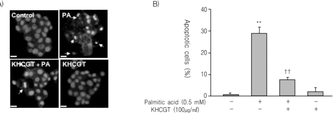
Apoptotic cells (%)
**
††
B) A)
Fig. 3. The protective effect of KHchunggan-tang (KHCGT) against palmitateinduced apoptotic cell death was confirmed again by DAPI staining.
HepG2 cells pretreated with KHCGT (100 μg/ml) for 1 h were incubated with palmitate. Following incubation for 21 h, apoptotic cells were visualized by DAPI staining. Condensed and deformed apoptotic nuclei appear as bright blue spots (A, white arrows).
Cells in at least four random fields of view were scored (n ≥ 100 each) and quantitative analyses were performed (B). The means
± SE (n=4) are presented. **P<0.01, compared to untreated cells; ††P<0.01, compared to palmitate-treated cells.
기 위해 PI staining 후 Sub-G1 analysis를 시행하였 다. Palmitate에 의해 sub-G1 부분이 증가하였으며, KH청간탕 물 추출물을 처리하였을 때 sub-G1 부분 이 농도 의존적으로 감소하였다(Fig. 2B). 이러한 결 과를 시각적으로 검증해보기 위해 DAPI staining을 시행하였다. Palmitate로 처리한 세포에서는 apoptosis 과정의 특징인 응축된 핵을 지닌 세포가 나타났으 며, KH청간탕 물 추출물을 함께 투여한 세포에서는
이러한 변화가 감소되는 것을 확인할 수 있었다(Fig.
3A and 3B).
3. KH청간탕 물 추출물이 palmitate에 의해 유발된 세포 내 ROS 생성과 지질의 과산화에 미치는 영향
지방산이 과다하게 되면, Free radical의 생성이 증가하게 되고 이로 인해 지질의 과산화가 유발되는
50 40 30 20 10 Palmitic acid (0.5 mM) 0
KHCGT (100㎍/㎖)
- -
+ -
+ +
- +
Fluorescence intensity
**
††
B)
3
2
1
0 Palmitic acid (0.5 mM)
KHCGT (100㎍/㎖)
- -
+ -
+ +
- +
TBARS (Fold)
**
††
C) Fluorescence Phase
contrast
Merge A)
Fig. 4. KHchunggan-tang (KHCGT) suppresses the palmitate-induced increase in intracellular ROS and reactive aldehydes.
HepG2 cells were pretreated with KHCGT for 1 h and then incubated with palmitate. After incubation for 21 h, the cells were treated with a peroxide-sensitive dye, H2DCF-DA (10 μM), during the final 30 min of the incubation. The intracellular ROS levels were detected (A) and quantified (B) by confocal laser scanning microscopy. (C) Levels of reactive aldehydes were estimated by quantifying TBARS in four independent samples. Data are representative of four separate determinations. The results are expressed as the means ± SE (n=4). **P<0.01, compared to untreated cells; †P<0.05, ††P<0.01 compared to palmitate-treated cells.
데, 이러한 과정은 비알코올성 지방간 질환에서 간 손상을 일으키는 주요 기전으로 알려져 있다15). KH 청간탕 물 추출물이 이러한 과정을 차단할 수 있는 지 확인해보기 위해 H2DCF-DA를 fluorescent probe 로 사용하여 세포 내 ROS 생성량의 변화를 살펴보 았다. Palmitate만으로 처리하였을 때는 세포 내 peroxide 량이 뚜렷하게 증가하였으나, KH청간탕 물 추출물을 함께 투여하자 세포 내 peroxide 량이 뚜렷하게 감소하였다(Fig. 4A and 4B). 한편, reactive aldehyde 량은 지질 과산화 정도를 반영하는 것으로 알려져 있으며16), reactive aldehyde 량을 측정하기 위해 thiobarbituric acid reactive substances(TBARS) 를 측정하였다. TBARS 량도 palmitate만 처리한 HepG2 세포에서는 뚜렷히 증가하였으나, KH청간 탕 물 추출물을 함께 투여한 세포에서는 현저하게
감소하였다(Fig. 4C).
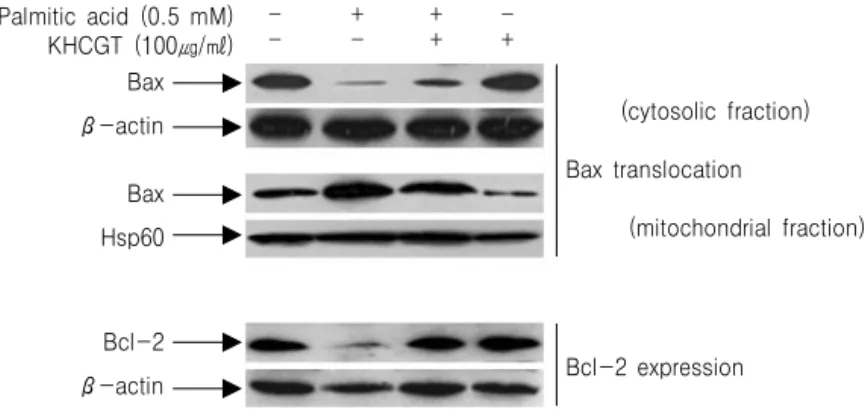
4. KH청간탕이 Bax의 translocation과 Bcl-2의 발현에 미치는 영향
KH청간탕 물 추출물이 나타낸 상기한 효과들의 분자 생물학적 기전을 알아보기 위해, apoptosis 과 정에서 관여하는 것으로 알려져 있는 key protein들 의 변화를 조사하였다. Palmitate로 처리하면 Bax의 cytosolic fraction이 감소하고, mitochondrial fraction 이 증가하는 translocation 양상이 뚜렷히 나타났으 며, KH청간탕 물 추출물을 함께 투여한 경우 이러 한 translocation이 현저히 감소하였다(Fig. 5). 또, KH청간탕 물 추출물을 함께 투여한 경우 palmitate 처리에 의해 감소되었던 Bcl-2 발현도 현저히 증가 하였다(Fig. 5).
(cytosolic fraction)
Bax translocation
(mitochondrial fraction)
Bcl-2 expression Bax
β-actin
Bax Hsp60
Bcl-2 β-actin
- -
+ -
+ +
- + Palmitic acid (0.5 mM)
KHCGT (100㎍/㎖)
Fig. 5. KHchunggan-tang (KHCGT) modulates the palmitate-induced translocation of Bax and expression of Bcl-2.
HepG2 cells pretreated with KHCGT for 1 h were incubated with palmitate for 21 h. Translocation of Bax was analyzed by quantifying the amount of Bax in the mitochondrial and cytosolic fractions using the indicated antibodies. Whole-cell lysates of HepG2 cells were analyzed by western blotting with the indicated antibodies to quantify the level of Bcl-2 expression. Similar results were obtained from three independent experiments.
A) B)
pJNK
JNK
- -
+ -
+ +
- + Palmitic acid (0.5 mM)
KHCGT (100㎍/㎖)
120 100 80 60 40 20 0 Palmitic acid (0.5 mM) KHCGT (100㎍/㎖) SP600125 (50μM)
- - -
Cell Viability (%)
**
††
+ - -
+ + -
+ + +
+ - +
- - +
- + -
§
††
††
Fig. 6. KHchunggan-tang (KHCGT) inhibits palmitate activation of the JNK signaling cascade.
(A) HepG2 cells were pretreated with KHCGT for 1 h and then stimulated with palmitate for 6 h. Aliquots of protein were analyzed by immunoblotting with an activation-specific antibody (anti-p-JNK), while parallel immunoblots were analyzed for total JNK levels.
(B) Cells pretreated with KHCGT in the presence or absence of SP600125 for 1 h were treated with palmitate for 21 h and then analyzed using the MTT assay. The means ± SE (n=4) are presented. **P<0.01 compared to untreated cells; ††P<0.01 compared to palmitate-treated cells; §P<0.05 compared to palmitate + KHCGT-treated cells.
5. KH청간탕이 c-Jun N-terminal kinase(JNK) 활성화에 미치는 영향
JNK signaling cascade는 비알코올성 지방간 질환 의 병리적 변화에 관여하며, 특히 지방산에 의한 apoptosis 과정에 관여하는 것으로 알려져 있기에17), JNK signaling cascade가 KH청간탕 물 추출물에 의 한 간 세포 보호 작용에도 관여하는지 확인해보았
다. HepG2 세포에 palmitate만을 처리하거나 KH청 간탕 물 추출물과 함께 처리한 후 phospho-JNK antibody로 immunoblotting하였다. 총 JNK 량도 immunoblotting하였다. Palmitate를 처리한 경우 phospho-JNK 량이 상당히 증가하였으며, KH청간탕 물 추출물을 함께 투여한 경우 phospho-JNK 증가가 뚜렷히 감소하였다(Fig. 6A). 또, JNK의 활성화를 억제시키는 SP600125를 투여한 경우 KH청간탕 물
A) B)
300
200
100
0 Palmitic acid (0.5 mM) KHCGT (100㎍/㎖) SP600125 (50μM)
- - -
Fluorescenceintensity
**
††
+ - -
+ + -
+ + +
+ - +
- - +
- + -
§§
††
††
Fig. 7. KHchunggan-tang (KHCGT) and a JNK inhibitor synergistically inhibit palmitate-induced ROS generation.
HepG2 cells pretreated with KHCGT for 1 h in the presence or absence of SP600125 were incubated with palmitate for 21 h.
ROS generation was detected (A) and quantified (B) by confocal laser scanning microscopy. The means ± SE (n=4) are presented.
**P<0.01 compared to untreated cells; ††P<0.01, compared to palmitate-treated cells; §§P<0.01 compared to palmitate + KHCGT- treated cells.
A) B)
500 400 300 200 100 0 Palmitic acid (0.5 mM) KHCGT (100㎍/㎖) SP600125 (50μM)
- - -
Fluorescenceintensity
**
††
+ - -
+ + -
+ + +
+ - +
- - +
- + -
§§
††
††
Fig. 8. KHchunggan-tang (KHCGT) and a JNK inhibitor synergistically inhibit palmitate-induced cellular steatosis.
HepG2 cells pretreated with KHCGT for 1 h in the presence or absence of SP600125 were incubated with palmitate for 21 h.
Nile red staining was performed (A) and cellular steatosis was quantified (B). The means ± SE (n=4) are presented. **P<0.01, compared to untreated cells; ††P<0.01, compared to palmitate-treated cells; §§P<0.01, compared to palmitate + KHCGT-treated cells.
A) B)
30
20
10
0 Palmitic acid (0.5 mM) KHCGT (100㎍/㎖) SP600125 (50μM)
- - -
Apoptoticcells (%)
**
††
+ - -
+ + -
+ + +
+ - +
- - +
- + -
§§
††
††
Fig. 9. KHchunggan-tang (KHCGT) and a JNK inhibitor synergistically inhibit palmitate-induced apoptosis.
HepG2 cells pretreated with KHCGT for 1 h in the presence or absence of SP600125 were incubated with palmitate for 21 h.
Apoptotic cells were visualized by DAPI staining (A) and quantified (B). The means ± SE (n=4) are presented. **P<0.01 compared to untreated cells; ††P<0.01, compared to palmitate-treated cells; §§P<0.01, compared to palmitate + KHCGT-treated cells.
추출물을 투여한 경우에서와 같이 palmitate에 의한 세포 활성도의 감소가 유의하게 억제되었으며, SP600125와 KH청간탕 물 추출물을 함께 투여한 경 우 이러한 효과가 더욱 뚜렷하게 나타났다(Fig. 6B).
이러한 결과로 볼 때 KH청간탕이 JNK signaling을 억제함으로써 palmitate에 의한 세포 손상을 억제하 는 것으로 추정할 수 있다.
HepG2 세포에 대한 KH청간탕의 작용에서 JNK signaling의 역할을 보다 구체적으로 파악하기 위해, HepG2 세포에 palmitate를 처리할 때 KH청간탕 물 추출물 또는 SP600125을 함께 투여하며 ROS의 생 성, 세포 내 지방 축적, apoptosis의 변화를 관찰하였 다. 세포 활성도 측정에서와 같이 SP600125를 투여 한 경우 palmitate에 의한 ROS의 생성이 억제되었 고, 세포 내 지방 축적이 감소하였으며, apoptosis 또 한 감소하였다. 또, KH청간탕과 SP600125를 함께 투여한 경우 이러한 효과는 더욱 증대되었다(Fig. 7, Fig. 8, Fig. 9). 따라서, KH청간탕 물 추출물이 palmitate에 의한 간 세포 손상을 보호하는 기전이, 최소한 부분적으로는 JNK signaling을 억제하는 방 법으로 나타나는 것으로 추정할 수 있다.
고찰 및 결론
비알코올성 지방간 질환(Non-alcoholic fatty liver disease, NAFLD)은 단순지방증(simple steatosis), 지 방간염(steatohepatitis), 섬유화(fibrosis) 및 간경변증 (cirrhosis)을 포함하는 간질환으로, 전세계적으로 비 만과 제2형 당뇨병의 유병률이 증가하면서 NAFLD 는 만성 간질환의 주요한 원인이 되고 있다18). 우리 나라에서도 유병률이 일반성인의 18%까지 보고되 고 있고, 서구화되는 식습관, 운동부족, 생활양식의 변화, 비만인구의 증가 등으로 인해 NAFLD는 추후 만성 간질환의 중요한 원인으로 자리 잡을 것으로 생각된다19).
비만 등으로 인해 지방 세포가 증가하고, 인슐린 저항이 유발되면 혈 중 유리 지방산(free fatty acid, FFA)의 농도가 증가하게 되며20), 이에 비례하여 간
의 유리 지방산 흡수도 증가하게 된다21). 간에서 흡 수된 유리 지방산은 산화 과정(fatty acid oxidation) 을 거쳐 에너지원으로 사용되거나, 에스테르화 과정 을 통해 중성지방(triglyceride)으로 변환되어진 후, very low density lipoprotein(VLDL) 입자에 포함되 어 혈중으로 이동하거나 간내에 저장되어지는데, 이 러한 과정이 비정상적으로 이루어질 경우 지방간증 (hepatic steatosis)을 유발하게 된다22). 간 세포 내 지방산 축적이 증가하면, 지방산의 산화가 더욱 증가 하게 되며, 이러한 과정에서 reactive oxygen species (ROS)의 생성도 증가하게 된다23-26). 과다해진 ROS 는 ATP와 NAD를 고갈시키고, DNA 손상을 일으키 며, 단백의 안정성과 세포막을 파괴한다27,28). 또, 항 산화작용을 담당하는 glutathione을 고갈시키고, TNF-α나 IL-8 같은 염증 관련 cytokine들의 생성을 촉진시키며, 염증 세포의 침윤을 유도하고, 성상 세 포(stellate cell)를 자극하여 섬유화(fibrosis)를 유발
한다29,30). 한편, 지방산에 의해 유발된 이러한 간 손
상은 주로 c-Jun N-terminal kinase(JNK) signaling pathway를 통해 매개되는 것으로 알려져 있다31-33).
본 연구에서는 한의학적 이론을 바탕으로 임상적 경험과 선행 실험연구 결과7)를 반영하여 입방된 KH청간탕이 상기한 비알코올성 지방간질환의 병리 적 과정에 어떠한 영향을 미치는지를 관찰하고자 하 였다. HepG2 세포에 palmitate를 처리하여 지방간질 환 세포 모델을 설정하고, 다양한 방법으로 세포 내 지질 축적, reactive oxygen species(ROS) 생성, apoptosis 등를 관찰하였다. KH청간탕 물 추출물을 투여한 HepG2 세포에서는 세포 내 지방 축적이 감 소하였으며, palmitate에 의해 유도된 apoptosis가 감 소되었고, reactive oxygen species(ROS) 생성이 억 제되었다. 또, apoptosis에 관여하는 protein으로 알 려진 Bax 및 Bcl-2를 조절하여 HepG2 세포의 apoptosis를 억제하였으며, 이러한 반응이 c-Jun N-terminal kinase(JNK) signaling pathway를 통해 매개될 개연성이 높은 것을 확인하였다.
이상의 결과에서 KH청간탕은 비알코올성 지방간 실험 모델에 있어서 유의한 간보호작용이 있는 것으
로 확인되었으며, 실제 비알코올성 지방간질환자에 서도 지방증을 완화시키고, ROS의 생성과 지질의 과산화를 감소시키며, 간세포의 apoptosis를 억제시 켜줄 수 있을 것으로 추정된다. 향후 추가적인 연구 를 통하여 임상에서 비알코올성 지방간질환에 다양 하게 활용할 수 있는 치료제로의 개발이 필요할 것 으로 사료된다.
감사의 글
본 연구는 2010년도 부산대학교 교내학술연구비 (신임교수연구정착금)에 의한 연구임.
참고문헌
1. Bacon BR. Genetic, metabolic, and infiltrative diseases affecting the liver. In: Fauci AS, Braunwald E, Isselbacher KJ, Wilson JD, Martin JB, Kasper DL, Hauser SL, Longo DL, et al.
Harrison’s principles of internal medicine. 17th ed. NewYork:McGraw-Hill. 2008:1980-3.
2. Angulo P. Nonalcoholic fatty liver disease. N Engl J Med. 2002;346:1221-31.
3. Marchesini G, Brizi M, Morselli-Labate AM, Bianchi G, Bugianesi E, McCullough AJ, et al.
Association of nonalcoholic fatty liver disease with insulin resistance. The American journal of medicine. 1999;107:450-5.
4. Sung TS, Son GM. Effect of Cnidi rhizoma boiling extract solution on enzyme and hormone of plasma, and liver in the fatted rats induced by high fat dietary. Korean J Food & Nutrition.
1994;7(2):108-13.
5. Yun KS, Woo HJ, Lee JH, Kim YC. Effect of Injinchunggan-tang & Injinsaryung-san on NASH induced by MCD-diet in A/J mice.
Korean J Orient Int Med. 2009;30(2):410-21.
6. Choi MY, Woo HJ, Kim YC, Lee JH. Effects of gamisaenggan-tang on high fat diet-induced
nonalcoholic fatty liver disease. Korean J Orient Int Med. 2009;30(2):365-74.
7. Yoo JY, Lee JH. Effects of Saenggantanggami- bang on nonalcoholic fatty liver disease model induced by fatty acids. Korean J Orient Int Med.
2010;31(2):143-57.
8. Pyo IM, Lee JH, Woo HJ, Kim BW. Effects of Injinsaryung-san on experimental liver damage in rats. J Korean Oriental Med. 1995;16(2):
281-98.
9. Shin SM, Lee JH, Woo HJ. Experimental study of the effect of Injinsaryungsan and Sosihotang on cholestatic liver injury induced by ANIT (-naphthylisothiocyanate). J Korean Oriental Med.
1996;17(2):214-26.
10. Ko H, Lee, JH, Woo HJ. The effects of 5 kinds of Injinsaryung-san fractions on cell viability, cell cycle progression and Fas-mediated apoptosis of HepG2 Cells. J Korean Oriental Med. 2000;
21(3):174-85.
11. Kim CR. Enhancement of liver function by Curcuma extract on acute hepatotoxicity in rat.
Korean J Food Sci Ani Resour. 2006;26(3):
386-93.
12. Ban SS, Yoon HD, Shin OC, Shin YJ, Park CS, Park JH, et al. The effects of Artemisiae capillaris, Ponciri fructus and Cartaegi fructus in obese rats Induced by High Fat Diet. Kor J Herboloy. 2006;21(3):55-67.
13. Kim HC, Kim HW, Cho SI, Kim YS, Lee JS, Kwon JN, et al.Effects of Salviae miltiorrhizae Radix(SMR) on Serum Lipid Level in Hyperlipidemic Rats. Kor J Herbology. 2007;22(4):239-45.
14. Wu HT, He XJ, Hong YK, Ma T, Xu YP, Li HH. Chemical characterization of Lycium barbarum polysaccharides and its inhibition against liver oxidative injury of high-fat mice. Int J Biol Macromol. 2010;46(5):540-3.
15. Nakamura S, Takamura T, Matsuzawa-Nagata N, Takayama H, Misu H, Noda H, et al. Palmitate
induces insulin resistance in H4IIEC3 hepatocytes through reactive oxygen species produced by mitochondria. J Biol Chem. 2009;284(22):14809-18.
16. Aruoma OI, Halliwell B, Laughton MJ, Quinlan GJ, Gutteridge JM. The mechanism of initiation of lipid peroxidation. Evidence against a requirement for an iron(Ⅱ)-iron(Ⅲ) complex.
Biochem J. 1989;258(2):617-20.
17. Malhi H, Bronk SF, Werneburg NW, Gores GJ.
Free fatty acids induce JNK-dependent hepatocyte lipoapoptosis. J Biol Chem. 2006;281(17):12093-101.
18. Yun JW, Cho YK. Clinical impact of non- alcoholic fatty liver disease on other medical diseases. Korean J Intern Med. 2009;76(1):25-9.
19. Park SH, Jeon WK, Kim SH, Kim HJ, Park DI, Cho YK, et al. Prevalence and risk factors of non-alcoholic fatty liver disease among Korean adults. J Gastroenterol Hepatol. 2006;21(1):138-43.
20. Lewis GF, Carpentier A, Adeli K, Giacca A.
Disordered fat storage and mobilization in the pathogenesis of insulin resistance and type 2 diabetes. Endocr Rev. 2002;23(2):201-29.
21. Wahren J, Sato Y, Ostman J, Hagenfeldt L, Felig P. Turnover and splanchnic metabolism of free fatty acids and ketones in insulindependent diabetics at rest and in response to exercise. J Clin Invest. 1984;73(5):1367-76.
22. Browning JD, Horton JD. Molecular mediators of hepatic steatosis and liver injury. J Clin Invest.
2004;114(2):147-52.
23. Lieber CS. CYP2E1: from ASH to NASH.
Hepatol Res. 2004;28(1):1-11.
24. Mannaerts GP, Van Veldhoven PP, Casteels M.
Peroxisomal lipid degradation via beta-and alpha-oxidation in mammals. Cell Biochem Biophys. 2000;32(Spring):73-7.
25. Garcia-Ruiz C, Colell A, Morales A, Kaplowitz N, Fernandez-Checa JC. Role of oxidative stress generated from the mitochondrial electron transport chain and mitochondrial glutathione
status in loss of mitochondrial function and activation of transcription factor nuclear factor- kappa B: studies with isolated mitochondria and rat hepatocytes. Mol Pharmacol. 1995;48(5):
825-34.
26. Hensley K, Kotake Y, Sang H, Pye QN, Wallis GL, Kolker LM, et al. Dietary choline restriction causes complex I dysfunction and increased H(2)O(2) generation in liver mitochondria.
Carcinogenesis. 2000;21(5):983-9.
27. Robertson G, Leclercq I, Farrell GC. Nonalcoholic steatosis and steatohepatitis. II. Cytochrome P-450 enzymes and oxidative stress. Am J Physiol Gastrointest Liver Physiol. 2001;281(5):G1135-9.
28. Bergamini CM, Gambetti S, Dondi A, Cervellati C. Oxygen, reactive oxygen species and tissue damage. Curr Pharm Des. 2004;10(14):1611-26.
29. Esterbauer H, Schaur RJ, Zollner H. Chemistry and biochemistry of 4-hydroxynonenal, malonaldehyde and related aldehydes. Free Radic Biol Med. 1991;11(1):81-128.
30. Yamauchi T, Kamon J, Waki H, Imai Y, Shimozawa N, Hioki K, et al. Globular adiponectin protected ob/ob mice from diabetes and ApoE-deficient mice from atherosclerosis. J Biol Chem. 2003;278(4):2461-8.
31. Puri P, Mirshahi F, Cheung O, Natarajan R, Maher JW, Kellum JM, et al. Activation and dysregulation of the unfolded protein response in nonalcoholic fatty liver disease. Gastroenterology.
2008;134(2):568-76.
32. Singh R, Wang Y, Xiang Y, Tanaka KE, Gaarde WA, Czaja MJ. Differential effects of JNK1 and JNK2 inhibition on murine steatohepatitis and insulin resistance. Hepatology. 2009;49(1):87-96.
33. Schattenberg JM, Singh R, Wang Y, Lefkowitch JH, Rigoli RM, Scherer PE, et al. JNK1 but not JNK2 promotes the development of steatohepatitis in mice. Hepatology. 2006;43(1):163-72.