․교신저자: 이장훈 서울시 동대문구 회기동 1번지 경희대학교 한의과대학 간계내과학교실 TEL: 02-958-9118 FAX: 02-958-9120 E-mail: [email protected]
비알코올성 지방간 세포 모델에 대한 택사, 산사, 구기자, 울금, 단삼, 인진의 효능 비교
한창우1, 주명수2, 이장훈3
1부산대학교 한의학전문대학원 내과학교실,2부산대학교 한의학전문대학원 응용의학부
3경희대학교 한의과대학 간계내과학교실
Comparison of the Therapeutic Efficacy of Rhizoma Alismatis, Fructus Crataegi, Fructus Lycii, Radix Curcumae, Radix Salviae Miltiorrhizae, Herba Artemisiae Scopariae
on the Experimental Cellular Model of Nonalcoholic Fatty Liver Disease
Chang-woo Han1, Myung-soo Joo2, Jang-hoon Lee3
1Dept. of Internal Medicine, School of Korean Medicine, Pu-San National University
2Division of Applied Medicine, School of Korean Medicine, Pu-San National University
3Dept. of Internal Medicine, College of Korean Medicine, Kyung-Hee University
ABSTRACT
Objectives :
We try to compared the efficacy of six herbal medicines, Rhizoma Alismatis (RA), Fructus Crataegi (FC), Fructus Lycii (FL), Radix Curcumae (RC), Radix Salviae Miltiorrhizae (RSM), and Herba Artemisiae Scopariae (HAS), constituting KHchunggan-tang which was previously proven to be hepatoprotective on non-alcoholic fatty liver disease with combined properties of cellular steatosis, ROS production, and cytoprotection.
Methods :
HepG2 cells were pretreated with aqueous extracts of the six herb medicines at concentrations of 1, 10, 50 and 100 μg/ml each, and treated with 0.5 mM palmitate consecutively. After 21 hrs, cell viability was assessed using MTT assay, and the percentage of cells with sub-G1 DNA content was measured using fluorescence-activated cell sorting after propidium iodide staining.
Results :
The first three extracts, RA, FC, and FL restored cell viability reduced by palmitate in MTT assay, and RA, FC, FL and RC inhibited palmitate-induced apoptosis in sub-G1 analysis. FL showed relatively weak potential only at tested maximal dose, and RA showed the greatest higher efficacy on this experimental cellular model of nonalcoholic fatty liver disease.
Conclusions :
According to this comparative experiment, Rhizoma Alismatis seems to have the most powerful potential among the six herbs constituting KHchunggan-tang, and consecutive further study seems to be required for more standardized and effective clinical application of KHchunggan-tang for treatment of non-alcoholic fatty liver disease.
Key words : nonalcoholic fatty liver disease, KHchunggan-tang, Rhizoma Alismatis
Ⅰ. 서 론
대사증후군이란 각종 심혈관 질환과 제 2형 당
뇨병의 위험 요인들이 서로 군집을 이루는 현상을
한 가지 질환군으로 개념화시킨 것으로서, 비만,
고지혈증, 인슐린 저항성 등을 특징으로 한다
1. 비
알코올성 지방간질환(nonalcoholic fatty liver disease, NAFLD)은 대사증후군의 한 형태로 중성지방이 간세포 내에 미만성으로 침윤한 경우를 말하며, 단 순 지방증(simple steatosis) 소견만을 나타내기도 하지만, 지방간염(steatohepatitis) 및 간경변증으로 까지 진행하기도 한다
2. 국내 유병률은 성인의 18%
까지 보고되고 있으며, 비만인구의 증가와 함께 향 후 만성 간질환의 원인에서 보다 큰 부분을 차지 할 것으로 생각된다
3.
이러한 중요성으로 인해 근래에는 비알코올성 지방간 질환의 병리 기전과 치료 약물에 대한 연 구가 활발히 이루어지고 있으며, 특히 인슐린 저항 성을 타깃으로 metformin이나 thiazolidinedione과 같은 2형 당뇨 치료제들에 대한 평가가 이루어지 고는 있으나, 비알코올성 지방간 질환에 대한 약물 치료로 공인된 것은 없다
4.
KH청간탕은 ○○대학교 한방병원에서 비알코올 성 지방간질환의 치료에 사용되어온 경험 처방으로 이전 연구를 통해 c-Jun N-terminal kinase(JNK) signaling pathway를 차단함으로써 비알코올성 지 방간질환에서 발생하는 지방증, 활성 산소 생성 및 세포손상을 회복시키는 효능이 확인된 바 있다
5.
이에 저자는 KH청간탕을 구성하는 택사, 산사, 구기자, 울금, 단삼, 인진에 대한 기존 연구를 조사 하여 이들 약재들이 모두 비알코올성 지방간 질환 을 개선시키는 효능이 있거나, 비만 및 고지혈증을 완화시키고 혈당을 낮추는 등의 대사증후군을 개 선시키는 효능이 있음을 확인하였고, 따라서 본 연 구에서는 일정 정도의 약리 작용을 가진 것으로 확인된 이들 한약재들의 상대적 약효 차이를 비교 측정해보기 위해 실험적으로 유도한 비알코올성 지방간 세포 모델에 개별 한약재 추출물을 처리하 여 lipotoxicity와 lipoapoptosis에 미치는 영향을 관 찰하고 그 결과를 보고하는 바이다.
Ⅱ. 재료와 방법
1. 실험에 사용된 약재
본 실험에서는 KH청간탕을 구성하는 한약재 중 택사(tuber of Alisma orientale Juzepzuk), 산사 (ripe fruit of Crataegus pinnatifida Bunge), 구기 자(ripe fruit of Lycium barbarum Linné), 울금 (root tuber of Curcuma wenyujin Y. H. Chen et C. Ling), 단삼(root of Salvia miltiorrhiza Bunge), 인진(aerial part of Artemisia capillaris Thunberg) 을 대한약전 및 대한약전외 한약규격집에 근거하여 경희대 부속 한방병원에서 구입하여 사용하였다.
2. 시료의 조제
각각의 한약재 200 g에 3차 증류수 1000 ml를 붓고 환류추출기에서 100 ℃ 온도로 2 시간 동안 가열 추출한 후 여과하였다. 여과한 잔사에 다시 증류수 1000 ml를 붓고 동일한 전탕의 과정을 거 쳐 여과한 다음, 처음 얻은 전탕액과 합하여 감압 농축한 후 동결 건조하였다. 택사, 산사, 구기자, 울 금, 단삼, 인진에 대하여 각각 37 g(수율 18.5%), 60 g(수율 30%), 62 g(수율 31%), 14 g(수율 7%), 53 g(수율 26.5%), 22 g(수율 11%)의 물 추출물을 얻었다.
3. 재 료
Palmitate, 3 (4,5-dimethylthiazol-2-yl)-2,5 diphenyltetrazolium bromide(MTT), propidium iodide (PI)는 Sigma-Aldrich Co.(St. Louis, MO, USA)에 서 구입하였다.
4. 세포배양
Human hepatocellular carcinoma cell line인 HepG2
cell을 American Type Culture Collection(Manassas,
VA, USA)으로부터 구입하여 사용하였다. penicillin
100 U/ml, streptomycin 100 μg/ml 및 fetal bovine
Eagle’s medium(DMEM)을 배양액으로 사용하고, 37 ℃, CO
25% 로 설정된 incubator에서 배양하였다.
5. 지방산 처리
Palmitate를 isopropanol에 50 mM 농도가 되도 록 녹인 다음, HepG2 cell에 처리할 때는 배양액에 대하여 0.5 mM(1%) 농도가 되도록 투약하였다.
배양액은 1 % bovine serum albumin(BSA)를 함유 한 Dulbecco’s modified Eagle’s medium(DMEM) 을 사용하였다.
6. 세포 활성도(cell viability)의 측정
MTT assay를 통해 세포 활성도를 측정하였다.
먼저 HepG2 cell을 24-well plate에 5×10
4cells/well 의 밀도로 seeding하였다. 다음 날, 각각의 한약재 추출물(1, 10, 50, 100 μg/ml)과 palmitate(0.5 mM) 를 1시간의 간격을 투고 투약한 다음 21시간 동안 배양하였다. 배양 후 세포 활성도를 측정하기 위해 각 well에 MTT(final concentration, 0.1 mg/ml)를 처리하였다. 4시간 후 medium을 모두 제거하고 활성 이 있는 세포들이 MTT를 환원하여 생성한 formazan crystal을 DMSO을 사용하여 용해하였다. background subtraction을 630 nm로 하고 570 nm에서의 흡광 도를 측정하였다.
7. 세포 사멸(apoptosis) 측정
HepG2 세포를 60 mm dish에 배양하여 80%
confluence 상태가 되면, 1시간 전 각각의 한약재 추출물을 투약하고, palmitate 0.5 mM로 21 시간 처 리하였다. Ice-cold phosphate-buffered saline(PBS) 로 세척 후, 세포를 고정하기 위해 70%(v/v) ethanol 에서 4 ℃ 냉장 상태로 하룻밤 처리하였다. Ice-cold PBS로 다시 세척한 후, propidium iodide(PI) staining solution(10 mM Tris-Cl, 1 mM NaCl, 0.1% NP-40, 0.7 μg/ml RNase A, and 50 μg/ml Propidium iodide, pH 8.0)을 처리하고 실온에서 차광 상태로
30분간 두었다. FACSCalibur flow cytometer(Becton Dickinson Immunocytometry Systems, San Jose, CA, USA)를 이용하여 세포 주기 상태를 분석하였다.
각 측정 조건마다 최소 20,000개 이상의 세포를 분 석하였다.
8. 통계분석
One-way analysis of variance(ANOVA) test 로 그룹 간의 측정값을 비교하였고, Turkey test로 사 후분석을 시행하였다. 모든 검정에서 P <0.05 인 경우 통계적으로 유의한 차이가 있는 것으로 판단 하였다. PASW Statistics Data Editor v18 Korean (SPSS Inc, Chicago, IL, USA) 프로그램을 사용하 였고, 모든 측정값은 평균(mean)±표준오차(SEM)로 표시하였다.
Ⅲ. 결 과
1. 택사 물 추출물이 palmitate에 의한 lipotoxicity 와 lipoapoptosis에 미치는 영향
택사 물 추출물이 지방산에 의해 유발된 세포 손상에 미치는 영향을 평가하기 위해, HepG2 cell 에 택사 물 추출물과 palmitate를 처리하고 배양한 다음 MTT assay를 통해 HepG2 세포의 세포 활성 도를 측정한 결과, palmitate에 의해 세포 활성도가 뚜렷이 감소하는 것을 확인하였으며, 이러한 세포 활성도의 저하가 택사 물 추출물에 의해 용량 의 존적으로 회복되는 경향을 관찰하였다(Fig. 1A).
택사 물 추출물이 지방산에 의해 유발된 apoptosis
에 미치는 영향을 평가하기 위해, HepG2 cell에 위
와 같이 택사 물 추출물과 palmitate를 처리하고 배
양한 다음, Sub-G
1부분을 측정한 결과, palmitate
에 의해 sub-G
1부분이 증가하였으며, 택사 물 추
출물을 전처리하였을 때 sub-G
1부분의 증가가 농
도 의존적으로 억제되었다(Fig. 1B).
A) B)
Fig. 1. Effect of of Rhizoma Alismatis (RA) on palmitate induced lipotoxicity in HepG2 cells.
HepG2 cells were pretreated with several different doses of aqueous extract of Rhizoma Alismatis (RA) for 1h and treated with 0.5 mM palmitate for 21 hrs in succession. After that, cell viability was assessed through MTT colorimetric assay (A). The percentage of cells with sub-G1 DNA content was measured using fluorescence-activated cell sorting after propidium iodide staining (B). The means±SE (n=4) are presented. *
P
<0.01, compared to untreated control;†P
<0.01, compared to palmitate-treated cells.2. 산사 물 추출물이 palmitate에 의한 lipotoxicity 와 lipoapoptosis에 미치는 영향
산사 물 추출물이 지방산에 의해 유발된 세포 손상에 미치는 영향을 평가하기 위해, HepG2 cell에 산사 물 추출물과 palmitate를 처리하고 배양한 다음 MTT assay를 통해 HepG2 세포의 세포 활성도를 측정한 결과, palmitate에 의해 세포 활성도가 뚜렷 이 감소하는 것을 확인하였으며, 산사 물 추출물이 10 μg/ml 이상의 농도로 투여된 경우 이러한 세포
활성도의 저하가 일부 회복되는 경향을 관찰하였다 (Fig. 2A). 산사 물 추출물이 지방산에 의해 유발된 apoptosis에 미치는 영향을 평가하기 위해, HepG2 cell에 위와 같이 산사 물 추출물과 palmitate를 처 리하고 배양한 다음, Sub G
1부분을 측정한 결과, palmitate에 의해 sub-G
1부분이 증가하였으며, 산 사 물 추출물을 전처리하였을 때 sub-G
1부분의 증가가 부분적으로 억제되었다(Fig. 2B).
A) B)
Fig. 2. Effect of of Fructus Crataegi (FC) on palmitate induced lipotoxicity in HepG2 cells.
HepG2 cells were pretreated with several different doses of aqueous extract of Fructus Crataegi (FC) for 1h and treated with 0.5 mM palmitate for 21 hrs in succession. After that, cell viability was assessed through MTT colorimetric assay (A). The percentage of cells with sub-G1 DNA content was measured using fluorescence-activated cell sorting after propidium iodide staining (B). The means±SE (n=4) are presented. *
P
<0.01, compared to untreated control;†P
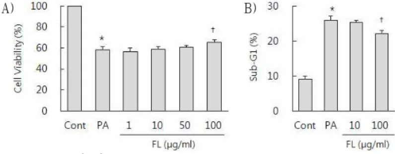
<0.01, compared to palmitate-treated cells.3. 구기자 물 추출물이 palmitate에 의한 lipotoxicity 구기자 물 추출물이 지방산에 의해 유발된 세포
HepG2 cell에 구기자 물 추출물과 palmitate를 처 리하고 배양한 다음 택사 및 산사 물 추출물을 테 스트한 방법과 동일한 방법으로 MTT assay와 Sub-G
1analysis를 시행하였다. 구기자 물 추출물
은 100 μg/ml로 투여된 경우에만 palmitate에 의해 저하된 세포 활성도를 증가시키고 증가한 sub-G
1부분을 감소시키는 효과를 나타냈다(Fig. 3).
A) B)
Fig. 3. Effect of of Fructus Lycii (FL) on palmitate induced lipotoxicity in HepG2 cells.
HepG2 cells were pretreated with several different doses of aqueous extract of Fructus Lycii (FL) for 1h and treated with 0.5 mM palmitate for 21 hrs in succession. After that, cell viability was assessed through MTT colorimetric assay (A). The percentage of cells with sub-G1 DNA content was measured using fluorescence-activated cell sorting after propidium iodide staining (B). The means±SE (n=4) are presented. *
P
<0.01, compared to untreated control;†P
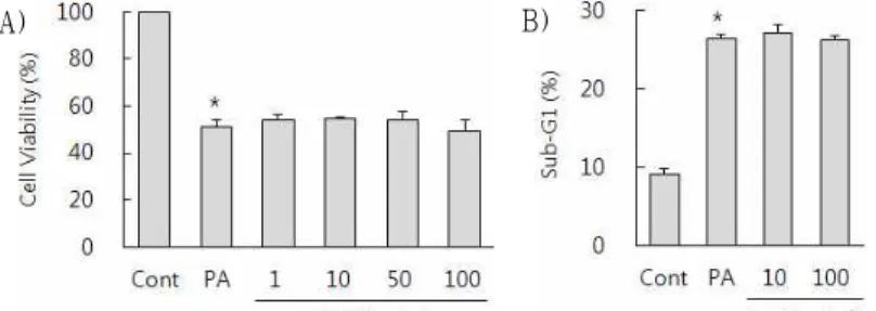
<0.01, compared to palmitate-treated cells.4. 울금 물 추출물이 palmitate에 의한 lipotoxicity 와 lipoapoptosis에 미치는 영향
울금 물 추출물이 지방산에 의해 유발된 세포 손상 및 apoptosis에 미치는 영향을 평가하기 위해, HepG2 cell에 울금 물 추출물과 palmitate를 처리 하고 배양한 다음 상기한 시료들과 동일한 방법으
로 MTT assay와 Sub-G
1analysis를 시행하였다.
울금 물 추출물은 palmitate에 의해 증가한 sub-G
1부분을 감소시키는 효과는 있었으나, 저하된 세포 활성도를 증가시키지 못했고, 오히려 세포 활성도 를 더욱 저하시키는 결과가 관찰되었다(Fig. 4).
A) B)
Fig. 4. Effect of of Radix Curcumae (RC) on palmitate induced lipotoxicity in HepG2 cells.
HepG2 cells were pretreated with several different doses of aqueous extract of Radix Curcumae (RC) for 1h and treated with 0.5 mM palmitate for 21 hrs in succession. After that, cell viability was assessed through MTT colorimetric assay (A). The percentage of cells with sub-G1 DNA content was measured using fluorescence-activated cell sorting after propidium iodide staining (B). The means±SE (n=4) are presented. *
P
<0.01, compared to untreated control;†P
<0.01, compared to palmitate-treated cells.5. 단삼 물 추출물이 palmitate에 의한 lipotoxicity 와 lipoapoptosis에 미치는 영향
단삼 물 추출물이 지방산에 의해 유발된 세포 손상 및 apoptosis에 미치는 영향을 평가하기 위해, 상기한 시료들과 동일한 조건 및 동일한 방법으로
MTT assay와 Sub-G
1analysis를 시행하였다. 단삼 물 추출물은 palmitate에 의해 저하된 세포 활성도 를 증가시키지 못했고, 증가한 sub-G
1을 감소시키 는 효과도 나타나지 않았다(Fig. 5).
A) B)
Fig. 5. Effect of of Radix Salviae Miltiorrhizae (RSM) on palmitate induced lipotoxicity in HepG2 cells.
HepG2 cells were pretreated with several different doses of aqueous extract of Radix Salviae Miltiorrhizae (RSM) for 1h and treated with 0.5 mM palmitate for 21 hrs in succession. After that, cell viability was assessed through MTT colorimetric assay (A). The percentage of cells with sub-G1 DNA content was measured using fluorescence-activated cell sorting after propidium iodide staining (B). The means±SE (n=4) are presented. *
P
<0.01, compared to untreated control;†
P
<0.01, compared to palmitate-treated cells.6. 인진 물 추출물이 palmitate에 의한 lipotoxicity 와 lipoapoptosis에 미치는 영향
인진 물 추출물이 지방산에 의해 유발된 세포 손상 및 apoptosis에 미치는 영향을 평가하기 위해, 상기한 시료들과 동일한 조건 및 동일한 방법으로
MTT assay와 Sub-G
1analysis를 시행하였다. 인진 물 추출물도 단삼 물 추출물과 같이 palmitate에 의해 저하된 세포 활성도를 증가시키지 못했고, 증 가한 sub-G
1을 감소시키지 못했다(Fig. 6).
A) B)
Fig. 6. Effect of of Herba Artemisiae Scopariae (HAS) on palmitate induced lipotoxicity in HepG2 cells.
HepG2 cells were pretreated with several different doses of aqueous extract of Herba Artemisiae Scopariae (HAS) for 1h and treated with 0.5 mM palmitate for 21 hrs in succession. After that, cell viability was assessed through MTT colorimetric assay (A). The percentage of cells with sub-G1 DNA content was measured using fluorescence-activated cell sorting after propidium iodide staining (B). The means±SE (n=4) are presented. *