J. Exp. Biomed. Sci. 13 (2007) 349–354
Effects of Vanillic Acid on the Cell Viability and Melanogenesis in Cultured Human Skin Melanoma Cells Damaged by
ROS-Induced Cytotoxicity
Dae-Ho Ha1, Yong-Ja Choi2 and Sun-Mi Yoo3,†
1Sanbon Medical Center, Wonkwang University, Gunpo 435-040, Korea.
2Deprtment of Radiology, Wonkwang Health Science College, Iksan 570-750, Korea.
3Department of Cosmetology, Dongkang College, Kwangju 500-714, Korea
The purpose of this study was to examine the effect of vanillic acid on the cell viability and melanogenesis in melanocytes damaged by reactive oxygen species (ROS). The human skin melanoma cells (SK-MEL-3) were cultured with various concentrations of hydrogen peroxide (H2O2). The cell viability for H2O2-induced cytotoxicity or vanillic acid against H2O2 was measured by XTT assay in these cultures. For the effect of vanillic acid on the melanogenesis, the tyrosinase inhibitory activity was measured by colorimetric assay at a wavelength of 490 nm, and melanin synthesis activity were assessed after cells were cultured in the media with or without various cencentrations of vanillic acid. In this study, H2O2 decreased cell viability dose- and time-dependent manners and XTT50 was determined at a concentration of 80 µM, H2O2. Vanillic acid increased the cell viability dose dependently in human skin melanoma cells damaged by H2O2-induced cytotoxicity. In the tyrosinase inhibitory activity, vanillic acid supresssed tyrosinase activity in dose- dependent manner, and also decreased significantly melanin synthesis activity compared with H2O2-treated group. From these results. It is suggested that H2O2-mediated cytotoxicity was highly by the toxic criteria of Borenfreund and Puerner and also, vanillic acid has the protective effect on ROS-induced cytotoxicity and melanogenesis in these cultures.
Key Words: Cytotoxicity, ROS, Melanogenesis, Vanilic acid
서 론
활성산소 (reactive oxygen species, ROS)는 강력한 산화 작용을 가지고 있어 세포의 -SH를 비롯한 지질과산화 및 아민의 니트로화와 같은 산화적 손상을 통해 세포의 고사를 초래한다고 알려져 있다 (Chung et al., 2000). ROS 는 주로 세포내 사립체 (mitochondria)를 비롯하여 cyclo- oxygenase (COX) 및 cytochrome P450 등에서 생성되며 (Boczkowski et al., 1999), 특히 이들은 nitric oxide (NO)와 같은 활성질소 (reactive nitrogen species, RNS)와도 강력히 반응하여 peroxynitrite (ONOO-)라는 독성이 매우 강한 물 질을 형성한다. 근래 ROS는 노화를 비롯한 암, 관절염,
동맥경화 등과 같은 각종 질환의 병리적 요인으로 작용 한다고 알려져 있으며 (Haenen et al., 1997; Pannala et al., 1997), 이같은 현상은 세포대사 과정에 ROS가 직접적으 로 이상을 유발함으로서 세포손상에 의한 병변을 초래 한다고 한다 (Pellegrini-Giampietro et al., 1990). 특히, ROS 는 광노화 (photoaging)의 주된 요인으로서 피부노화와 관련이 깊다고 보고된 바 있다 (Chung et al., 1995). 광노 화는 햇빛속의 자외선 (ultraviolet light, UV)과 밀접한 관 련이 있는데 이는 피부에 조사된 UV에 의해 생성된 O-2, H2O2 및 OH-와 같은 ROS가 피부세포를 공격함으로서 콜라겐의 감소를 비롯하여 모세혈관 감소 및 멜라닌세포 활성 증가와 같은 현상을 초래하기 때문에 이를 일명 프 리라디칼설이라 한다 (Harman, 1956; Kim et al., 2006). 따 라서 피부노화를 비롯하여 ROS에 의해 유발되는 각종 질환에 항산화물질이나 자유기제거제 등을 투여함으로 서 노화나 병변의 치료적 접근을 위하여 많은 연구가 시 도되고 있다 (Heilmann et al., 2000; Furuya et al., 2001; Kim et al., 2006). 피부노화의 하나중 색소침착은 피부미용과
*논 문 접 수: 2007년 11월 28일 수정재접수 : 2007년 12월 12일
†교신저자: 유선미, (우) 570-714 광주광역시 북구 두암동 771, 동강대학 피부미용과
Tel: 062-520-2391, Fax: 062-520-2392 e-mail: [email protected]
밀접한 관련이 있으며 특히, 햇빛속의 UV에 의하여 형 성된 ROS가 멜라닌세포로 하여금 과도히 멜라닌의 합성 을 촉진시켜 그 결과 기미나 주근깨, 흑자, 반점 등 다양 한 색소침착을 유발한다고 알려져 있다 (Hill et al., 1997;
Lee et al., 2003). 멜라닌의 합성은 멜라닌세포내에 위치 하고 있는 멜라노좀 (melanosome)에서 이루어지는데 멜 라노좀내의 tyrosinase가 tyrosin으로 변환된 후 이는 다시 DOPA 퀴논에서 멜라닌으로 합성되어 진다 (Lerner and Fitzpateick, 1984). 따라서 현재 색소침착증의 예방이나 치 료를 위한 목적으로 항산화 측면에서 ROS와 멜라닌세 포간의 작용기전을 밝히려는 연구가 활발히 진행중이다 (Lee et al., 2003; Kang et al., 2007). 최근 천연추출물중 식 물성분에 항암이나 항산화 효과가 뛰어난 물질들이 다량 포함되어 있다고 보고되면서 이에 대한 연구가 관심의 대상이 되고 있다 (Soung et al., 1999; Shahrzad et al., 2001).
현재까지 알려진 대표적인 몇몇 성분들을 살펴보면 페놀 화합물을 중심으로 이소프렌 유도체, 터펜, 스테로이드 및 카로틴과 같은 수많은 물질들이 순수 분리 정제되었 다 (Duthie and Alan, 2000). 이중 phenol 화합물은 수산기 (OH)를 갖는 방향족 고리모양의 구조를 가지고 있는 물 질로서 과일을 비롯한 채소, 버섯 등에 풍부히 들어 있 으며, 금속의 착화를 비롯하여 항암이나 항산화 및 활성 산소형성의 효소저해와 같은 기능을 가지고 있다고 알려 져 있다 (Krizkova et al., 2000; Wu et al., 2000). 지금까지 알려진 페놀화합물로는 vanillic acid를 비롯하여 cafferic acid, gallic acid 등 다양한 성분들이 분리 추출되었다 (Wu et al., 2000; Shahrzad et al., 2001). 이중 vanillic acid는 다른 페놀화합물 처럼 shikimic acid pathway에 의하여 합성되어 지며 또한 방향족 고리구조에 치환될 수 있는 수산기와 카르복실기, 메톡시기로 구성되어 있다. 이러한 잔기들 중 항산화작용은 수산기와 메톡시기가 밀접하게 관련되 어 있음이 제시된 바 있다 (Heilmann et al., 2000).
본 연구는 ROS에 의하여 손상된 인체피부흑색종세포 (SK-MEL-3)에 대한 vanillic acid의 영향을 알아보기 위 하여 colorimetric assay를 이용한 세포생존율의 분석과 tyrosinase 활성 및 멜라닌합성에 미치는 영향을 조사하 였다.
재료 및 방법
1. 시약 제조
본 실험에 사용한 hydrogen peroxide (H2O2, Sigma)는 혈
청이 포함되어 있지 않은 MEM (minimun essential medium, Sigma) 배양액에 최종 농도가 각각 10 µM, 50 µM, 100 µM, 500 µM, 1 mM이 되도록 저장액을 만들어 냉장고에 보관 한 후 실험 당일 일정 농도로 희석하여 사용하거나 또는 직접 필요한 양을 배양액에 넣어 첨가하여 사용하였다.
XTT(2,3-bis-[2-methoxy-4-nitro-5-sulfophenyl]-2H-tetrazolium -5-caboxanilide, disodium salt)는 실험 전날 500 µg/ml의 저 장액을 만들어 냉암소에 보관한 후 실험 당일 300 µg/ml, 100 µg/ml, 50 µg/ml, 1 µg/ml 농도로 희석하거나 필요한 양만큼 배양액에 넣어 사용하였다.
2. 세포 배양
본 실험에 사용한 인체피부흑색종세포 (SK-MEL-3)는 변형된 Mosmann (1983)의 방법에 따라 일정 시간 동안 배양용기에 자란 세포는 trypsin에 의한 효소분리에 의 하여 용기로 부터 분리한 후 Eagle's minimum essential medium (MEM, Gibco)에 10% fetal bovine serum (FBS, Gibco)이 포함된 배양액에 부유시켰다. 부유된 세포들은 1×10⁵ cells/well의 밀도로 산정하여 96 배양용기 (well plate)에 넣은 후 48시간 동안 배양하였으며 배양이 완료 된 세포는 본 실험의 분석에 사용하였다.
3. ROS 처리
배양이 완료된 세포에 70 µM 부터 90 µM 까지의 H2O2
가 농도별로 각각 포함된 배양액에서 6시간 동안 배양한 후 H2O2가 인체피부흑색종에 미치는 영향을 분석하였다.
4. 약재 처리
H2O2에 대한 vanillic acid의 영향을 조사하기 위하여 H2O2를 배양세포에 처리하기 전에 vanillic acid가 140~
180 µg/ml의 농도로 각각 포함된 배양액에서 인체피부흑 색종세포를 2시간 동안 처리한 후 대조군과 비교 조사하 였다.
5. 세포생존율 분석
XTT에 의한 분석은 Borenfreund와 Puerner (1984)의 방 법에 의하였다. 즉 약재를 처리하지 않은 배양 인체피부 흑색종세포를 5×10⁶ 세포수로 하여 laminin으로 코팅한 배양용기에서 24시간 동안 배양하였다. 배양이 완료된 후 약재를 처리하여 배양하였으며 처리된 세포를 PBS (phosphate buffered saline)로 3회 세척한 다음 실험 전날 제조한 XTT를 well당 200 µl씩 넣어 37℃, 5% CO2/95%
air로 조절된 정온기에서 3시간 동안 배양하였다. 배양완 료 후 효소산물추출액으로 처리한 다음 spectrophotometer 로 450 nm에서 흡광도를 측정하여 대조군과 비교 조사 하였다.
6. Tyrosinase 활성저해능
Tyrosinase 활성저해능의 측정은 Choi 등 (1998)의 변 형된 방법에 따라 0.1 M sodium phosphate buffer에 1.5 mM tyrosinase 용액과 0.3 ml의 시료액 및 tyrosinase 효소액 (1250 U/ml)을 넣은 후 37℃에서 15분간 반응시킨 다음 490 nm에서 spectrophotometer로 흡광도를 측정하여 대조 군과 비교 조사하였다. Tyrosinase 활성저해능은 시료용액 의 첨가군과 무첨가군의 흡광도 감소율에 대한 백분율로 나타냈다.
7. 멜라닌합성능 분석
Hosoi 등 (1985)의 변형된 방법에 따라 분석하였다. 즉, 약재를 처리한 용기 부착세포를 수확하여 1,000 × g에서 15분간 원침한 후 얻어진 침전물에 1 ml의 homogenization buffer를 넣어 용해시켰다. 용해 완료 후 다시 원침하여 얻은 pellet에 DMSO가 포함된 1N NaOH를 첨가한 다음 405 nm에서 spectrophotometer로 흡광도를 측정하여 대조 군과 비교 조사하였다. 실험군의 멜라닌합성량은 대조군 에 대한 백분율로 나타냈다.
8. 통계 처리
대조군과 실험군 사이의 통계학적 유의성 검정은 Student's t-test를 사용하였으며 P가 0.05 이하인 것만 유 의한 것으로 하였다.
결 과
1. H2O2의 독성효과
1) 세포생존율 분석
(1) 농도에 따른 H2O2의 영향
배양중인 인체피부흑색종세포를 PBS로 3회 세척한 후 70 µM에서 90 µM까지의 H2O2가 각각 포함된 배양액에서 6시간 배양한 다음 흡광도에 의한 세포생존율을 대조군 과 비교 조사하였다. 70 µM에서 처리에서 세포생존율은 대조군인 100% (10.75±1.42)에 비하여 74.5% (8.01±0.57) 로 나타났으며 80 µM의 경우 54.0% (5.80±0.32)로 나타났 다 (P<0.05). 또한, 90 µM에서는 25.4% (2.73±0.16)로 나 타나 이는 대조군에 비하여 유의하게 감소하였으며 (P<
0.01), 이 때 XTT50 값은 80 µM에서 나타났다 (Table 1).
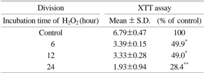
(2) 시간에 따른 H2O2의 영향
인체피부섬유모세포에 H2O2를 처리한 시간에 따른 세 포생존율을 조사하기 위하여 H2O2의 XTT50 농도에서 6~
24시간 동안 처리한 결과 6시간 처리에서 세포생존율은 대조군인 100% (6.79±0.47)에 비하여 49.9% (3.39±0.15) 로 나타났으며 12시간 배양에서는 49.0% (3.33±0.28)로 나타났다 (P<0.05). 또한, 24시간 배양에서는 28.4% (1.93
±0.94)로 나타나 이는 대조군에 비하여 유의하게 감소하 였다 (P<0.01) (Table 2).
2. H2O2에 대한 vanillic acid의 영향
배양중인 인체피부흑색종세포를 PBS로 3회 세척한 후 140 µM에서 180 µM의 vanillic acid가 각각 포함된 배 양액에서 2시간 전배양한 다음 세포생존율을 조사하였 다. XTT50 농도의 H2O2만을 처리한 경우 대조군인 100%
(5.45±0.42)에 비하여 37.1% (2.02±0.19)로 나타났으나 Table 2. Time-response relationship of hydrogen peroxide (H2O2) for its cytotoxic effect on cultured human skin melanoma cells (SK-MEL-3)
Division XTT assay
Incubation time of H2O2 (hour) Mean ± S.D. (% of control)
Control 6.79±0.47 100
6 3.39±0.15 49.9*
12 3.33±0.28 49.0*
24 1.93±0.94 28.4**
The human skin melanoma cells were cultured in the media con- tainning hydrogen peroxide (H2O2) for 6, 12, and 24 hours, respectively. Cell viability was determined by XTT assay. The data indicate the mean ± SD for triplecate experiments. Significantly different from control. *P<0.05, **P<0.01
Table 1. Cell viability of cultured human skin melanoma cells (SK-MEL-3) treated with hydrogen peroxide (H2O2) by XTT assay
Division XTT assay
Concentration of H2O2 (µM) Mean ± S.D. (% of control)
Control 10.75±1.42 100
70 8.01±0.57 74.5
80 5.80±0.32 54.0*
90 2.73±0.16 25.4**
The XTT values of cultured human skin melanoma cells treated with various concentrations of hydrogen peroxide (H2O2) for 6 hours. The data indicate the mean ± SD for triplecate experiments.
Significantly different from control. *P<0.05, **P<0.01
140 µM vanillic acid를 처리한 경우 세포생존율은 52.8%
(2.88±0.17)로 나타났다. 또한, 180 µM의 처리에서는 세포 생존율이 66.8% (3.64±0.23)로 나타나 이는 H2O2만의 처 리에 비하여 유의하게 증가하였다 (P<0.05) (Table 3).
3. Tyrosinase 활성저해능 분석
Vanillic acid가 tyrosinase 활성저해능에 미치는 영향을 조사하기 위하여 40~100 µM 농도의 vanillic acid에 대 한 tyrosinase 활성저해능을 조사하였다. 그 결과 40 µM vanillic acid를 처리한 경우 tyrosinase 활성저해능은 시료 의 무첨가군과 동일하게 나타난데 비하여 70 µM의 처리 에서는 tyrosinase 활성저해능이 26.7%로 나타났다. 또한, 100 µM의 처리에서는 60.0%로 나타나 유의하게 증가하 였다 (P<0.05) (Table 4).
4. 멜라닌합성능 분석
Vanillic acid가 멜라닌합성능에 미치는 영향을 알아보 기 위하여 XTT50 농도의 H2O2를 처리하기 전에 vallic acid가 70~100 µM의 농도로 각각 포함된 배양액에서 2
시간 동안 처리한 후 이에 대한 합성량을 측정하였다. 그 결과 80 µM 농도의 H2O2만의 처리에서는 대조군에 비 하여 멜라닌합성량이 108.1%로 나타난데 비하여 70 µM vanillic acid의 처리에서는 멜라닌합성량이 대조군에 비 하여 85.3%로 감소하였다. 또한, 100 µM의 처리에서는 61.2%로 나타나 이는 대조군에 비하여 유의하게 감소하 였다 (P<0.05) (Table 5).
고 찰
식물성 천연추출물에 들어 있는 페놀화합물은 항노화 를 비롯하여 항암작용이나 항돌연변이 및 항산화능이 있 다고 보고되었다 (Duthie and Alan, 2000). 최근, vanillic acid를 비롯한 페놀화합물이 ROS에 대한 항산화능이 있 다고 알려지면서 산화적 손상에 의한 피부노화와도 깊은 연관성이 있다고 제시된 바 있다 (Heilmann et al., 2000).
본 연구에서는 vanillic acid가 ROS의 세포독성에 미치는 영향을 인체피부흑색종세포를 재료로 조사하였으며 또 한 vanillic acid가 ROS에 의한 멜라닌세포의 tyrosinase의 활성과 멜라닌합성에 미치는 영향을 조사하였다. 본 연 구의 ROS 세포독성에 있어서, H2O2를 인체피부흑색종세 포에 처리한 결과 처리 농도와 처리시간에 따라 세포생 존율을 감소시켰으며 특히, XTT50 값으로 나타난 80 µM 의 H2O2 처리에서는 대조군에 비하여 유의한 세포생존율 의 감소를 나타냈다 (P<0.05). 또한, H2O2는 Borenfreund 와 Puerner (1984)의 독성판정기준에 의하여 인체피부흑 색종세포에 고독성인 것으로 나타났는데 이 같은 원인은 H2O2의 산화적 손상이 세포내 신호전달체계나 칼슘체널 에 이상을 초래하여 항상성을 파괴하였을 가능성도 있 겠으나 (Kaneko et al., 1989), XTT 분석이 세포소기관과 밀접한 관련이 있는 것으로 미루어 볼 때 H2O2가 세포내 Table 5. The Effect of vanillic acid on melanin synthesis activity
in cultured human skin melanoma cells (SK-MEL-3) Melanin synthesis activity Concentration of vanillic
acid (µM) % of control
Control 100
80H2O2 108.1±8.5
70 85.3±6.4
100 61.2±4.7*
The melanin synthesis activity at vanillic acid, 70~100 µM was determined at a wavelength of 405 nm. The data was expressed as % of control and indicates the mean ± SD for triplecate experi- ments. Significantly different from H2O2-treated group. *P<0.05 Table 3. Cell viability of cultured human skin melanoma cells
(SK-MEL-3) treated with vanillic acid by XTT assay
Division XTT assay
Concentration of vanillic
acid (µM) Mean ± S.D. (% of control) Control 5.45±0.42 100
80H2O2 2.02±0.19 37.1
140 2.88±0.17 52.8
180 3.64±0.23 66.8*
Cultured human skin melanoma cells were preincubated with 140
~180 µM vanillic acid, respectively. The values were determined by XTT assay in cultured human skin melanoma cells. The data indicate the mean ± SD for triplecate experiments. Significantly different from H2O2-treated group *P<0.05
Table 4. The tyrosinase inhibitory activity of vanillic acid on cultured human skin melanoma cells (SK-MEL-3)
Tyrosinase inhibitory activity Concentration of vanillic
acid (µM) % of control
40 0
70 26.7±1.4
100 60.0±4.7*
Cultured human skin melanoma cells were preincubated with 40
~100 µM vanillic acid, respectively. The values were determined at a wavelength of 490 nm. The data was expressed as % of control and indicates the mean ± SD for triplecate experiments.
Significantly different from control. *P<0.05
단백질합성이나 전자전달계에 관여하는 세포소기관을 손 상시켰을 가능성이 더 클 것으로 생각된다. 한편, vallinic acid가 ROS에 미치는 영향을 세포생존율 측면에서 분석 함에 있어서는 vallinic acid의 처리 농도에 비례하여 H2O2
에 의하여 감소된 세포생존율을 증가시켰으며 특히, 180 µM vanillic acid의 처리에서는 유의한 세포생존율의 증 가를 나타냈다 (P<0.05). 본 결과는 vallinic acid가 H2O2
의 독성으로 부터 세포를 보호하는 효과가 있다는 것을 말해주고 있으며 동시에 vallinic acid가 항산화능을 가지 고 있음을 증명하고 있다 (Duthie and Alan, 2000). 한편, vallinic acid가 tyrosinase의 활성저해에 미치는 영향을 알 아보기 위하여 40~100 µM의 농도에서 vallinic acid를 분 석한 결과 농도가 증가할수록 tyrosinase의 활성저해능이 증가하였으며 이중 100 µM 처리에서는 유의하게 증가한 것으로 나타났다 (P<0.05). 이는 아마도 vanillic acid가 항 산화작용과 같은 약리활성에 의하여 melanosome내에서 tyrosinase가 tyrosin으로 변환되는 것을 방해함으로서 효 소활성을 저해한 것으로 생각된다 (Lerner and Fitzpateick, 1984). 한편, vanillic acid가 멜라닌합성에 미치는 영향을 알아보기 위하여 vallinic acid가 70~100 µM이 포함된 배 양액에서 세포를 전처리한 결과 XTT50 농도인 H2O2만의 처리에 비하여 멜라닌합성량이 감소하였으며 특히, 100 µM의 vanillic acid의 처리에서는 유의하게 감소하였다 (P<0.05). 위 결과는 vanillic acid가 tyrosinase의 활성을 억 제하여 melanin 합성을 방어한 결과로서 이는 vanillic acid 가 tyrosinase 활성을 억제했다는 본 실험의 결과와 일치 하였다. 멜라닌세포에서 멜라닌의 과도한 합성은 피부노 화를 비롯한 각종 피부암이나 피부착색증의 주요 요인일 뿐아니라 또한 피부미백과도 밀접한 관련이 있어 관심의 대상이 되고 있다. 따라서 본 실험의 결과는 vanillic acid 와 같은 페놀화합물들이 멜라닌합성의 조절물질로서의 가능성을 제시하고 있다고 생각된다. 그러나 ROS의 세 포독성에 대한 vanillic acid의 영향에 대한 자세한 기전 규명을 위하여서는 항산화 측면에서의 연구는 물론이며 vanillic acid의 구조식에서 볼 수 있는 수산기를 비롯하여 알데하이드기 및 메톡시기와 같은 잔기들에 대한 정확한 작용현상을 PKC와 같은 신호전달체계나 NMDA와 같은 칼슘통로와 같은 다양한 측면에서 종합적인 연구가 이루 어져야 할 것으로 생각된다.
감사의 글
이 논문은 2005년도 원광대학교의 교비 지원에 의해서
수행됨.
REFERENCES
Boczkowski J, Lisdero CL, Lanone S, Samb A, Carreras MC, Boveris A, Aubier M, Poderoso JJ. Endogenous peroxynitrite mediates mitochondrial dysfunction in rat diaphragm during endotoxemia. FASEB J. 1999. 13: 1637-1646.
Borenfreund E, Puerner JA. A simple quantitative procedure using monolayer culture for cytotoxicity assay (HTD/NR-90). J Tiss Cult Meth. 1984. 9: 7-9.
Choi BW, Lee BH, Kang KJ, Lee ES, Lee NH. Screening of the tyrosinase inhibitors from marine algae and medicinal plants.
Kor J Pharmacogn. 1998. 29: 237-242.
Chung HY, Yokozawa T. Study on Antioxidative and Antimu- tagenic Mechanism of Epicatechin 3-O-gallate Isolated from Green Tea. Kor Soc Food Sci Technol. 1995. 3: 65-81.
Chung HY, Soung DY, Kim A, Choin HR, Kim HJ, Choin JS, Yang R, Lee KH, Yu BP. Generatio, Toxicity and Scavenging of ONOO-: Its Involvement in the Aging Process. Kor J Gerontol. 2000. 10: 46-59.
Duthie Garry C, Alan. Plant-derived phenolic antioxidants. Current opinion in Clinical Nutrition and Metabolic Care. Lippincott Williams and Wilkins. 2000. 3: 447-451.
Furuya S, Takayama F, Mimaki Y, Sashida Y, Satoh K, Sakagami H. Cytotoxic activity of saponins from Camassia leichtlinii against human oral tumor cell lines. Anticancer Res. 2001.
21: 959-964.
Haenen GR, Paquay JB, Korthouwer RE, Bast A. Peroxynitrite scavenging by flavonoids. Biochem Biophys Res Commun.
1997. 236: 591-593.
Harman D. Aging: Theory based on free radical and radiation chemistry. J Gerontol. 1956. 11: 298-300.
Heilmann J, Calis I, Kirmizibekmez H, Schuhly W, Harput S, Sticher O. Radical scavenger activity of phenylethanoid glycosides in FMLP stimulated human polymorphonuclear leukocytes: structure-activity relationships. Planta Med. 2000.
66: 746-748.
Hill HZ, Li WXinP, Mitchell DL. Melanin: a two edged sword?
Pigment Cell Res. 1997. 10: 158-161.
Hirota A, Taki S, Kawaii S, Yano M, Abe N. 1,1-Diphenyl-2- picrlhydrazyl radical-scavenging compounds from soybean miso and antiproliferative activity of isoflavones from soybean miso toward the cancer cell lines. Biosci Biotechnol Biochem.
2000. 64: 1038-1040.
Hosoi J, Abe E, Suda T, Kuroki T. Regulation of melanin synthesis og B16 mouse melanoma cells by 1 alpha, 25-dihydroxy- vitamin D3 and retinoic acid. Cancer Res. 1985. 45: 1474 -1478.
Kaneko M, Elmban V, Dhalla NS. Mechanism for depression of heart sarcolemma Ca2+ pump by oxygen free radiclas. Am J Physiol. 1989. 261: 4948-4955.
Kang JR, Lee JY, Whang WK. Antioxidant Activity and Tyro- sinase Inhibitory Activity of Elscholtziae Splendense. J Kor Soc Cosm. 2007. 13: 163-170.
Kim EJ, Ahn SY, Nam GW, Lee HK, Moon SJ, Kim YM, Oh MS, Kim NS. The Anti-aging Effect of the Cosmetic Products Containing the Needles of Red pine on Human Skin. Kor J Herbol. 2006. 21: 25-31.
Kizkova L, Nagy M, Polonyi J, Dobias J, Belicova A, Grancai D, Krajcovic J. Phenolic acids inhibit chloroplast mutagenesis in Euglena gracilis. Mutat Res. 2000. 469: 107-114.
Lee JK, Jung HM, Kim SY. 1,8-Dihydroxynaphthalene (DHN)- melanin biosynthesis inhibitors increase erythritol production in Torula corallina, and DHN-melanin inhibits erythrose reductase. Appl Envir Microbiol. 2003. 69: 3427-3434.
Lerner AB, Fitzpateick TB. Biochemstry of melanin formation.
Physiol Rev. 1984. 30: 92-126.
Mosmann T. Rapid colorimetric assays for cellular growth and survival: Application to proliferation and cytotoxicity assays.
J Immunol Methods. 1983. 65: 55-63.
Pannala AS, Razaq R, Halliwell B, Singh S, Rice-Evans CA.
Inhibition of peroxynitrite dependent tyrosine nitration by hydroxycinnamates: nitration or electron donation?. Free Radic Biol Med. 1998. 24: 594-606.
Pellegrini-Giampietro DE, Cherici G, Alesiani M, Carla V, Morroni F. Excitatory amino acid and free radical formation may cooperate in the genesis of ischemia-induced neuronal damage.
J Neurosci. 1990. 10: 1035-1041.
Shahrzad S, Aoyagi K, Winter A, Koyama A, Bitsch I. Phar- macokinetics og gallic acid and its relative bioavailability from tea in healthy humans. J Nutr. 2001. 131: 1207-1210.
Soung DY, Kim JS, Chung HY, Jung HA, Park JC, Choi JS.
Flavonoids and chlorogenic acid from Eriobotrya japonica scavenge peroxynitrite. Nat Pro Sci. 1999. 5: 80-84.
Wu H, Haig T, Pratley J, Lemerle D, An M. Allelochemicals in wheat (Triticum aestivum I.): variation of phenolic acids in root tissues. J Agric Food Chem. 2000. 48: 5321-5325.