Loganin Inhibits α-MSH and IBMX-induced Melanogenesis by Suppressing the Expression of Tyrosinase in B16F10 Melanoma Cells
Hee Jin Jung
1, EunJin Bang
2, Byeong Moo Kim
2, Seong Ho Jeong
2, Gil Han Lee
2and Hae Young Chung
1,2*
1
Longevity Life Science and Technology Institutes, Pusan National University, Busan 46241, Korea
2
Department of Pharmacy, College of Pharmacy, Pusan National University, Busan 46241, Korea Received September 27, 2019 /Revised October 29, 2019 /Accepted November 14, 2019
Ultraviolet radiation exposure is a major cause of extrinsic skin aging, which leads to skin hyper- pigmentation. Loganin, a major iridoid glycoside obtained from Corni fructus, has anti-inflammatory, anti-diabetic, and neuroprotective effects. In this study, we investigated the mechanisms underlying the anti-melanogenic effects of loganin in B16F10 melanocytes treated with α-melanocyte stimulating hormone (α-MSH) and 3-isobutyl-1-methylxanthine (IBMX). Anti-melanogenic activity was measured by treating cells with loganin at concentrations between 1 and 20 μM. Cell viability assays confirmed that doses of loganin up to 20 μM were not cytotoxic. Loganin significantly and dose-dependently de- creased intracellular melanin production. We also investigated potential molecular signaling pathways for the anti-melanogenesis effects of loganin. Western blotting showed that treatment with α-MSH and IBMX increased the phosphorylation of cAMP response element-binding protein (CREB) and the gene expressions of microphthalmia-associated transcription factor (MITF) and tyrosinase. Addition of loga- nin suppressed these increases, while promoting the phosphorylation of extracellular signal regulated kinase (ERK) and the anti-melanogenesis response. Our data therefore indicated that loganin could at- tenuate the increased melanin synthesis induced by α-MSH and IBMX treatment of B16F10 melanocytes.
This attenuation appears to occur by downregulation of CREB phosphorylation and MITF and ty- rosinase gene expression and upregulation of ERK phosphorylation. These finding suggests that loga- nin could be a valuable candidate for treatment of skin diseases related to hyperpigmentation.
Key words : Anti-melanogenesis, iridoid glycoside, loganin, tyrosinase
*Corresponding author
*Tel : +82-51-510-2814, Fax : +82-51-518-2821
*E-mail : [email protected]
This is an Open-Access article distributed under the terms of the Creative Commons Attribution Non-Commercial License (http://creativecommons.org/licenses/by-nc/3.0) which permits unrestricted non-commercial use, distribution, and reproduction in any medium, provided the original work is properly cited.
Journal of Life Science 2019 Vol. 29. No. 11. 1200~1207 DOI : https://doi.org/10.5352/JLS.2019.29.11.1200
Introduction
Melanin is produced and formed by melanocytes in the layer of epidermis outermost in the skin, and protects against damage due to ultraviolet (UV) radiation [44].
However, dysregulated activation of melanin synthesis can induce skin diseases associated with hyperpigmentation, in- cluding melasma, melanoma, freckles, age spots, and senile lentigines [18]. Therefore, numerous studies have focused on the development of effective skin whitening agents.
Melanin synthesis is activated by several important mole- cules and environmental conditions, including α-melanocyte stimulating hormone (α-MSH), cyclic AMP (cAMP) elevating agents, such as forskolin, glycyrrhizin, and 3-isobutyl-1-
methylxanthine (IBMX); and UV radiation, and cAMP-medi- ated pathway, which plays a pivotal role in regulating mela- nogenesis [8, 22, 23], binds and activates the melanocortin 1 receptor (MC1R) in the epidermis layer of the skin, which subsequently activates cAMP-dependent protein kinase A (PKA) and other regulatory protein molecules [8]. PKA sub- sequently activates the cAMP response element-binding pro- tein (CREB), which is a transcriptional activator of the micro- phthalmia-associated transcription factor (MITF) gene [4, 5].
The increase of CREB phosphorylation induces the tran- scription of MITF, which is a core transcription factor that induces the expression of tyrosinase, consequently increas- ing melanin synthesis [47, 55]. Furthermore, inhibition of ex- tracellular signal-regulated kinase (ERK) signaling mediates melanin biosynthesis by increasing the activity of tyrosinase [25, 28, 30].
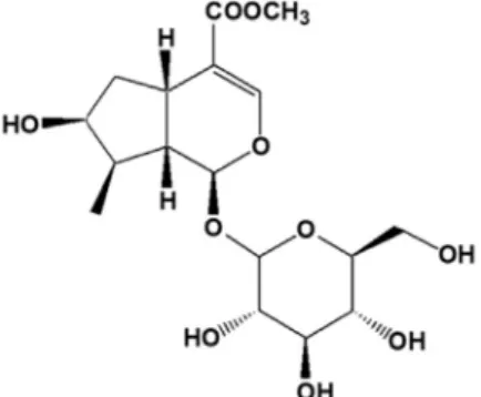
Loganin, an iridoid glycoside, is a biologically active com- pound obtained from Corni fructus (Cornus officinalis et Zucc), and one of the most popular plant products used in the tra- ditional medicines in Japan, China, and Korea [12, 40, 53].
Previous studies have demonstrated that loganin possesses
Fig. 1. Chemical structure of loganin.
several biological activities, including immune regulatory, anti-inflammatory properties related to pulmonary compli- cations [32] anti-atherosclerotic [39], hepatoprotective [46, 59], anti-Alzheimer’s [6, 24, 63], anti-Parkinson’s [56], neuro- protective [15, 31, 33, 52, 61], and anti-diabetic [26, 35, 42, 62] properties. A previous study reported that hot aqueous extracts and ethanolic extracts of C. officinalis inhibit UVB-in- duced pigmentation via radical scavenging activity in B16 melanoma cells, and are responsible for the inhibitory effects of loganin on melanogenesis [43]. Recently, An et al. [1] re- ported that methanolic extract of C. officinalis stimulates mel- anogenesis in Melan-a cells by increasing the levels of ty- rosinase-related protein (TRP)-1, TRP-2 and MITF.
Despite of numerous studies, the mechanism by which loganin suppresses melanin production induced by α-MSH and IBMX is known. Therefore, our aim was to investigate the anti-melanogenic property of loganin against in α-MSH and IBMX-induced hyper pigmentation and the underlying molecular mechanism.
Materials and Methods
Materials
Loganin was isolated from C. officinalis according to the procedure described by Yamabe and coworkers [57-59]. The structure of loganin is depicted in Fig. 1. Kojic acid, α-MSH, and IBMX were purchased from Sigma-Aldrich (St. Louis, MO, USA). RIPA buffer was obtained from Biosesang (Sun- gnam, Gyeonggi-do, Korea). Dulbecco’s modified Eagle’s medium (DMEM), fetal bovine serum (FBS), streptomycin, and amphotericin were purchased from Welgene Inc.
(Gyeongsan-si, Korea). The CREB (9197s) antibody was pur- chased from Cell Signaling Technology (Danvers, MA, USA).
Antibodies against p-CREB (sc-81486), p-ERK (sc-7383), ty- rosinase (sc-7833), MITF (sc-11002), ERK (sc-514302), and β-
actin (sc-47778) were acquired from Santa Cruz Biotechnol- ogy (Dallas, TX, USA). All other reagents were purchased from Merck (Frankfurt Str., Darmstadt, Germany), Fluka (St.
Louis, Mo, USA), or Sigma Aldrich Co., unless stated otherwise.
Cell culture
Murine melanoma B16F10 cells were purchased from the Korean Cell Line Bank (Seoul, Korea) and cultured in DMEM supplemented with 10% FBS and 1% streptomycin, and in- cubated at 37℃, in a humidified atmosphere with 5% CO
2.
Cell viability assay
Cell viability assays were performed as previously de- scribed [27]. Briefly, the cells were seeded at a density of 1×10
4cells/well in a 96-well plate for 24 hr. On the following day, the cells were exposed to different concentrations of loganin and incubated for 24 hr or 48 hr, respectively.
Subsequently, 10 μl of EZ-Cytox solution was added to each well, and the cells were incubated for 2-4 hr at 37℃.
Absorbance was determined using a spectrophotometric mi- croplate reader (Tecan, Mannedorf, Switzerland) at a wave- length of 450 nm. Each assay was performed in triplicate.
Assay for melanin contents
Melanin content was determined as previously described [7]. B16F10 melanoma cells (2×10
5cells/well) were seeded in 6-well culture plates. In order to determine the inhibitory effect of loganin on melanogenesis, fresh medium was re- placed with media supplemented with 1, 5 and 20 μM loga- nin or 20 μM kojic acid, which served as the positive control, and incubated for 2 hr. The cells were subsequently stimu- lated with 500 nM α-MSH and 200 μM IBMX for 48 hr. After washing twice with PBS, the cells were detached by incubat- ing in trypsin/EDTA, and the pellets were dissolved in 100 μl of 1N NaOH incubated at 60℃ for 1 hr, and pipetting to solubilize the melanin. The melanin content was de- termined by measuring the absorbance at 405 nm by an ELISA reader (Tecan, Mannedorf, Switzerland). The melanin content was calculated using the following equation: (Δ sam- ple/Δ control) ×100%. All measurements were performed in triplicate.
Western blot analysis
Western blotting was performed as previously described
[21]. B16F10 cells were treated with the samples for 2 hr,
following which 500 nM α-MSH and 200 μM IBMX were
added. After 6 hr, the cells were collected and lysed in a
A B
Fig. 2. Cell viability of loganin in B16F10 melanoma cells. Cells (1×10
4cells/well) were preincubated using various concentrations of loganin, up to 20 μ M, for 24 hr (A) or 48 hr (B). Cell viability was determined by the EZ-Cytox assay and expressed as the percentage of absorbance values relative to those of the control group. Data are represented as the mean ± S.E.M.
of experiments performed in triplicate.
RIPA cell lysis buffer containing 50 mM Tris-HCl (pH 7.4), 150 mM NaCl, 1 mM EDTA, 1 mM EGTA, 1.2% Triton X-100, 0.5% sodium deoxycholate, and 0.1% sodium dodecyl sulfate (SDS). The total protein-equivalents were separated by 8-10% SDS-PAGE and subsequently transferred onto PVDF membranes at 25 V for 10 min in a semi-dry transfer system (ATTO, Tokyo, Japan). The membranes were immediately placed in blocking buffer (5% non-fat milk) in 0.1% Tween 20 and were blocked at 25℃ for 1 hr. The membrane was incubated overnight at 4℃ with the appropriate specific pri- mary antibodies, and then treated with horseradish perox- idase conjugated anti-mouse, anti-rabbit, or anti-goat anti- bodies at 25℃ for 1 hr. The immunoblots were visualized using Western Bright Peroxide solution (Advansta, CA, USA) and Davinch-Chemi CAS-400 (Davinch-K, Seoul, Korea) ac- cording to the manufacturer’s instructions. Pre-stained pro- tein markers were used for determining the molecular weight.
Statistical analyses
Statistical significance was analyzed by one-way analysis of variance for determining the differences within treat- ments, and subsequently analyzed by the Bonferroni test (GraphPad Prism 5 software, La Jolla, CA, USA). Values of
*
p<0.05 were considered to be statistically significant. All ex-
periments were carried out in triplicate and represented as the mean ± standard errors of mean (S.E.M.) (n=3).
Results
Cell viability of loganin in the murine B16F10 mela- nocyte
To investigate the anti-melanogenic properties of loganin in the cell culture system, we investigated its cytotoxic ef- fects on B16F10 cells. The cells were treated with several concentrations of loganin for 24 hr or 48 hr, respectively, and cell viability was determined using the EZ-Cytox assay.
As shown in Fig. 2, loganin had no cytotoxic effect on B16F10 cells up to a concentration of 20 μM (Fig. 2A, Fig. 2B). There- fore, subsequent experiments were performed with loganin concentrations up to 20 μM.
Inhibitory effect of loganin on melanin biosynthesis induced by α-MSH and IBMX in B16F10 melanoma cells
Melanogenesis can be reduced by inhibiting the formation of melanin precursors or by converting them into their re- duced forms [54]. In order to investigate the inhibitory ef- fects of loganin on the elevation of cAMP levels and sub- sequent hyperpigmentation, B16F10 cells were stimulated with α-MSH and IBMX prior to treatment with 1, 5, and 20 μM loganin or 20 μM kojic acid for 48 hr, and the melanin content was determined. Melanin content was found to be significantly lower in the cells that were treated with α-MSH, IBMX, and loganin than in the cells that were treated with α-MSH and IBMX. Additionally, loganin inhibited the pro- duction of melanin in a dose-dependent manner (Fig. 3).
Effects of loganin on the CREB phosphorylation and expression levels of MITF/tyrosinase
The α-MSH-induced melanogenesis signaling pathway is
mediated by MC1R, which triggers cAMP/PKA, sub-
sequently resulting in CREB phosphorylation and regulation
Fig. 3. Effects of loganin on the melanin content of B16F10 mela- noma cells. Melanin content of the cells was determined following incubation with1, 5, or 20 μM loganin or 20 μM kojic acid for 48 hr. Each experiment was performed in triplicate, and the data are represented as the mean
± S.E.M.
#p<0.05 compared with the control;
*p<0.05 and
**
p<0.01, compared with the cells treated with α-MSH and IBMX.
A B C
Fig. 4. Loganin decreased the levels of CREB phosphorylation (A), MITF (B) and tyrosinase (C) proteins in B16F10 cells. Equal protein loading was checked by reaction with β-actin and phosphorylation-independent CREB antibodies. The protein levels of p-CREB, MITF, and tyrosinase were quantified by CS analyzer software. A representation of three experimental replicates yielded similar results. One-factor ANOVA:
#p<0.05 versus vehicle treated controls;
*p<0.05 and
**p<0.01 versus cells treated with 500 nM α-MSH and 200 μM IBMX. The bars indicate the S.E.M.
of MITF [14, 49, 54]. The CREB phosphorylation and ex- pression of MITF transcription factor and tyrosinase in B16F10 cells were evaluated by Western blotting. As ex- hibited in Fig. 4A, CREB phosphorylation (Ser133) increased after treatment with α-MSH and IBMX, and significantly de- creased following treatment with loganin. As depicted in Fig. 4B, MITF expression increased following treatment with α-MSH and IBMX, and the significant changes in MITF ex- pression were attenuated in the cells that were pretreated with 20 μM loganin. Tyrosinase is a rate-limiting enzyme for melanin synthesis, and inhibition of its activity or ex- pression is frequently engaged to treat hyper-pigmentation [36]. As shown in Fig. 4C, tyrosinase expression slightly de-
creased following treatment with 20 μM loganin, compared to that in the control group. These results suggested that loganin has the potential to be developed into a skin de- pigmenting agent.
Effects of loganin on the ERK expression levels ERK phosphorylation has been reported to inhibit ty- rosinase expression, which subsequently results in reduction in melanogenesis [17, 37, 38, 45]. We therefore proceeded to investigate the effects of loganin on ERK expression levels in B16F10 melanoma cells. The results demonstrated that treatment with loganin induced expression levels of ERK phosphorylation (Fig. 5). This indicated that the suppression of α-MSH and IBMX-induced melanogenesis could be re- lated to expression of ERK phosphorylation.
Discussion
In order to identify an effective whitening agent from a
plants source, we evaluated the anti-melanogenic activity of
loganin in cultured B16F10 cells, in which melanin synthesis
was induced by α-MSH and IBMX, which are inducer of
cAMP. Loganin had no significant cytotoxic effects up to a
concentration of 20 μM, when treated for a period of 48 hr
(Fig. 2). Therefore, we used this concentration of loganin for
all experiments. There results of the melanin production as-
say revealed that loganin reduced the melanin content in
a dose-dependent manner (Fig. 3). In order to understand
the molecular mechanism underlying the anti-melanogenic
activity of loganin, we examined the effects of loganin on
the expression levels of proteins involved in the melanogenic
Fig. 5. Loganin increased the levels of ERK phosphorylation in B16F10 cells. Equal protein loading was checked by reaction with the phosphorylation-independent ERK antibody. The protein levels of p-ERK was quantified by CS analyzer software. A representation of three ex- periments yielded similar results. One-factor ANOVA:
#
p<0.05 versus vehicle treated controls;
*p<0.05 and
**
p<0.01 versus cells treated with 500 nM α-MSH and 200 μM IBMX. The bars indicate the S.E.M.
Fig. 6. Possible mechanism underlying the anti-melanogenic ef- fect of loganin.
signaling pathway, including CREB phosphorylation, MITF and tyrosinase, by western blotting.
Tyrosinase is a critical regulatory enzymes necessary for melanin biosynthesis because it acts as an initiating catalyst for the rate-limiting reaction in melanin synthesis [3].
Transcription of the tyrosinase enzyme is regulated by MIFT, a major transcription factor in the melanogenic signaling pathways [16, 19]. The results of this study demonstrated that loganin significantly suppressed the expression levels of MIFT and tyrosinase in a dose-dependent manner (Fig.
4). These results indicated that loganin exerts its inhibitory effect by regulating tyrosinase by inhibiting the gene ex- pression of MITF. Interestingly, we observed that loganin had no inhibitory effect on the activity of mushroom ty- rosinase in a cell-free system (data not shown). Based on these data, we supposed that the loganin-induced reduction in pigmentation should be attributed to the action of loganin on the signaling pathways that regulate the expression of tyrosinase.
MIFT plays a key role in accelerating hyperpigmentation by upregulating the cAMP-mediated melanogenic signaling pathway [50]. The activation of cAMP upregulates CREB phosphorylation by activating PKA, and CREB activation has been reported to induce the expression of MITF gene [13] However, several studies have reported that melanin synthesis is controlled via upregulation of ERK signaling
pathways in human melanocytes [60] and B16F10 melanoma cells [34, 48]. On the other hand, the results of this study demonstrated that the inhibition of melanogenesis by loga- nin was significantly related to the expression of ERK phos- phorylation (Fig. 5). Generally, ERK phosphorylation has been reported to be a negative regulator of melanogenesis, which induces the double phosphorylation of MITF on Ser73 and Ser409 through proteasome-mediated MITF protein deg- radation, followed by a reduction in the tyrosinase activity of G361 melanoma and B16 melanoma cells [9, 29, 41, 51].
These findings indicated that ERK phosphorylation by loga- nin may contribute to the loganin-mediated melanogenesis by downregulating the CREB phosphorylation and ex- pression of MITF and tyrosinase (Fig. 6). Although this study dose not explain the detailed underlying the mechanism of loganin, it is necessary to study the signaling pathways in- volved in loganin-mediated anti-melanogenic in vivo study.
Loganin, a major iridoid glycoside obtained from Corni fructus, is also distributed in the plants Lonicerae japonica Flos [10], Neonauclea reticulata (Havil.) Merr [11], and Stychonos nux vomica Linne [20]. In addition, Skihisa and coworkers [2] previously reported that iridoid-, hemiterpene-, and fatty acid-glycosides isolated from the fruits of Morinda citrifolia reduce the melanin content in α-MSH-induced B16 melano- ma cells, and are potent inhibitors of melanin production.
This finding is supported by previous studies on iridoid gly- cosides that reported their anti-melanogenic activity.
In conclusion, this study demonstrated the anti-melano- genic effect of loganin in B16F10 cells and the underlying mechanisms. These findings indicated that loganin could be an effective agent in treating hyperpigmentation disorders.
Acknowledgements
This work was supported by "Cooperative Research
Program for Agriculture Science & Technology Develop-
ment (Project No. PJ006522132013)" Rural Development Administration, Republic of Korea. This study was sup- ported by the Korean National Research Foundation (NRF), funded by the Korean government (NRF-2015M3A9B8029074 and NRF-2018R1A6A3A11047399).
References
1. Ah, A. Y., Hwang, J. Y., Lee, J. S. and Kim, Y. C. 2015. Cornus officinalis methanol extract upregulates melanogenesis in melan-a cells. Toxicol. Res. 31,165-172.
2. Akihisa, T., Seino, K., Kaneko, E., Watanabe, K., Tochizawa, S., Fukatsu, M., Banno, N., Metori, K. and Kimura, Y. 2010.
Melanogenesis inhibitory activities of iridoid-, hemiterpene-, and fatty acid-glycosides from the fruits of Morinda citrifolia (Noni). J. Oleo. Sci. 59, 49-57.
3. Ando, H., Kondoh, H., Ichihashi, M. and Hearing, V. J. 2007.
Approaches to identify inhibitors of melanin biosynthesis via the quality control of tyrosinase. J. Invest. Dermatol. 127, 751-761.
4. Bertolotto, C., Abbe, P., Hemesath, T. J., Bille, K., Fisher, D. E., Ortonne, J. P. and Ballotti, R. 1998. Microphthalmia gene product as a signal transducer in cAMP-induced dif- ferentiation of melanocytes. J. Cell Biol. 142, 827-835.
5. Bertolotto, C., Bille, K., Ortonne, J. P. and Ballotti, R. 1996.
Regulation of tyrosinase gene expression by cAMP in B16 melanoma cells involves two CATGTG motifs surrounding the TATA box: implication of the microphthalmia gene product. J. Cell Biol. 134, 747-755.
6. Bhakta, H. K., Park, C. H., Yokozawa, T., Min, B. S., Jung, H. A. and Choi, J. S. 2016. Kinetics and molecular docking studies of loganin, morroniside and 7-O-galloyl-D-sedo- heptulose derived from Corni fructus as cholinesterase and beta-secretase 1 inhibitors. Arch. Pharm. Res. 39, 794-805.
7. Bilodeau, M. L., Greulich, J. D., Hullinger, R. L., Bertolotto, C., Ballotti, R. and Andrisani, O. M. 2001. BMP-2 stimulates tyrosinase gene expression and melanogenesis in differ- entiated melanocytes. Pigment Cell Res. 14, 328-336.
8. Busca, R. and Ballotti, R. 2000. Cyclic AMP a key messenger in the regulation of skin pigmentation. Pigment Cell Res. 13, 60-69.
9. Buscà, R., Bertolotto, C., Ortonne, J. P. and Ballotti, R. 1996.
Inhibition of the phosphatidylinositol 3-kinase/p70(S6)-kin- ase pathway induces B16 melanoma cell differentiation. J.
Biol. Chem. 271, 31824-31830.
10. Cai, Z., Wang, C., Zou, L., Liu, X., Chen, J., Tan, M., Mei, Y. and Wei, L. 2019. Comparison of multiple bioactive con- stituents in the flower and the caulis of Lonicera japonica based on UFLC-QTRAP-MS/MS combined with multi- variate statistical analysis. Molecules 24, 1936.
11. Chang, F. P., Chao, W., Wang, S. Y., Huang, H. C., Sung, P. J., Chen, J. J., Cheng, M. J., Huang, G. J. and Kuo, Y.
H. 2018. Three new iridoid derivatives have been isolated from the stems of Neonauclea reticulata (Havil.) Merr. with cytotoxic activity on hepatocellular carcinoma cells. Mole-
cules 23, 2297.
12. Chang, J. S., Chiang, L. C., Hsu, F. F. and Lin, C. C. 2004.
Chemoprevention against hepatocellular carcinoma of Cor- nus officinalis in vitro. Am. J. Chin. Med. 32, 717-725.
13. Chang, T. S. 2012. Natural melanogenesis inhibitors acting through the down-regulation of tyrosinase activity. Materials 5, 1661-1685.
14. Costin, G. E. and Hearing, V. J. 2007. Human skin pigmenta- tion: melanocytes modulate skin color in response to stress.
FASEB J. 21, 976-994.
15. Cui, Y., Wang, Y., Zhao, D., Feng, X., Zhang, L. and Liu, C. 2018. Loganin prevents BV-2 microglia cells from Aβ
1-42