Introduction
Temporomandibular joint (TMJ) is a bilateral synovial articulation formed between the mandibular fossa and mandibular condyle. It is important for normal mouth opening and closing function.
Anatomically, the closing function is achieved by medial pterygoid, masseter, and temporalis muscles.
On the contrary, opening function of mouth is per- formed through lateral pterygoid and suprahyoid (SH) muscles. The SH and infrahyoid (IH) muscles are attached to the hyoid bone. To open the mouth, IH muscle depresses the hyoid bone and the SH muscle acts as a coupled force with inferior head of the lateral pterygoid. Thus, the biomechanics of the SH and IH muscles influence the position of the hy-
oid bone in the hyo-mandibular system (Muto and Kanazawa, 1994; Thurow, 1977; Winnberg et al, 1988).
It was well known that poor head and neck pos- ture can negatively impact the masticatory muscles and related structures. Moreover, poor positioning of the head and neck might lead to temporomandibular disorder (TMD) (Catanzariti et al, 2005; Thilander et al, 2002). Forward head posture (FHP), one of the most common postural problems, has been associated with increased load in the cervical spine (Bonney and Corlett, 2002) and changes in the length and strength of cervical muscles (Gonzalez and Manns, 1996). In FHP, the resting position of the mandible is pulled posteriorly due to increased passive tension in the SH and IH muscles (Neumann, 2010; Ohmure et al, 2008).
Corresponding author: Hye-seon Jeon [email protected]
This work was supported by the National Research Foundation of Korea Grant funded by the Korean Government (NRF-2013S1A5B8A01055336).
Influence of Forward Head Posture on Electromyography Activity of Hyoid Muscles During Mouth Opening
Jae-ik Song1, BPT, PT, Sun-young Kang1, MSc, PT, Joo-hee Park1, MSc, PT, Heon-seock Cynn2,3, PhD, PT, Hye-seon Jeon2,3, PhD, PT
1Dept. of Physical Therapy, The Graduate School, Yonsei University
2Dept. of Physical Therapy, College of Health Science, Yonsei University
3Dept. of Ergonomic Therapy, The Graduate School of Health and Environment, Yonsei University
Abstract
1)Although the relationship between temporomandibular disorder and forward head posture (FHP) is controversial, it is generally accepted that altered head posture can affect mandible position and masticatory muscles activity. Because suprahyoid (SH) and infrahyoid (IH) muscles are stretched by increased passive tension in FHP, this study investigated their activity during mouth opening in FHP compared to neutral head posture (NHP). Twenty healthy subjects (10 males and 10 females) participated in this study. Head postures were evaluated with a cervical range of motion instrument.
Electromyography (EMG) activity of bilateral SH and IH muscles was measured while an open mouth was maintained at each head posture. Paired t-test was used to identify significant differences in normalized EMG activity between head postures. Statistical significance was set at .01. Results showed the normalized EMG activity of SH and IH muscles were significantly lower in FHP compared to NHP.
This finding indicates that FHP affects the EMG activity of hyoid muscles when they are stretched.
Key Words: Forward head posture; Hyoid muscles; Temporomandibular disorder.
Previous studies have shown that FHP is more often seen in patients with TMD than in those with- out TMD (Huggare and Raustia, 1992; Watson and Trott, 1993). However, other studies indicated that it was considered not to be clinically significant (Armijo-Olivo et al, 2011; Hackney et al, 1993).
Although the relationship between TMD and FHP is still controversial (Rocha et al, 2013), it is generally accepted that altered head posture can affect man- dible position (Forsberg et al, 1985).
A previous study reported that the electro- myography (EMG) activity of the masticatory mus- cles including masseter and anterior temporalis mus- cles are influenced by head posture (Ballenberger et al, 2012). Also, the influence of FHP on SH muscle at mandibular resting position was described by Milidonis et al (1993). However, no previous re- searches have studied effects of FHP on IH muscle activity or relationship between FHP and hyoid mus- cles during mouth opening. Therefore, the purpose of this study is to investigate the influence of FHP on EMG activity of hyoid muscles during mouth opening. It was hypothesized that the EMG activity of hyoid muscles in FHP would be different in neu- tral head posture (NHP) during mouth opening.
Methods
Subjects
This study included 20 volunteers (10 males and 10 females) who had no perceived symptoms in the cervical or TMJ area for at least six months.
Subjects were recruited from college students in Wonju according to the inclusion/exclusion criteria described as follows: subjects with any cranio- mandibular dysfunction assessed by axis 1 of the re- search diagnostic criteria for temporomandibular dis- orders (Dworkin and LeResche, 1992) were excluded from the study. Craniomandibular dysfunctions as- sessed by axis 1 include (1) myofascial pain with or without reduced mouth opening; (2) disc displacement
with or without disc reduction; and (3) arthralgia and osteoarthritis. Other exclusion criteria were den- tal disease, tumors, mental disorders, and rheumatic diseases. Each participant received a detailed ex- planation of the research content and purpose before providing written informed consent. Study protocols were approved by the Yonsei University Wonju Institutional Review Board (IRB: 1041849-201408-BM- 036-02).
Instrumentation
Cervical range of motion
Head posture was measured using the cervical range of motion (CROM) instrument (Performance Attainment Associates, St Paul, MN, USA). Previous study has reported excellent intra-rater [intraclass correlation coefficient (ICC)=.93] and inter-rater (ICC=.83) reliability with this instrument in measure- ment of FHP (Garrett et al, 1993). The CROM in- strument was used to measure the degree of NHP or FHP in this study.
Electromyography
The TeleMyo 2400T surface EMG device with a wireless telemetry system (Noraxon Inc., Scottsdale, AZ, USA) was used to measure activity of SH and IH muscles. EMG data were collected bilaterally from the SH and IH muscles. Before positioning the elec- trodes over the muscles, the electrode sites were shaved, swabbed, and exfoliated with alcohol-soaked cotton to decrease skin resistance. Disposable Ag/AgCl surface electrodes were placed on the SH muscle in the direction of the anterior belly of the digastric muscle fibers, according to a technique described in a previous study (Criswell, 2010; Pancherz et al, 1986). For IH EMG activity recordings, the electrodes were placed on the prominent anterior part of the thyroid cartilage, 1 ㎝ lateral to the anterior median line (Ding et al, 2003; Valenzuela et al, 2006). EMG data were recorded at a 1000 ㎐ sampling rate and analyzed with Myo-Research Master Edition 1.06 XP
software (Noraxon Inc., Scottsdale, AZ, USA). The raw signal was filtered using a digital band-pass fil- ter (Lancosh FIR) between 20 and 400 ㎐ to elimi- nate movement artifacts, and a 60 ㎐ notch filter was used to reduce electrical noise. Root-mean-square values were considered with a moving window of 50 ㎳.
Procedures
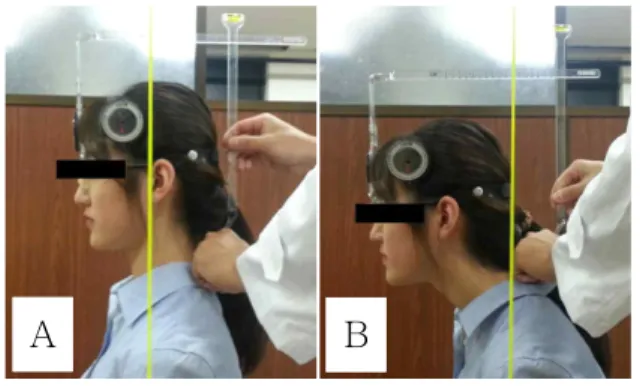
All measurements were conducted by two phys- ical therapists; one placed the patient in the meas- urement position and the other recorded maximal mouth opening (MMO) and EMG activity. After the CROM instrument was placed, the subject was seated in an upright position on a chair, arms hanging down at the sides, and feet flat on the floor. While keeping the sacrum and thoracic spine in contact with the back of the chair, subjects were asked to maintain a vertical head position. NHP was defined as the position in which the tragus of the ear and acromion were bisected by the plumb line (La Touche et al, 2011) (Figure 1A). FHP was defined as anterior cervical translation with upper cervical extension and lower cervical flexion (La Touche et al, 2011) (Figure 1B). To attain maximal protracted head position, subjects were asked to slide their jaws forward as far as they could from NHP (Table 1).
Prior to EMG data collection, subjects were asked to open their mouth as wide as possible with less cervical extension. The vertical mouth opening was
measured between the incisal edges of the upper and lower central incisors using a millimeter ruler (Higbie et al, 1999). Of the three vertical mouth opening measurements performed in NHP, the greatest value was considered the MMO (Table 2).
This was used as a reference point for EMG data collection of SH and IH muscles during mouth opening in both NHP and FHP.
After the EMG electrodes were attached, EMG signals of the SH and IH muscles were recorded while the subject performed 5-second maximum isometric resistive mandible depressions (depression of the jaw against manual resistance) to normalize EMG data to the maximal voluntary isometric con- traction (MVIC) (Juul-Kristensen et al, 2004). We used the central 3 seconds of collected EMG data to determine the mean amplitude of MVIC. The nor- malized activity of each muscle is presented as a percentage of MVIC.
EMG data were collected while the subjects maintained MMO for 5 seconds. There were three trials for each neck posture. A practice trial was allowed for each head posture so participants felt familiar with the procedure. A mirror was located in front of the subjects to provide visual feedback.
After attaining the given head position. To avoid fatigue, a 2 minutes break was provided between postures. After each mouth opening, subjects rested for 10 seconds.
Statistical analysis
Kolmogorov-Smirnov Z-tests were performed to
A B
Figure 1. Measurement of head posture with the cervical range of motion and plumb line (A: neutral head posture, B: forward head posture).
Mean±SDa Range
MMOb (㎝) 4.63±.48 1.90
amean±standard deviation,bmaximal mouth opening.
Table 2. Maximal mouth opening (N=20) Head posture (㎝) Mean±SDa Range
FHPb 6.35±1.11 4.00
amean±standard deviation, bforward head posture.
Table 1. Anterior cervical translation at rest (N=20)
assess the normality of distribution. p-values<.01 were considered statistically significant. Mean EMG activity during MMO of the bilateral SH and IH muscles were compared between NHP and FHP us- ing paired t-tests. Statistical analyses were per- formed with PASW ver. 21.0 software (SPSS Inc., Chicago, IL, USA).
Results
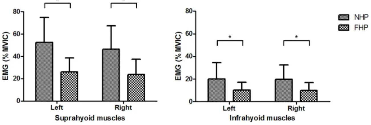
All of the continuous variables were found to ap- proximate a normal distribution (Kolmogorov-Smirnov Z-test, p>.05). There was a significant bilateral de- crease in normalized EMG activity of the SH muscle during mouth opening in FHP compared to NHP (both: p<.001). A similar significant difference was noted in the IH muscle during mouth opening (right:
p=.001, left: p=.003) (Figure 2).
Discussion
The purpose of this study was to determine the influence of FHP on EMG activity of the hyoid muscles during mouth opening. The result of this study showed that there was significantly decreased EMG activity of SH and IH muscles during mouth
opening in FHP compared to NHP. Milidonis et al (1993) investigated EMG activity of the genioglossus muscle (one of the SH muscles) between NHP and FHP when the mouth was closed. They found that genioglossus muscle activity was greater in FHP than in NHP. While the previous study investigated activity of the SH muscle at mandibular resting po- sition, our study focused on SH muscle at man- dibular depressed position. Therefore, the position of the mandible explains the difference in SH muscle activity.
In regard to mouth opening, Higbie et al (1999) found that vertical mouth opening was greater in FHP than NHP. To control for this potentially con- founding variable, we measured MMO in the NHP.
Mean MMO of this study is 4.63±.48 ㎝, which is nearly consistent with values (4.15±.48 ㎝) from a previous study. For both NHP and FHP, EMG data had collected while the subject maintained MMO which was obtained in the NHP.
There is possible explanation for the decrease in SH and IH muscles EMG during mouth opening in FHP. Based on the interrelationship of muscles in the craniocervical region, when the mouth is closed, there may be increased passive tension to the IH muscle in FHP. The stretched IH muscle could cre- ate an inferior and posterior pull on the hyoid bone.
The traction is transferred to the mandible through
Figure 2. Comparison of normalized EMG activities in NHP vs. FHP (NHP: neutral head posture, FHP:
forward head posture, EMG : electromyography, MVIC : maximal voluntary isometric contraction).
the SH muscle (Neumann, 2010). The position of the mandible was changed by different stretches of soft tissues and muscles in the cervical area. This is due to alterations of head posture caused by changes in length of the muscle fibers. In the literature, it has been demonstrated that an alteration of muscle length results in a change of EMG activity (Babault et al, 2003; Heckathorne and Childress, 1981;
Kennedy and Cresswell, 2001; Kubo et al, 2004;
Lunnen et al, 1981). Therefore, mouth opening with relatively low EMG activity in FHP compared to NHP could be indirect evidence that SH and IH muscles are lengthened in FHP.
Ohmure et al (2008) demonstrated that the man- dibular condyle is positioned more posteriorly in FHP than that in NHP. When the condyle is positioned posteriorly, an additional force might be applied to the posterior region of the TMJ. Theoretically, a posteriorly displaced condyle could compress the weak retrodiscal tissues, creating inflammation and muscle spasm. Moreover, the anterosuperior structure of the TMJ is a durable load-bearing site, while the posterior structure of the TMJ is not (Radu et al, 2004). It has been reported that posterior displace- ment of the condyle possibly might be a one of the contributing factors to TMD including TMJ disc dis- placement (Hibi and Ueda, 2005; Imai et al, 2001;
Pullinger et al, 1986). A previous study revealed that the head tended to be positioned more forward in the group with TMD than in the healthy group (Lee et al, 1995). However, it is difficult to find supporting literature that unequivocally proves this cause-and-ef- fect relationship between FHP and TMD.
This study has some limitations. First, in this study, EMG activity of the hyoid muscles was eval- uated only at the point the mouth was fully opened, rather than during the gradual mouth opening process. To assess the sequential influence of neck posture on EMG activity of the hyoid muscles ac- cording to mouth position in a more functional way, we suggest collecting EMG data through the entire mouth opening process rather than just at a fixed
position. Second, this research was performed on on- ly healthy subjects. Therefore, a further research is necessary to examined the EMG activity of patients with FHP who have had lengthened SH and IH muscles, and to evaluate the influence of the chroni- cally lengthened SH and IH muscles in patients with FHP on TMJ.
Conclusion
To examine the relationship between FHP and TMJ, SH and IH muscles which are highly sugges- tible on TMJ were selected to be investigated during mouth opening with FHP and NHP. The results of the current study showed that EMG activity of SH and IH muscles were significantly decreased during mouth opening with FHP than with NHP. This find- ing supports that FHP might affect TMJ indirectly through alteration of the hyomandibular system.
References
Armijo-Olivo S, Rappoport K, Fuentes J, et al. Head and cervical posture in patients with tempor- omandibular disorders. J Orofac Pain. 2011;25(3):
199-209.
Babault N, Pousson M, Michaut A, et al. Effect of quadriceps femoris muscle length on neural acti- vation during isometric and concentric contractions. J Appl Physiol (1985). 2003;94(3):
983-990.
Ballenberger N, von Piekartz H, Paris-Alemany A, et al. Influence of different upper cervical positions on electromyography activity of the masticatory muscles. J Manipulative Physiol Ther. 2012;35 (4):308-318. http://www.dx.doi.org/10.1016/j.jmpt.
2012.04.020
Bonney RA, Corlett EN. Head posture and loading of the cervical spine. Appl Ergon. 2002;33(5):415-417.
Catanzariti JF, Debuse T, Duquesnoy B. Chronic
neck pain and masticatory dysfunction. Joint Bone Spine. 2005;72(6):515-519.
Criswell E. Cram’s Introduction to Surface Electromyography. 2nd ed. Sudbury, Jones and Bartlett Publishers, 2010:262-265.
Ding R, Logemann JA, Larson CR, et al. The effects of taste and consistency on swallow physiology in younger and older healthy individuals: A sur- face electromyographic study. J Speech Lang Hear Res. 2003;46(4):977-989.
Dworkin SF, LeResche L. Research diagnostic cri- teria for temporomandibular disorders: Review, criteria, examinations and specifications, critique.
J Craniomandib Disord. 1992;6(14):301-355.
Forsberg CM, Hellsing E, Linder-Aronson S, et al.
EMG activity in neck and masticatory muscles in relation to extension and flexion of the head.
Eur J Orthod. 1985;7(3):177-184.
Garrett TR, Youdas JW, Madson TJ. Reliability of measuring forward head posture in a clinical setting. J Orthop Sports Phys Ther. 1993;17(3):
155-160.
Gonzalez HE, Manns A. Forward head posture: Its structural and functional influence on the sto- matognathic system, a conceptual study. Cranio.
1996;14(1):71-80.
Hackney J, Bade D, Clawson A. Relationship be- tween forward head posture and diagnosed in- ternal derangement of the temporomandibular joint. J Orofac Pain. 1993;7(4):386-390.
Heckathorne CW, Childress DS. Relationships of the surface electromyogram to the force, length, ve- locity, and contraction rate of the cineplastic human biceps. Am J Phys Med. 1981;60(1):1-19.
Hibi H, Ueda M. Body posture during sleep and disc displacement in the temporomandibular joint: A pilot study. J Oral Rehabil. 2005;32(2):85-89.
Higbie EJ, Seidel-Cobb D, Taylor LF, et al. Effect of head position on vertical mandibular opening. J Orthop Sports Phys Ther. 1999;29(2):127-130.
Huggare JA, Raustia AM. Head posture and cervico- vertebral and craniofacial morphology in patients
with craniomandibular dysfunction. Cranio. 1992;
10(3):173-177; discussion 178-179.
Imai H, Sakamoto I, Yoda T, et al. A model for in- ternal derangement and osteoarthritis of the temporomandibular joint with experimental trac- tion of the mandibular ramus in rabbit. Oral Dis. 2001;7(3):185-191.
Juul-Kristensen B, Laursen B, Pilegaard M, et al.
Physical workload during use of speech recog- nition and traditional computer input devices.
Ergonomics. 2004;47(2):119-133.
Kennedy PM, Cresswell AG. The effect of muscle length on motor-unit recruitment during iso- metric plantar flexion in humans. Exp Brain Res. 2001;137(1):58-64.
Kubo K, Tsunoda N, Kanehisa H, et al. Activation of agonist and antagonist muscles at different joint angles during maximal isometric efforts.
Eur J Appl Physiol. 2004;91(2-3):349-352.
La Touche R, París-Alemany A, von Piekartz H, et al. The influence of cranio-cervical posture on maximal mouth opening and pressure pain threshold in patients with myofascial tempor- omandibular pain disorders. Clin J Pain. 2011;27 (1):48-55. http://www.dx.doi.org/10.1097/AJP.0b01 3e3181edc157
Lee WY, Okeson JP, Lindroth J. The relationship between forward head posture and tempor- omandibular disorders. J Orofac Pain. 1995;9(2):
161-167.
Lunnen JD, Yack J, LeVeau BF. Relationship be- tween muscle length, muscle activity, and torque of the hamstring muscles. Phys Ther. 1981;61 (2):190-195.
Milidonis MK, Kraus SL, Segal RL, et al. Genioglossi muscle activity in response to changes in ante- rior/neutral head posture. Am J Orthod Dentofacial Orthop. 1993;103(1):39-44.
Muto T, Kanazawa M. Positional change of the hy- oid bone at maximal mouth opening. Oral Surg Oral Med Oral Pathol. 1994;77(5):451-455.
Neumann DA. Kinesiology of the Musculoskeletal
System: Foundations for rehabilitation. 2nd ed.
St Rouis, Mosby, 2010:431-433, 436-438, 451.
Ohmure H, Miyawaki S, Nagata J, et al. Influence of forward head posture on condylar position. J Oral Rehabil. 2008;35(11):795-800. http://www.dx.
doi.org/10.1111/j.1365-2842.2007.01834.x
Pancherz H, Winnberg A, Westesson PL. Masticatory muscle activity and hyoid bone behavior during cyclic jaw movements in man. A synchronized electromyographic and videofluorographic study.
Am J Orthod. 1986;89(2):122-131.
Pullinger AG, Solberg WK, Hollender L, et al.
Tomographic analysis of mandibular condyle po- sition in diagnostic subgroups of tempor- omandibular disorders. J Prosthet Dent. 1986;55 (6):723-729.
Radu M, Marandici M, Hottel TL. The effect of clenching on condylar position: A vector analy- sis model. J Prosthet Dent. 2004;91(2):171-179.
Rocha CP, Croci CS, Caria PH. Is there relationship between temporomandibular disorders and head and cervical posture? A systematic review. J Oral Rehabil. 2013;40(11):875-881. http://www.dx.
doi.org/10.1111/joor.12104
Thilander B, Rubio G, Pena L, et al. Prevalence of
temporomandibular dysfunction and its associa- tion with malocclusion in children and adoles- cents: An epidemiologic study related to speci- fied stages of dental development. Angle Orthod.
2002;72(2):146-154.
Thurow RC. Atlas of Orthodontic Principles. 2nd ed.
St Louis, Mosby, 1977:373-375.
Valenzuela S, Baeza M, Miralles R, et al. Laterotrusive occlusal schemes and their effect on supra- and infrahyoid electromyographic activity. Angle Orthod.
2006;76(4):585-590.
Watson DH, Trott PH. Cervical headache: An inves- tigation of natural head posture and upper cer- vical flexor muscle performance. Cephalalgia.
1993;13(4):272-284.
Winnberg A, Pancherz H, Westesson PL. Head pos- ture and hyo-mandibular function in man. A synchronized electromyographic and videofluoro- graphic study of the open-close-clench cycle.
Am J Orthod Dentofacial Orthop. 1988;94(5):
393-404.
This article was received December 8, 2014, was reviewed December 9, 2014, and was accepted January 12, 2015.