Received: November 18, 2013, Revised: November 29, 2013, Accepted: December 1, 2013 ISSN 1598-4478 (Print) / ISSN 2233-7679 (Online)
†
Correspondence to: Do-Seon Lim
Department of Dental Hygiene, College of Health Science, Eulji University, 553 Sanseong-daero, Sujeong-gu, Seongnam 461-713, Korea Tel: +82-31-740-7229, Fax: +82-31-740-7352, E-mail: [email protected]
*Moon-Jin Jeong and Ji-Jyang Lim contributed equally to this article.
Copyright © 2013 by the Korean Society of Dental Hygiene Science
Comparison of Anticariogenic Effect after Applying Fluoride Varnish on Sound and Artificial Caries Enamel
Moon-Jin Jeong*, Ji-Hyang Lim 1 *, Ji-Hye Min 1 , Soon-Jeong Jeong, Jung-Hui Son 2 and Do-Seon Lim 1†
Department of Oral Histology and Developmental Biology, School of Dentistry, Chosun University, Gwangju 501-759,
1 Department of Dental Hygiene, College of Health Science, Eulji University, Seongnam, 461-713, 2 Department of Dental Hygiene, Daewon University College, Jecheon 390-702, Korea
정상법랑질과 인공우식법랑질에 불소바니쉬 도포 후 항우식 효과 비교
정문진*ㆍ임지향
1
*ㆍ민지혜1
ㆍ정순정ㆍ손정희2
ㆍ임도선1†
조선대학교 치과대학 구강조직발생학교실,
1을지대학교 보건과학대학 치위생학과,
2대원대학교 치위생과
In order to examine the anticariogenic effect after fluoride varnish was applied to sound enamel and artificial caries enamel, anterior teeth of healthy cattle were used and divided into four groups such as group 1 (sound enamel), group 2 (application of fluoride varnish to sound enamel), group 3 (artificial caries enamel) and group 4 (application of fluoride varnish to artificial caries enamel). Remineralization on the surface of enamel and changes of crystalline structure after demineralization were observed by using a field emission scanning electron microscope (FE-SEM). Quantitative analysis of Ca and P was measured by using the energy dispersive X-ray spectrophotometer (EDS). The following conclusions were obtained:
1)Surface pattern of enamel was the roughest in group 3 due to the defects caused by porosity and microcracks. Group 4, group 1 and group 2 were followed in order; 2) It was found that pattern of crystalline structures in a group of application of fluoride varnish and a group of no application showed bigger change in artificial caries enamel than that in sound enamel. In other words, groups 4 and 1 showed a similar pattern; 3) The contents of Ca and P were higher in groups of application of fluoride varnish (group 2 and group 4) than in groups of no application of fluoride varnish (group 1 and group 3). Taken results of this study together, in the case of application of fluoride varnish, crystalline structure was changed by remineralization even in the sound enamel. In particular, porous structures showed a smooth and uniform pattern due to the recalcification in the artificial caries enamel. In addition, according to results of EDS analysis, the contents of Ca and P were increased and it had great anticariogenic effects which inhibit decalcification of sound enamel and artificial caries enamel.
Key Words: Anticariogenic effect, Energy dispersive X-ray spectrophotometer, Field emission scanning electron microscope, Fluoride varnish
Introduction
Teeth in the mouth have experienced continuous loss of minerals and re-deposition processes. If good condition for loss of minerals is formed due to the failure to clean oral hygiene, the process of dental caries begins to proceed. Because incipient caries enamel lesions known as
ʻwhite spotʼ are not easy to be clinically identified, they are often neglected. It may be developed to a deeper caries lesion. However, as incipient caries enamel lesions can be remineralized by improvement on the oral hygiene state or application of fluoride, the pathological process cannot be only prevented but it can also be restored to the original condition
1)
.Preventive mechanism for dental caries by fluoride includes the increase in the resistance of tooth structure, the promotion of remineralization, the inhibition of en- zymatic action of microorganism and the deposition of calcium fluoride by high concentration of fluoride
2-4)
. Prevention methods for dental caries using fluoride include the systemic and topical administrations. The systemic administration includes the administration of fluoride supplements and the intake of fluoride water. The topical administration includes tooth brushing with flu- oride, use of fluoride toothpaste and application of flu- oride by experts. The topical application largely consists of application by experts and self application. Marinho et al.5)
investigated and analyzed multiple studies on topical application of fluoride. They reported that average D(M) FT was lower in application by experts by 10% than that in application of fluoride at home. Thus, they claimed that application of fluoride by experts was more effective.Among applications of fluoride by experts, topical application of fluoride using APF gel is widely being used in the clinics. The preventive effect of APF gel containing 1.23% fluoride on dental caries has been proven through previous studies
6,7)
. However, upon topical application of fluoride, children may swallow fluoride, which is pointed out as the problem. The amount of swallowing fluoride varies depending on physical properties of materials con- taining fluoride, application methods and usage and age of the children8)
. In particular, in the case of infants/ toddlers or disabled children, it is highly likely to excessively swa- llow the gel, while fluoride gel is applied. Thus, children are likely to be exposed to the side effects of fluoride. In fact, some studies reported that children swallowed 14∼31 mg of fluoride upon topical application of 1.23% APF gel
9-11)
. In addition, since this APF gel is water soluble and the contact time with tooth structure is short, the effect is not sustained and it is the disadvantage. The possibilities for discomfort such as temporary nausea and vomiting as well as systemic toxicity exist due to the intake of fluoride during the application period for four minutes. It has been reported that the roughness of surface of esthetic resto- rative materials such as glass ionomer or composite resin present in the mouth is increased by the acidic APF gel and then aesthetics are influenced8,12)
. Thus, the use of nonwater soluble fluoride varnishes is proposed as one of methods for safe and effective application of fluoride.
Since Duraphat (Colgate-Palmolive, Canton, MA, USA) product was developed and sold for the first time in Europe in the late 1960s, the second generation varnishes such as Fluor Protector (Ivoclar/Vivadent, Schaan, Liech- tenstein) was developed in the early 1990s and the third generation varnishes which are individually packaged in the form of stick such as Cavity Shield (OMNII Oral Parmaceuticals, West Palm Beach, FL, USA) are recently developed and sold
13)
. It has been known that fluoride varnish has the high preventive effect for dental caries by putting the fluoride at high concentration into contact with teeth for a long period of time by combining the fluoride into the natural resin with good adhesive property. Since the method is simple and convenient, patients easily take it and it is unlikely to swallow fluoride excessively. In addition, the time for procedure is short compared to that in traditional fluoride gel or fluoride foam14)
. It has been reported that preventive effect of fluoride varnish on dental caries varies from 30% to 70%15)
. Moreover, it has been confirmed that fluoride varnish prevents the decal- cification like the initial symptom of dental caries and delays the progression of pre-existing enamel lesions throu- gh a variety of studies16-18)
.Upon looking at literature comparing the preventive effect of other fluoride materials different from fluoride varnish on dental caries, Seppa et al.
18)
conducted the study in which children at 12∼13 years old were divided into two groups and fluoride varnishes and APF gel were applied twice in a year. According to results of comparison for 3 years, it was found that there was no significant difference between two groups. In addition, approximately one hun- dred relevant research papers were reviewed in order to exa- mine the effect of topical application of fluoride into chi- ldren and adolescents. It was reported that fluoride varnish, fluoride gel, fluoride tooth brushing and fluoride too- thpaste had preventive effect for dental caries in 40%, 21%, 26% and 24%, respectively. Thus, fluoride varnish showed the highest preventive effect for dental caries5)
. Likewise, results of clinical research on effect of fluoride varnishes are quite different depending on research methods, subjects and methods to compare other fluoride materials and effects.Fluoride varnishes currently being sold in the world are Duraphat, Fluor protector, Duraflor (Pharma Science, Mon- treal, QC, Canada), Cavity Shield, Bifluoride 12 (Voco, Cuxhaven, Germany) and Carex (Voss, Norway). Fluor Protector containing 1% difluorsilane and Cavity Shield containing 5% sodium fluoride are being sold in Korea.
Compared with products of fluoride in forms of gel or rinse which are being previously used, varnish is not lost immediately after application and it remains on tooth structure for a long time. It gradually dissociates fluoride and it can be applied easily and rapidly. Moreover, since curing is made fast, chair time can be shortened. Thus, the amount of swallowing could be less
16,19)
. Such clinical ease is thought to be especially effective for very young patients, disabled children and patients who have diffi- culty to control behaviors. However, preventive method for dental caries by application of fluoride varnish is the most recently developed, but most of previous studies are using fluoride varnish only in healthy teeth without caries or with incipient caries lesions.Thus, this study observed changes in surface shape of enamel after applying fluoride varnish to sound enamel and artificial caries enamel by using a field emission scan- ning electron microscope (FE-SEM), and compared and analyzed the anticariogenic effects based on the changes in Ca and P by using the energy dispersive X-ray spec- trophotometer (EDS).
Materials and Methods
1. Materials
Among extracted bovine anterior teeth, teeth with no caries and healthy surface of enamel were selected and specimens were prepared. Fluoride materials used in this study was Cavity Shield (5% sodium fluoride/2.26% F) fluoride varnish. ‘Taliva’ (Hanlim Pharm Co. Ltd., Seoul, Korea) which was artificial saliva was used as the artificial saliva.
2. Methods
1) Preparation of specimen
Among bovine anterior teeth, teeth with healthy surface
of enamel were selected and the dental calculus was removed by using the ultrasonic scaler. After that, colo- ration was removed by performing the scaling for 30 se- conds with pumice which does not contain fluoride. After that, it was stored in 2% NaOCl solution for 24 hours and residual organic substance was removed. And then, ultrasonic cleaning was performed for 10 minutes and washed again with distilled water. They were dried. After drying them, six teeth were cut into tooth crowns and tooth roots. After that, tooth roots were removed. Approxi- mately 2 mm of tooth crowns showing the wear pattern were cut and the tooth crown was divided into four parts.
Thus, a total of 24 specimens were used. In addition, 24 teeth were randomly divided into four groups with 6 teeth for each group such as group 1 (sound enamel), group 2 (application of fluoride varnish to sound enamel), group 3 (artificial caries enamel) and group 4 (application of flu- oride varnish to artificial caries enamel). In order to pre- vent the dehydration, they were stored in triple distilled water.
2 ) Induction of artificial caries
Artificial caries solution used in the experiment was acetate buffer solution at pH 4.2 containing 2.2 mM cal- cium chloride (CaCl
2
), 2.2 mM phosphate (NaH2
PO4
) and 50 mM acetic acid according to the methods of Gon- zalez-Ede et al.20)
In order to distinguish sound enamel and artificial caries enamel, specimens of groups 1 and 2 were stored in distilled water and specimens of groups 3 and 4 were incubated in the artificial caries solution for 72 hours at 37o
C in the incubator to form artificial caries on the surface of enamel.
3) Application of fluoride varnish
Specimens with induced artificial caries were taken out and washed with sufficient amount of distilled water.
After drying them, Cavity Shield (5% sodium fluori- de/2.26% F) was applied to specimens of groups 2 and 4 according to the manufacturer's instructions. After that, the surface of specimens containing residual fluoride varnish was washed with distilled water and tooth surface was kept in a clean state.
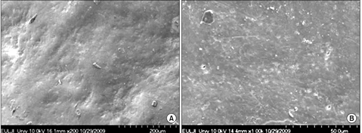
Fig. 1. Scanning electron micrograph of sound enamel surface (A, 200×; B, 1,000×).
4) Stored in artificial saliva
Each specimen was put in the container containing 50 ml of artificial saliva and stored in the incubator at 37
o
C for 72 hours. Taliva’ artificial saliva (Hanlim Pharm Co.Ltd) which was commercially available was used as the artificial saliva.
5) Observation through a FE-SEM
After exposed specimens were washed with distilled water, they were dehydrated with a graded ethanol series for 15 minutes in each step. After that, they were dried with a critical point dryer (HCP-2; Hitachi, Tokyo, Japan).
After that, they were coated with platinum in thickness of 20 nm by using the ion sputter (E-1030; Hitachi) and observed at 10 kV with a FE-SEM (S-4700; Hitachi).
6) Quantitative analysis of Ca and P
After all teeth specimens were dehydrated, the surface of subjects was coated with platinum. The contents of Ca and P were quantified in EDS with 15 kV acceleration voltage and 250 fold magnification .
Results
1. Findings of a field FE-SEM
1) Group 1
The shape of pit was partially observed at low magni-
fication on the surface of sound enamel in which fluoride varnish was not applied. Smooth and uniform structural shape was shown as the whole (Fig. 1A). Smooth pattern was shown on surface of sound enamel at high magni- fication, but some deeply indented pit was observed (Fig.
1B).
2) Group 2
The pits which were observed on sound enamel were not almost observed by remineralization on the surface of enamel in which fluoride varnish was applied to sound enamel. A very smooth pattern was shown. In addition, it was shown that visible crystalline structures containing fluoride varnish composition were scattered on the entire surface (Fig. 2).
3) Group 3
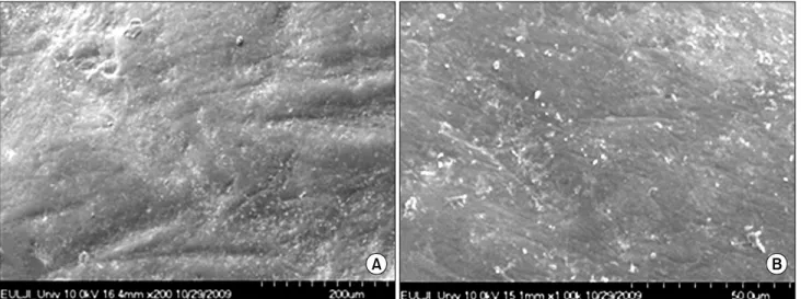
Porosity of surface was generally increased in artificial caries enamel compared with that in sound enamel. Since the surface was irregularly fractured, a very rough pattern was shown. In addition, crack area was partially observed due to microcracks and cracks (Fig. 3).
4) Group 4
In the case of low magnification, since the structures of porosity which were observed in artificial caries enamel were mostly remineralized, they were almost not observed on the surface of enamel in which fluoride varnish was
Fig. 2. Scanning electron micrograph of sound enamel surface treated with fluoride varnish (A, 200×; B, 1,000×).
Fig. 4. Scanning electron micrograph of artificial carious enamel surface treated with fluoride varnish (A, 200×; B, 1,000×).
Fig. 3. Scanning electron micrograph of artificial carious enamel surface (A, 200×; B, 1,000×).
Element Value of sound enamel (%)
Value of artificial carious enamel (%)
Weight Atomic Weight Atomic
P K +3.74 +3.87 +5.05 +4.61
Ca K +7.17 +5.37 +7.10 +4.95
Table 5. Comparative of the Ca and P Amount in Sound and Artificial Carious Enamel Surface Treated with Fluoride Varnish
Element Weight (%) Atomic (%)
C K 13.77 23.36
O K 38.95 49.61
P K 20.07 13.20
Ca K 27.20 13.83
Total 100.00
Table 4. Ca and P Amount of Artificial Carious Enamel Treated with Fluoride Varnish (Group 4)
Element Weight (%) Atomic (%)
C K 29.08 42.89
O K 35.80 39.64
P K 15.02 8.59
Ca K 20.10 8.88
Total 100.00
Table 3. Ca and P Amount of Artificial Carious Enamel (Group 3)
Element Weight (%) Atomic (%)
C K 14.36 27.16
O K 23.76 33.74
P K 24.09 17.68
Ca K 37.79 21.43
Total 100.00
Table 2. Ca and P Amount of Sound Enamel Treated with Fluoride Varnish (Group 2)
Element Weight (%) Atomic (%)
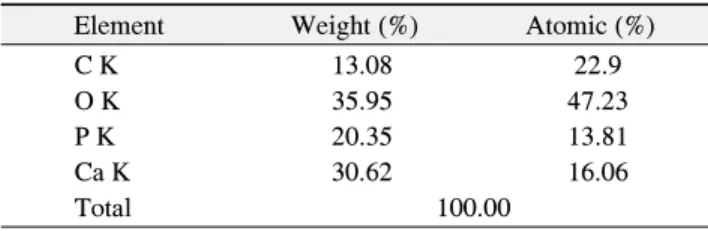
C K 13.08 22.9
O K 35.95 47.23
P K 20.35 13.81
Ca K 30.62 16.06
Total 100.000
Table 1. Ca and P Amount of Sound Enamel (Group 1)
applied to artificial caries enamel. Smooth and universal pattern was represented similar to that on the surface of sound enamel (Fig. 4A). However, a smooth pattern was shown in general through high magnification observation, but the pits caused by artificial caries were partially shown (Fig. 4B).
2. Quantitative analysis of Ca and P using EDS
1) Group 1
The amount of Ca per unit area on the surface of sound enamel was 30.62% and the amount of P was 20.35%
(Table 1).
2) Group 2
In the case of application of fluoride varnish to sound enamel, the amount of Ca per unit area on the surface of enamel was 37.79% and the amount of P was 24.09%
(Table 2).
3) Group 3
The amount of Ca per unit area on the surface of artificial caries enamel was 20.10% and the amount of P was 15.02% (Table 3).
4) Group 4
In the case of application of fluoride varnish to artificial caries enamel, the amount of Ca per unit area on the
surface of enamel was 27.20% and the amount of P was 20.07% (Table 4).
5) Comparison of amounts of Ca and P in sound enamel and artificial caries enamel by application of fluo- ride varnish
The amounts of Ca and P per unit area on the surface of sound enamel with application of fluoride varnish were increased by 7.17% and 3.74%, respectively, compared with those in sound enamel. In addition, the amounts of Ca and P per unit area on the surface of artificial caries enamel with application of fluoride varnish were increased by 7.10% and 5.05%, respectively, compared with those in artificial caries enamel (Table 5).
Discussion
Methods to prevent the dental caries include plaque control, dietary control, pit and fissure sealant and the use of fluoride. Among them, the use of fluoride is divided into the systemic use such as water fluoridation and
administration of fluoride supplements and the topical use such as use of fluoride toothpaste, tooth brushing with fluoride solution and application of fluoride by experts
21)
.It has been known that fluoride can prevent the tooth caries by increasing the hardness of the surface of the teeth and the acid resistance of tooth structures or promoting the remineralization of demineralized tooth structures as it is deposited on the tooth structures in the mouth
3,22)
. In addition, since it inhibits the actions of enzymes involved in metabolism of bacteria inducing the dental caries at high concentration, the production of acid and formation of adhesive polysaccharide of bacteria are inhibited23)
. Among the use of fluoride in various forms, Maia et al.24)
reported that the absorption of fluoride was noticeable and the increase in the surface strength of teeth was the highest when fluoride toothpaste was used. White25)
recommen- ded that the use of fluoride toothpaste several times in a day was the most simple and excellent method to obtain the topical application effect. In clinical practice, the scope of the selection of products is wide, and the fluoride toothpaste and fluoride brushing solution without additi- onal prescription is frequently used. However, since these methods require cooperation of patients in most cases, it is often difficult to obtain the sufficient performance when they are used for children or adolescents who have less cooperation. Thus, the method to get the long-term effect with one time application regardless of the cooperation of patients is required. Fluoride varnish can be considered as one of materials close to it26)
.Fluoride varnish was developed about 30 years ago and it began to be widely used in Europe in the 1980s. It was approved as hypersensitivity treatment agent and cavity liner by Food and Drug Administration in the United States in 1994
27)
. However, fluoride varnish is widely used for the prevention of enamel demineralization as the topi- cal fluoride application agent13,28)
. In particular, fluoride varnish is believed to have excellent preventive effect for the dental caries since it is easily attached on the surface of teeth and it continues to dissociate fluoride compared to other fluoride materials29)
.It is important to have a small amount of fluoride in the mouth all the time in terms of prevention of the dental caries by fluoride. In particular, fluoride present in the oral
biofilm and saliva is involved in the process of enamel remineralization if the acid is formed and acidity is decreased in the mouth
30)
. Twetman et al.31)
measured the concentration of fluoride in saliva after 3 kinds of fluoride varnishes were applied. After 1 hour, the concentration of fluoride was increased in groups of application of Biflu- orid (6% F) and Duraphat (2.26% F), but it was not significantly increased in a group of application of Fluor Protector (0.1% F). Skold-Larsson et al.32)
applied the same fluoride varnish and measured the concentration of fluoride in dental plaque. In groups of application of Bifluorid (6% F) and Duraphat (2.26% F), the concen- tration of fluoride was significantly increased in 3 and 7 days and 3 days, respectively. However, it was not signi- ficantly increased in a group of Fluor Protector (0.1% F).According to results of two studies, it was shown that the concentration of fluoride was increased in saliva and oral biofilm with increasing concentration of fluoride varnish.
In addition, it was observed that the concentration of fluoride continued to be increased for a long period of time in groups of Bifluorid and Fluor Protector.
The purpose of this study is to examine the difference in the anticariogenic effect when fluoride material used in this study is Cavity Shied (5% sodium fluoride/2.26% F) which is commercially available is applied to sound ena- mel and artificial caries enamel. There are several methods to compare the anticariogenic effect and the reminerali- zation effect such as the method to directly measure the changes of crystalline structure and trace atoms in the teeth structures by using a FE-SEM and methods to ob- serve the pattern of caries lesions and indirectly obtain the changes in minerals by using the Vickers Hardness Num- ber. However, a FE-SEM is used in this experiment.
As the results of this study, the surface pattern of enamel was porous and irregularly fractured. It was observed that it was the roughest in a group of artificial caries enamel with partial defect caused by microcracks and cracks. A group of application of fluoride varnish to artificial caries enamel, a group of sound enamel and a group of application of fluoride varnish to sound enamel were followed in the order. Son
33)
compared the changes in surface of caries lesions based on application of fluoride through observation of a FE-SEM. In the artificial cariesenamel, the porosity of surface was generally increased and the surface was irregularly fractured. Thus, rough pattern was shown and defect parts such as microcracks were frequently observed. In the case of artificial caries enamel treated with fluoride varnish, rough and irregularly fractured pattern was relatively decreased compared to that before the treatment. This result was similar to that. In addition, the pattern of crystalline structure in groups of application of fluoride varnish and no application was significantly changed in artificial caries enamel compared to that in sound enamel. In other words, a group of application of fluoride varnish to artificial caries enamel showed a similar pattern in the sound enamel. Lee
34)
conducted the research on surface change of enamel by application of fluorides and showed that the density of surface structure was increased by deposition of calcified materials. It was consistent with results of this study.EDS is the device attached to the electron microscope used for quantitative and qualitative analysis for ingre- dients composed of materials. In particular, it is a very useful method to elucidate the contents of Ca and P which has high atomic weight composed of hard tissue. In this study, as the results of quantitative analysis of Ca and P contained in enamel by using the EDS, the contents were measured higher in a group of sound enamel than those in a group of artificial caries enamel. It was measured higher in a group of application of fluoride varnish than that in a group of no application of fluoride varnish. In addition, the increased amount of Ca and P after application of fluoride varnish was slightly higher in the artificial caries enamel than that in sound enamel. The result of this study showed that the amount of absorption of fluoride calculated by the enamel biopsy method was the highest in a group of application of fluoride varnish. It was consistent with results of other studies. Kidd et al.
35)
, Wei36)
and Tveit et al.37)
reported that absorption of fluoride was dependent on the time in contact with the surface of teeth and the reactivity of enamel. In addition, Lee et al.38)
studied the anticariogenic effect of fluoride varnish and chlorhexidine varnish and its result was similar to that in this study.Since it is thought that fluoride is dissociated and fluro- apatite which has higher acid resistance than hydroxy- apatite is formed and affects the histological crystalline
structure of enamel surface in a group of application of fluoride varnish, it takes anticariogenic action and plays a role to increase the resistance against caries. In addition, it is thought to be caused by higher reactivity on the surface of artificial caries enamel than that of sound enamel. The results are consistent with those of Hicks and Silver- stone
39,40)
. The reason why the surface of enamel treated with anti-corrosion is restored to the original state by saliva or fluoride within a short period of time in the clinics could be such reactions on the surface.Taken results of this study together, in the case of application of fluoride varnish, crystalline structure was changed by remineralization even in the sound enamel. In particular, porous structures showed a smooth and uniform pattern due to the recalcification in the artificial caries enamel. In addition, according to results of EDS analysis, the contents of Ca and P were increased and it had great anticariogenic effects which inhibit the decalcification of sound enamel and artificial caries enamel.
Summary
In order to examine the anticariogenic effect after flu- oride varnish was applied to sound enamel and artificial caries enamel, anterior teeth of healthy cattle were used and divided into four groups. Remineralization on surface of enamel and changes of crystalline structure after demineralization were observed by using a FE-SEM.
Quantitative analysis of Ca and P was measured by using EDS. As the results, in the case of application of fluoride varnish, irregular and very rough surface and porous structure were shown in a smooth and uniform pattern due to the remineralization. It was found that the increase in contents of Ca and P greatly promoted the anticariogenic effect of sound enamel and artificial caries enamel.
요 약
정상법랑질과 인공우식법랑질에서 불소바니쉬 도포 후 의 항우식 효과를 알아보기 위하여 건전한 소의 전치를 사 용하였고, 네 군으로 분류하여 실험하였다. 법랑질 표면의 재광화 및 탈회 후 결정구조의 변화를 FE-SEM으로 관찰하 였고, Ca과 P의 정량적 분석은 EDS를 사용하여 측정하였
다. 그 결과 불소바니쉬 도포한 경우, 재석회화로 인하여 불 규칙하고 매우 거친 표면과 다공성의 구조물이 평활하고 균 일한 양상으로 나타났으며, Ca 및 P의 함량이 증가되어 정 상법랑질과 인공우식법랑질의 항우식 효과를 크게 증진시 키는 것으로 확인되었다.
Acknowledgements
This study was supported by research fund from Chosun University, 2013.