For many years patients with medial compartment osteoarthritis and varus deformity have been successfully treated with high tibial osteotomy (HTO).1) Recently, opening-wedge osteotomy has been mentioned to be more advantageous than closing wedge technique.2) Open wedge HTO has been more popular and with good results due the development of locking plate.3)
However the disadvantage of open wedge HTO is the empty space on the proximal tibia which is needed to be filled in with a spacer to gain union (autograft, allograft, etc.).4) Recently, hydroxy- apatite (HAp) and beta-tricalcium phosphate (TCP) blocks are be- ing used to fill the vacant space and through studies, TCP ceramics were shown to have superior absorption rate and osteoconductivity.1) The authors report two patients who had a non-union at the os- teotomy site after the HTO using a TCP block.
CASE REPORT
1. Case 1
A 53-year-old woman was admitted due to left knee pain. The range of knee motion was 0o to 115o and medial joint line tenderness was positive.
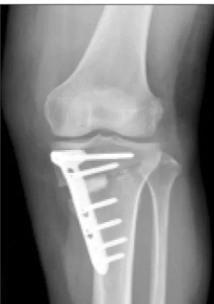
The patient received arthroscopic partial meniscectomy for me- dial meniscus posterior root tear, and arthroscopic chondroplasty for medial femoral condyle chondral lesion. Correction of 11o was made in order to move weight bearing line to 62% lateral of the proximal tibia. To achieve 11o correction, 13 mm is required, which is the os- teotomy site length (6.2 cm) multiplied by the tangent 11o. Opening- wegde HTO with TOMOFIX (Synthes, Oberdorf, Switzerland), and 13 mm TCP block (Synthes) insertion was performed (Fig. 1).
For the initial 6 weeks, partial weight bearing with crutch was
Copyright © 2015 by The Korean Orthopaedic Association
“This is an Open Access article distributed under the terms of the Creative Commons Attribution Non-Commercial License (http://creativecommons.org/licenses/by-nc/4.0/) which permits unrestricted non-commercial use, distribution, and reproduction in any medium, provided the original work is properly cited.”
The Journal of the Korean Orthopaedic Association Volume 50 Number 5 2015 Received April 6, 2015 Revised May 5, 2015 Accepted July 3, 2015
Correspondence to: Ju Seon Jeong, M.D.
Department of Orthopedic Surgery, Bumin Hospital, 59 Mandeok-daero, Buk-gu, Busan 46555, Korea
TEL: +82-51-330-3082 FAX: +82-51-330-5041 E-mail: [email protected]
Interference of Union after the Use of Beta-Tricalcium Phosphate Block in High Tibial Osteotomy
Man Seok Ko, M.D., Ju Seon Jeong, M.D. , and Dong Wook Jung, M.D.
Department of Orthopedic Surgery, Bumin General Hospital, Busan, Korea
High tibial osteotomy (HTO) is a commonly used treatment for genu varum and medial compartment osteoarthritis. Recently open wedge HTO has been the preferred method due to its facilitated technique, fewer neurovascular and joint injuries, etc. In open wedge HTO materials such as autogenous, allogenous bone graft and tricalcium phosphate (TCP) are used to help with bone union and have a role in filling in the empty space. However the authors of this study report on two cases of nonunion 1 year after HTO using TCP block.
Key words: high tibial osteotomy, tricalcium phosphate, nonunion
Figure 1. Open wedge high tibial osteotomy performed using a 13 mm tricalcium phospate block with locking plate.
425
Interference of Union after the Use of β-Tricalcium Phosphate Block in High Tibial Osteotomy
permitted and range of motion exercise was performed. After 6 weeks, tolerable weight bearing was allowed and muscle strength- ening exercise was done.
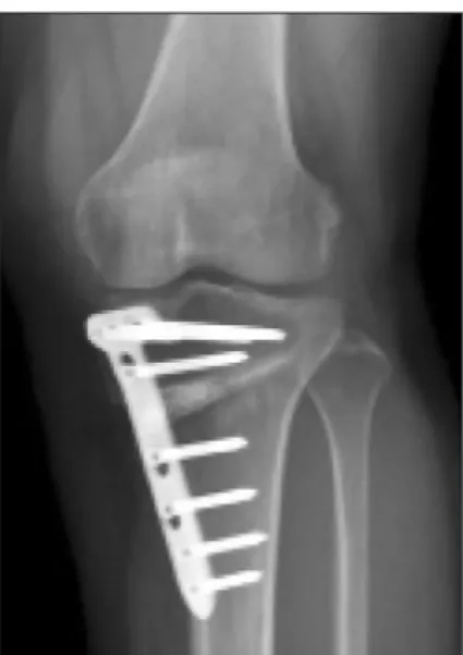
At 12 months after surgery, on the plain radiograph, nonunion of the wedge was seen (Fig. 2). Implant removal was performed. Most of the TCP blocks were nonabsorbed. Remaining TCP was re- moved, and then a 3×2 cm vacant space was observed intraopera- tively (Fig. 3). The space due to nonunion was untouched because the good union state of the other portions.
At 3 months after implant removal and anteroposterior radio- graph also showed a radiolucent space with a well maintained cor-
rection angle (Fig. 4).
2. Case 2
A 55-year-old woman was admitted due to right knee pain. The range of knee motion was 14o to 98o and medial joint line tenderness was positive. The patient had a varus angle of 8.1o and Kellgren- Lawrence grade 2 OA change.
The patient received arthroscopic partial menisectomy for me- dial meniscus tear, arthroscopic microfracture for medial femoral condyle chondral lesion, and HTO with TOMOFIX (Synthes), TCP block (10 mm) for genu varum (Fig. 5).
Figure 3. An intraoperative image of the nonunion shown in the wedge space.
Figure 2. Nonunion around the edges of the tricalcium phospate block after 1 year was shown in a simple radiograph.
Figure 5. An anteroposterior radiograph at 3 months after implant re- moval was checked and the well maintained space was observed.
Figure 4. An anteroposterior radiograph at 3 months after implant removal was checked and the well maintained space was observed.
At 12 months, radiolucency was seen around the spacer on the plain radiograph (Fig. 6). Implant removal was done and TCP from the nonunion site was removed. The space due to nonunion was untouched because the good union state of the other portions.
At 3 months after implant removal, standing anteroposterior radiograph also showed a radiolucent space and a well maintained angle (Fig. 7).
DISCUSSION
HTO is a procedure which is performed in patients with medial compartment degenerative disease, osteoarthritis, and varus defor- mity which delay the time until TKA is needed. Many studies have shown good results of HTO due to the development of TOMOFIX (Synthes).2,4-6) Open wedge HTO showed better results than closed wedge HTO.2)
The advantages of open wedge HTO includes less risk of pero- neal nerve injury and less instability of lateral knee ligament, facili- tated technique, maintenance of bone stock, and better correction achievement.1) It has brought satisfactory clinical results for patients with medial unicompartmental osteoarthritis.7)
The disadvantages include the need for filling in the free space after osteotomy (autogenous bone graft causing donor site pain), de- layed union and nonunion.2,8,9) TCP and HAp grafts are used to help with the union process and many studies have shown the efficacy of them both with TCP being a better choice.6) Onodera et al.6) reported that TCP had a higher osteoconductivity and greater absorbability than HAp spacer.
Saragaglia et al.10) reported the outcome of opening wedge HTO augmented with a Biosorb® (TCP) wedge in 124 patients with a mean of 10 years follow-up. There were only 7 knees with delayed union which required deferring weight-bearing. They reported the Biosorb® wedge to be completely integrated in all cases.
Present study demonstrated the 2 cases out of 38 cases (5.2%) from March 2012 to March 2014 of nonunion with the use of TCP block. Both patients are nonsmokers, and did not have a history of steroid use. Other complications, excluding nonunion of two cases, did not occur.
In both cases, correction angle was maintained at 3 months after implant removal. The reason why the angle was well maintained was because the area without TCP block showed good union.
The authors of this study reports that the use of TCP block in HTO procedure may interfere with the process of union. Therefore caution is thought to be needed when choosing the spacers when performing HTO.
CONFLICTS OF INTEREST
The authors have nothing to disclose.
REFERENCES
1. Tanaka T, Kumagae Y, Chazono M, Kitasato S, Kakuta A, Marumo K. A novel evaluation system to monitor bone for- mation and β-tricalcium phosphate resorption in opening wedge high tibial osteotomy. Knee Surg Sports Traumatol Figure 7. At 3 months after implant removal, a standing anteroposterior radiograph also showed a radiolucent space and well maintained angle.
Figure 6. An anteroposterior radiograph before implant removal was performed.
427
Interference of Union after the Use of β-Tricalcium Phosphate Block in High Tibial Osteotomy
Arthrosc. 2015;23:2007-11.
2. Poignard A, Flouzat Lachaniette CH, Amzallag J, Hernigou P. Revisiting high tibial osteotomy: fifty years of experience with the opening-wedge technique. J Bone Joint Surg Am.
2010;92 Suppl 2:187-95.
3. Niemeyer P, Koestler W, Kaehny C, et al. Two-year results of open-wedge high tibial osteotomy with fixation by medial plate fixator for medial compartment arthritis with varus malalignment of the knee. Arthroscopy. 2008;24:796-804.
4. Gouin F, Yaouanc F, Waast D, Melchior B, Delecrin J, Passuti N. Open wedge high tibial osteotomies: calcium-phosphate ceramic spacer versus autologous bonegraft. Orthop Trauma- tol Surg Res. 2010;96:637-45.
5. Floerkemeier S, Staubli AE, Schroeter S, Goldhahn S, Loben- hoffer P. Outcome after high tibial open-wedge osteotomy:
a retrospective evaluation of 533 patients. Knee Surg Sports Traumatol Arthrosc. 2013;21:170-80.
6. Onodera J, Kondo E, Omizu N, Ueda D, Yagi T, Yasuda K.
Beta-tricalcium phosphate shows superior absorption rate and osteoconductivity compared to hydroxyapatite in open-
wedge high tibial osteotomy. Knee Surg Sports Traumatol Arthrosc. 2014;22:2763-70.
7. Lee YS, Moon GH. Comparative analysis of osteotomy accu- racy between the conventional and devised technique using a protective cutting system in medial open-wedge high tibial osteotomy. J Orthop Sci. 2015;20:129-36.
8. van Houten AH, Heesterbeek PJ, van Heerwaarden RJ, van Tienen TG, Wymenga AB. Medial open wedge high tibial os- teotomy: can delayed or nonunion be predicted? Clin Orthop Relat Res. 2014;472:1217-23.
9. Meidinger G, Imhoff AB, Paul J, Kirchhoff C, Sauerschnig M, Hinterwimmer S. May smokers and overweight patients be treated with a medial open-wedge HTO? Risk factors for non-union. Knee Surg Sports Traumatol Arthrosc. 2011;
19:333-9.
10. Saragaglia D, Blaysat M, Inman D, Mercier N. Outcome of opening wedge high tibial osteotomy augmented with a Bio- sorb® wedge and fixed with a plate and screws in 124 patients with a mean of ten years follow-up. Int Orthop. 2011;35:1151-6.
근위 경골 절골술 시에 인산삼석회 블럭을 사용 후 발생한 불유합
고만석• 정주선 • 정동욱
부산부민병원 정형외과
근위 경골 절골술은 최근 내반슬과 내측 구획 골관절염 치료에 흔히 시행되고 있는 치료 방법이다. 특히 열린 쐐기 근위 경골 절골술 은 수술 술기가 쉽고, 신경과 혈관 손상이 적으며, 관절의 손상이 적어 자주 시행되고 있다. 열린 쐐기 근위 경골 절골술의 시행에 있 어 골유합과 빈공간을 채우기 위하여 자가골 이식, 동종골 이식, 인산삼석회 같은 물질이 종종 쓰인다. 하지만 이 논문의 저자들은 인 산삼석회 블럭을 이용한 근위경 골 절골술을 시행한 1년 후 발생한 불유합 2예를 경험하였기에 이를 문헌 고찰과 함께 보고하는 바이 다.
색인단어: 근위 경골 절골술, 인산삼석회, 불유합
접수일 2015년 4월 6일 수정일 2015년 5월 5일 게재확정일 2015년 7월 3일 책임저자 정주선
46555, 부산시 북구 만덕대로 59, 부산부민병원 정형외과
TEL 051-330-3082, FAX 051-330-5041, E-mail [email protected]
Copyright © 2015 by The Korean Orthopaedic Association
“This is an Open Access article distributed under the terms of the Creative Commons Attribution Non-Commercial License (http://creativecommons.org/licenses/by-nc/4.0/) which permits unrestricted non-commercial use, distribution, and reproduction in any medium, provided the original work is properly cited.”
대한정형외과학회지:제 50권 제 5호 2015