Copyright © 2018 Korean Neurotraumatology Society 39
Introduction
Vertebral collapse after a trivial injury in elderly patients with severe osteoporosis, associated with severe back pain and vertebral fractures, is a common clinical problem.
Although osteoporosis is the most common cause of ver- tebral fractures in elderly patients, the spine is also one of the most common sites of metastatic disease, accounting for up to 39% of all bony metastasis.1,5)
Magnetic resonance (MR) imaging is helpful in determin- ing the exact cause of vertebral collapse using morphologic and signal intensity criteria.
However, in the clinical setting, malignant spine metasta- sis can be difficult to differentiate from acute osteoporotic compression fracture, especially in cases with an accompa- nying trivial injury. Here, we present a confusing case of ma- lignant metastasis misdiagnosed as acute osteoporotic com- pression fracture.
Case Report
A 78-year-old man was referred to our emergency depart- ment for the evaluation of low back pain and progressive weakness of the left lower limb. The patient had undergone percutaneous vertebroplasty at the L3 level with the diag- nosis osteoporotic compression fracture 4 months earlier after a minor fall, and he had received a foraminotomy at the L3-4 level for foraminal stenosis 2 months before ad- mission.
Neurological examination showed motor weakness of left knee extension, grade III. Routine hematological and se- rum chemistry tests, including erythrocyte sedimentation rate and C-reactive protein, yielded normal results.
Malignant Metastasis Misdiagnosed as Osteoporotic Compression Fracture: A Case Report
Seul Gi Kim, Chang Il Ju, Hui Sun Wang, and Seok Won Kim
Department of Neurosurgery, College of Medicine, Chosun University, Gwangju, Korea
In cases of vertebral collapse after a trivial injury in elderly patients with severe osteoporosis, it can be a diagnostic chal- lenge to determine whether the cause is a benign compression fracture or malignant metastasis. A 78-year-old male pa- tient was referred to the emergency department for the evaluation of weakness of the left lower limb. He had undergone percutaneous vertebroplasty four months earlier after being diagnosed with L3 osteoporotic compression fracture. He was treated with foraminotomy at the L3-4 level after being diagnosed with foraminal stenosis two months earlier at a spine clinic. Magnetic resonance (MR) images showed significant signal change from the vertebral body to the posterior ele- ment, and widely spreading extraspinal extension of soft tissue at L3. Computed tomography scan revealed osteolytic changes in regions including the ventral body and pedicle. Emergent decompressive laminectomy and bone biopsy were performed, and the histologic evaluation showed metastatic squamous cell carcinoma. A retrospective review of previous MR images showed obvious pedicle and facet involvement, and paraspinal extension of soft tissue, which are highly sug- gestive of malignant metastasis.
(Korean J Neurotrauma 2018;14(1):39-42) KEY WORDS: Fracture ㆍMetastasis ㆍOsteoporosis.
Received: November 22, 2017 / Revised: March 22, 2018 Accepted: March 31, 2018
Address for correspondence: Seok Won Kim
Department of Neurosurgery, College of Medicine, Chosun Univer- sity, 365 Pilmun-daero, Dong-gu, Gwangju 61453, Korea Tel: +82-62-220-3126, Fax: +82-62-227-4575
E-mail: [email protected]
cc This is an Open Access article distributed under the terms of Cre- ative Attributions Non-Commercial License (http://creativecommons.
org/licenses/by-nc/4.0/) which permits unrestricted noncommercial use, distribution, and reproduction in any medium, provided the original work is properly cited.
CASE REPORT
Korean J Neurotrauma 2018;14(1):39-42
pISSN 2234-8999 / eISSN 2288-2243 https://doi.org/10.13004/kjnt.2018.14.1.39
40 Korean J Neurotrauma 2018;14(1):39-42
Malignant Metastasis Misdiagnosed as Osteoporotic Compression Fracture
Gadolinium-enhanced MR images taken on the day of transfer showed increased signal intensity from the verte- bral body to the posterior element (Figure 1A and B). An enhanced soft tissue mass in the left side of the L3 vertebral body was also observed. Computed tomography scan tak- en in the emergency department showed significant osteo- lytic change from the body to the pedicle (Figure 1C and D).
On the basis of the findings of typical pathologic com- pression fracture, emergent decompressive laminectomy of L3 and bone biopsy were performed including spinous pro- cess and vertebral body via left side pedicle. Histological
evaluation confirmed metastatic squamous cell carcinoma.
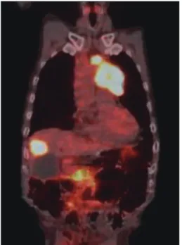
The patient was transferred to the hemato-oncological de- partment for adjuvant treatment and further diagnosis. He was diagnosed as having squamous cell carcinoma of the lung on the percutaneous biopsy with hepatic and spine me- tastasis (Figure 2).
In the retrospective review of MR images taken 2 months (Figure 3) and 4 months (Figure 4) previously, obvious fac- et and pedicle involvement, and paraspinal soft tissue ex- tension were noted. We believe that the doctors in the spine clinic should have suspected malignant metastasis and they should have investigated the radiological images more carefully during the patient’s first visit.
FIGURE 1. Magnetic resonance imaging and computed tomography (CT) scans at the time of admission. (A, B) The contrast-en- hanced T1-weighted sagittal and axial images of the L3 vertebral body and posterior element reveal an enhanced lesion in the ex- traspinal space, epidural space, and paravertebral muscle. (C, D) Sagittal and coronal CT scans show osteolytic change from the vertebral body to the pedicle of the L3 vertebral body, indicating the possibility of malignancy.
A B C D
FIGURE 2. Positron emission tomography-computed tomogra- phy scan showing hypermetabolic lesions in the left upper lung, L3 vertebrae, and right hepatic lobe.
FIGURE 3. (A, B) Magnetic resonance images taken 2 months before admission. T1-weighted sagittal and axial images show low signal intensity extending posteriorly to the articular facet joints and a paraspinal lesion with low signal intensity involving the vertebral body, pedicle, transverse process, and lamina.
A B
Seul Gi Kim, et al.
http://www.kjnt.org 41
Discussion
Differentiation between benign osteoporotic compression fractures and malignant pathologic fractures is clinically important, particularly in elderly patients with severe osteo- porosis; however, it can be difficult.
Although several recent studies have applied new MR techniques in assessing vertebral collapse, routine evaluation of vertebral collapse commonly uses conventional spin-echo MR imaging.2,7,9)
Benign osteoporotic compression fracture is a common disease in elderly patients. Conventional spin-echo MR im- aging findings suggestive of acute osteoporotic compression fracture include low-signal-intensity band on T1-weighted imaging (WI) and T2-WI, spared normal bone marrow sig- nal intensity of the vertebral body, retropulsion of a poste- rior bone fragment, and multiple compression fractures.3)
On the other hand, MR imaging findings suggestive of metastatic pathologic fracture include a convex posterior border of the vertebral body, abnormal signal intensity of the pedicle or posterior element, epidural mass, encasing epi- dural mass, focal paraspinal mass, and other spinal metas- tases.6)
Among the various findings suggesting malignant patho- logic fractures or benign compression fracture, pedicle in- volvement is controversial. Yuh et al.8) compared the conven- tional spin-echo MR imaging findings between 84 benign fractures and 25 malignant fractures on T1-WI and T2-WI.
They reported pedicle involvement in 22 of the 25 patients with malignant compression fractures but in none of 52 nontraumatic compression fractures. Therefore, involve- ment of the pedicle is an important clue for the diagnosis of
malignant pathologic fractures.
However, Ishiyama et al.4) reported that pedicle involve- ment, which had been accepted as a common indicator of malignant processes, was also frequent in patients with os- teoporotic compression fractures, particularly in the early phase, and was not specific for malignancy on new MR techniques, such as short inversion time inversion recovery (STIR) and gadolinium-enhanced T1-WI.
In this case, some clinical and MR imaging data may have misled the doctors of the local spine clinic to the diagnosis of benign compression fracture at L3, including the clini- cal history of minor trauma and severe osteoporosis based on bone mineral densitometry. Moreover, they performed conventional MR examination, as most spine doctors do.
In the retrospective review of T1- and T2-weighted MR imaging findings, it was determined that the obvious left pedicle involvement in the image taken 4 months previous- ly and the aggravation of the pedicle and facet with paraspi- nal soft tissue mass at L3 in the image taken 2 months ear- lier were highly suggestive of malignancy.
The spine clinicians had better to suspect malignant me- tastasis from the beginning, in case of pedicle involvement on T1, T2 and fat suppression view although they did not perform STIR or gadolinium-enhanced MR imaging.
Conclusion
Although differentiating between benign compression fractures and malignant pathologic fractures is possible in most cases, a diagnosis of malignant pathologic fracture should be assumed when there is an obvious pedicle in- volvement. In case of ambiguity, further tests should be performed for an exact diagnosis, including bone biopsy or short-term MR imaging follow-up.
■ The authors have no financial conflicts of interest.
REFERENCES
1) Baker LL, Goodman SB, Perkash I, Lane B, Enzmann DR. Be- nign versus pathologic compression fractures of vertebral bod- ies: assessment with conventional spin-echo, chemical-shift, and STIR MR imaging. Radiology 174:495-502, 1990
2) Chen WT, Shih TT, Chen RC, Lo HY, Chou CT, Lee JM, et al.
Blood perfusion of vertebral lesions evaluated with gadolinium- enhanced dynamic MRI: in comparison with compression frac- ture and metastasis. J Magn Reson Imaging 15:308-314, 2002 3) Cuénod CA, Laredo JD, Chevret S, Hamze B, Naouri JF, Chapaux
X, et al. Acute vertebral collapse due to osteoporosis or malignan- cy: appearance on unenhanced and gadolinium-enhanced MR im- ages. Radiology 199:541-549, 1996
4) Ishiyama M, Fuwa S, Numaguchi Y, Kobayashi N, Saida Y. Ped- icle involvement on MR imaging is common in osteoporotic com- pression fractures. AJNR Am J Neuroradiol 31:668-673, 2010 FIGURE 4. (A, B) Magnetic resonance images taken 4 months
before admission. The T1-weighted sagittal and axial images already show a low-signal-intensity area with a clear border on the body of L3 vertebrae. The low signal change was already spreading to the pedicle at that time (white arrow).
A B
42 Korean J Neurotrauma 2018;14(1):39-42
Malignant Metastasis Misdiagnosed as Osteoporotic Compression Fracture
5) Jeong JH, Roh SW, Rhim SC, Jeon SR. Effectiveness of surgical decompression and fixation in patients with metastatic spinal cord compression. Korean J Spine 4:210-214, 2007
6) Jung HS, Jee WH, McCauley TR, Ha KY, Choi KH. Discrimina- tion of metastatic from acute osteoporotic compression spinal frac- tures with MR imaging. Radiographics 23:179-187, 2003 7) Spuentrup E, Buecker A, Adam G, van Vaals JJ, Guenther RW. Dif-
fusion-weighted MR imaging for differentiation of benign frac- ture edema and tumor infiltration of the vertebral body. AJR Am J
Roentgenol 176:351-358, 2001
8) Yuh WT, Zachar CK, Barloon TJ, Sato Y, Sickels WJ, Hawes DR.
Vertebral compression fractures: distinction between benign and malignant causes with MR imaging. Radiology 172:215-218, 1989 9) Zampa V, Cosottini M, Michelassi C, Ortori S, Bruschini L, Bar-
tolozzi C. Value of opposed-phase gradient-echo technique in dis- tinguishing between benign and malignant vertebral lesions. Eur Radiol 12:1811-1818, 2002