www.jkfas.org pISSN 1738-3757 eISSN 2288-8551 J Korean Foot Ankle Soc 2015;19(4):197-200 http://dx.doi.org/10.14193/jkfas.2015.19.4.197
절면에 봉합사를 통과시키는 술식의 경우에는 관절 연골 손상의 가능성이 있을 것으로 생각된다.
이에 술자는 Akin 절골술을 비흡수성 봉합사로 고정하는 데 있 어 관절면을 지나지 않고 피질골 파괴가 적은 새로운 술식을 보고 하고자 한다.
술 기
외측 인대 유리술과 변형 마우 절골술을 이용한 중족골 절골술 을 시행하였고 발의 중립 자세에서 잔여 외반각과 회내 변형 정도 를 확인하였다. 무지 근위 지골 기저부의 약 5∼6 mm 원위부에 서 제 1중족지관절과 평행하도록 전동 톱을 이용하여 내측에서 외 측으로 절골을 시작하여 외측 피질골 1∼2 mm를 남겨 놓았다. 잔 여 외반각의 정도에 따라 2∼5 mm 크기로 내측 폐쇄성 쐐기 절골 술을 시행하였다(Fig. 1). 무지 외반의 교정을 확인하고 무지의 전 면에서 회내 변형 정도를 다시 확인한다. 1.6 mm 두께의 K-강선 을 이용해 절골면에서 근위 약 3 mm 위치에서 근위 족지골 내측 무지외반증의 수술적 치료에서 근위 지골 근위부의 쐐기 절골술
만 단독으로 시행하는 경우는 드물며 대개 중족골의 절골술과 병 행한다. Akin 절골술 부위를 별다른 고정 없이 두는 경우도 있으나 주로 K-강선(Kirschner wire)을 이용하여 고정을 한다. 그 외 금속 나사못, J-강선 술식, 금속 스테이플, PLLA (poly-L-lactic acid) 스테 이플과 봉합사를 사용한 고정 방법이 있다.1-3) 금속 고정물을 사용 한 경우에는 수술 후 6∼8주에 고정물 제거술을 시행해야 하는 반 면 흡수성 또는 비흡수성 봉합사를 이용하여 고정하는 경우에는 추가 제거술이 필요 없다. 그러나 고정 방법에서 절골면의 수직으 로 고정하는 경우 봉합사가 통과하는 피질골의 면적이 작아 단단 한 고정이 어렵고 피질골 파손이 될 수 있는 단점이 있다. 또한 관
Technical Report
This is an Open Access article distributed under the terms of the Creative Commons Attribution Non-Commercial License (http://creativecommons.org/licenses/CC
by-nc/4.0) which permits unrestricted non-commercial use, distribution, and reproduction in any medium, provided the original work is properly cited.
Copyright 2015 Korean Foot and Ankle Society. All rights reserved.ⓒ
The Akin osteotomy is a complimentary procedure in hallux valgus surgery. Surgical techniques may vary depending on the operators or fixation devices. Suture fixation, for which a removal procedure is not necessary, can often be recommended. However, there is a risk of failure due to the thin cortex of the phalanx. We describe a new technique using Ethibond suture fixation in Akin osteotomy, which can lower the risk of phalangeal cortical failure and articular cartilage irritation. First, the Akin osteotomy was performed on the proxi- mal phalanx 5 to 6 mm distal to the first metatarsophalangeal joint. Then bone holes were drilled from dorsum to plantar parallel to os- teotomy with the Kirschner wire. The final procedure involved passing the Ethibond sutures connected to a straight needle through the holes and tying it. This fixation method offers an effective and easy technique for performance of Akin osteotomy.
Key Words: Hallux valgus, Akin osteotomy, Suture fixation
Akin 절골술의 내측 횡 봉합사 고정: 술기 보고
윤영필, 김상환
대전우리병원 정형외과
Medial Horizontal Suture Fixation of the Akin Osteotomy:
A Technical Report
Young-Phil Yune, Sanghwan Kim
Department of Orthopedic Surgery, Daejeon Woori Hospital, Daejeon, Korea
Received August 13, 2015 Revised September 17, 2015 Accepted November 4, 2015 Corresponding Author: Young-Phil Yune
Department of Orthopedic Surgery, Daejeon Woori Hospital, 70 Munjeong-ro 48beon-gil, Seo-gu, Daejeon 35262, Korea
Tel: 82-42-829-0891, Fax: 82-42-478-9114, E-mail: [email protected] Financial support: None.
Conflict of interest: None.
198 Vol. 19 No. 4, December 2015
고 찰
1925년에 Akin4)이 소개한 절골술은 제 1중족골두의 내측 융기와 근위 지골 기저부 내측 돌출부를 제거하고 근위 지골에 내측 쐐기 절제술을 시행하는 방법이었다.
무지외반증 교정을 위해 Akin 절골술을 단독으로 시행하는 경우 는 매우 드물며 주로 원위 또는 근위 중족골 절골술과 함께 시행하 는 것이 보통이다. Akin 절골술도 여러 변형된 방법이 있으나 제 1중 족지관절면에 평행하게 절골을 하고, 잔여 무지 외반각에 따라 2∼
5 mm 정도의 두께로 내측 폐쇄성 쐐기 절골술을 시행한다.
절골술 부위 고정은 K-강선, 금속 나사못, 금속판, 금속 또는 흡 수성 스테이플, J-강선, 흡수성 또는 비흡수성 봉합사 고정, 수평 골 간 강선 고정 등 다양한 방법들이 사용되고 있다. Akin 절골술 고 의 족배에서 족저 방향으로 양측 피질골에 구멍을 뚫는다. 원위 골
편에서는 절골면과 3 mm 정도 원위로 떨어진 지점에서 회외 감염 (supination derotation) 교정된 위치로 1.6 mm K-강선을 족배에서 족저 방향으로 근위 족지골 내측의 양측 피질골에 구멍을 뚫는다 (Fig. 2). 직침에 달린 No. 2 Ethibond 봉합사(Ethicon Inc., Somer- ville, NJ, USA) 2개를 각각의 구멍으로 통과시킨 후 우선 족저면에 서 두 봉합사를 묶고 족배면에서 마저 매듭을 만들었다(Fig. 3, 4).
봉합사는 피질골 4군데를 지나게 되면서 최대한 많은 양의 골을 포 함하도록 하였다.
A B
Figure 2. (A, B) Two holes are placed horizontally on either side of os- teotomy using 1.6 mm Kirschner wire.
A B
Figure 3. (A, B) No. 2 Ethibond sutures (Et- hibond Inc.) on straight needle are passed into both side holes.
A B
Figure 1. (A, B) Osteotomy is performed on the proximal phalanx 5 to 6 mm distal to the first metatarsophalangeal joint. The size of medial closing wedge osteotomy was determined by remaining valgus angle.
www.jkfas.org 199 Young-Phil Yune, et al. Medial Horizontal Suture Fixation of the Akin Osteotomy
Schlefman9)은 평행 골간 강선 루프 고정을 시행하였다. Akin 절 골술을 하기 전에 강선 고정 구멍을 절골면 양측으로 뚫은 후 강선 을 내측에서 외측으로 먼저 통과시키고 내측 쐐기 절골술을 시행 한 후 완전 고정을 시행하였다. 이 술기는 외측 힌지(hinge) 골절이 잘 일어나지 않으며 피질골을 4군데 고정하여 단단한 고정이 이루 어지는 장점이 있다. 그러나 추가적으로 족배 절개를 해야 하고 회 외(supination) 교정이 힘든 단점이 있다.
이번 술기 보고에서 소개한 수술 기법은 금속 내고정의 추가 제 거 수술, 봉합사의 관절면 통과, 절골면과 봉합사 통과 구멍이 짧 아 발생하는 골절과 고정 실패의 단점을 피할 수 있다. 절골면에 수평으로 내측에서의 비흡수성 봉합사를 이용하여 피질골 4군데 정 방법은 절골 부위의 안정성, 회내 변형 교정, 추가적인 금속물
제거, 합병증 및 술기의 편리성 등을 고려하여 결정하여야 한다.
Chacon 등5)은 모형 뼈에 Akin 절골술을 가하여 5가지 방법으 로 내고정하고 생역학 비교 연구를 하였다. 금속판(2-hole lock- ing plate and locking screws), 감열성 형상기억 스테이플(heat- sensitive memory staple), 강선(28-gauge monofilament wire), 나 사못(2.7 mm bicortical screws) 및 교차 K-강선(crossed K-wire) 중 에서 교차 K-강선이 생역학적으로 가장 안정된 고정 방법이었으며 다른 고정 방법 사이에서는 통계적으로 차이가 없었다. K-강선을 이용한 고정은 강한 안정성이 있으나 피부 자극으로 인한 핀 감염, 관절 운동 범위의 감소, 관절 연골을 관통했을 때 퇴행성 변화 진 행의 위험도 증가와 강선을 제거해야 하는 단점이 있다.
Ahn 등6)은 무지 근위지골의 근위부 골 돌출이 있어 골편 절제술 을 시행하면 근위부 골편의 내측 피질골이 제거되면서 금속성 또 는 비흡수성 고정물을 사용한 고정이 불가능하여 흡수성 봉합사를 관절면을 관통하여 봉합하였다.
Roy와 Tan7)은 절골면에서 2∼3 mm 떨어진 곳에 구멍을 뚫어 수 직 방향으로 2개의 흡수성 봉합사를 이용하여 봉합하였다. Young 등8)은 수직 방향으로 2개의 비흡수성 봉합사로 고정한 경우와 1개 의 비흡수성 봉합사로 고정한 경우 교정각의 유지와 유합 시기의 차이는 없었다고 하였다.
그러나 절골면 양측으로 한쪽 피질골에만 구멍을 내어 실을 통 과시켜 매듭을 만드는 술식에서는 봉합사가 반대편 피질골을 잡지 못하고, 피질골을 2군데만 지나가서 상대적으로 골 면적이 작다.
봉합사의 매듭 시 너무 강한 힘으로 인해 근위 지골의 절골면과 봉 합사를 위한 구멍을 연결하는 부위의 골절이 발생하는 경우가 있 어 추가적인 고정을 필요로 하는 단점이 있다.
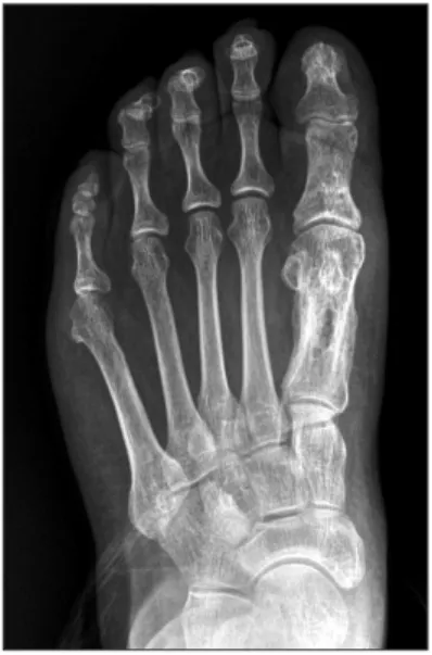
Figure 5. Preoperative weight-bearing anteroposterior radiograph of a 65-year-old woman shows hallux valgus in the left foot.
Figure 6. Radiograph at postoperative 1 week shows drill holes fixed with Ethibond (Ethibond Inc.) at the proximal phalanx of the great toe.
Figure 4. (A, B) Osteotomy site was closed and tied with Ethibond su- ture (Ethibond Inc.).
A B
200 Vol. 19 No. 4, December 2015
결론적으로 Akin 절골술 시 내측 평행 봉합사 고정 방법은 편리 성, 고정의 견고성, 재수술의 불필요성, 그리고 회외 변형 교정 면 에서 유용한 술식이라 생각된다.
REFERENCES
11 Murphy JS, Mozena JD, Walker RE. J-wire technique for fixation of the Akin osteotomy1 J Am Podiatr Med Assoc1 1989;79:291-31 21 Neumann JA, Reay KD, Bradley KE, Parekh SG. Staple fixation
for akin proximal phalangeal osteotomy in the treatment of hal- lux valgus interphalangeus1 Foot Ankle Int1 2015;36:457-641 31 Barca F, Busa R. Resorbable poly-L-lactic acid mini-staples for
the fixation of Akin osteotomies1 J Foot Ankle Surg1 1997;36:
106-111
41 Akin OF. The treatment of hallux valgus; a new operative pro- cedure and its results1 Med Sentinel1 1925;33:678-91
51 Chacon Y, Fallat LM, Dau N, Bir C. Biomechanical comparison of internal fixation techniques for the Akin osteotomy of the proximal phalanx1 J Foot Ankle Surg1 2012;51:561-51
61 Ahn SJ, Kim BH, Song MH, Kang SW, Oh KT, Yoo SH. Transar- ticular fixation of akin osteotomy on patients with hallux valgus after resection of medial protrusion of base of proximal pha- lanx1 J Korean Foot Ankle Soc1 2013;17:220-41
71 Roy SP, Tan KJ. A modified suture technique for fixation of the Akin osteotomy1 J Foot Ankle Surg1 2013;52:276-81
81 Young KW, Lee KT, Kim JY, Cha SD, Kim ES. Fixation with su- ture material in Akin osteotomy1 J Korean Foot Ankle Soc1 2004;
8:138-411
91 Schlefman BS. Akin osteotomy with horizontal interosseous wire-loop fixation1 J Am Podiatr Med Assoc1 1999;89:194-81 고정을 함으로써 골절과 관절면 손상의 위험성을 줄일 수 있었다.
절골면과 수평으로 고정을 하되 강선을 이용한 내외측의 고정 시 회외 변형 교정이 불편할 수 있으나, 이 술기 보고의 방식은 봉합 시 직침을 이용하여 회외 교정을 미리 예측하여 교정함으로써 보 다 편리하고 정확하게 교정할 수 있었다. 또한 절골면 고정 시 두 개의 매듭이 생기게 되는데, 족배부나 족저부에 위치한 매듭으로 연부조직 자극 증상은 관찰되지 않았다. 수술 전후 촬영한 단순 방 사선 촬영에서 합병증 없이 절골 부위의 골유합과 무지외반 교정 이 유지됨을 관찰할 수 있었다(Fig. 5∼7).
Figure 7. Radiograph at postoperative 12 months shows improved alignment of the hallux and union of Akin osteotomy.