Incidental Findings on Brain Magnetic Resonance Imaging in Children with Central Precocious Puberty
Ja Young Kim, Ji Hyen Lee, Hyun-Hae Cho
1, Hae Soon Kim
Departments of Pediatrics, 1Radiology, Ewha Womans University College of Medicine, Seoul, Korea
Objectives: To investigate brain magnetic resonance imaging (MRI) findings in pa- tients with central precocious puberty (CPP) by age at onset and sex.
Methods: We included 130 CPP patients with brain MRI findings of the pituitary gland treated at Ewha Womans University Mokdong Hospital between February 2007 and October 2013 and divided them by age and sex: boys, girls aged ≤6 years, and girls aged
>6 years. The control group comprised 224 patients who underwent brain MRIs, and we compared their incidental brain findings with those of the CPP group.
Results: In the CPP subgroups who underwent pituitary MRIs, the frequency of in- cidental brain lesions was 31.6% in boys, 47.1% in girls ≤6 years and 29.8% in girls >6 years. The incidence of pituitary abnormalities was 42.1% in boys, 64.7% in girls ≤6 years and 47.9% in girls >6 years. Among pituitary abnormalities, pituitary hypoplasia had a significantly higher incidence rate in girls ≤6 years (41.2%) than in boys (15.8%) or girls
>6 years (13.8%, P=0.027). Hypothalamic hamartomas were detected in one girl aged
≤6 years and in one boy, but not in girls aged 6 years (P=0.075). The incidence of pineal cysts was higher in the CPP groups and significantly higher in girls ≤6 years (47.1%) than in the control group (11.2%, P=0.001).
Conclusion: There was a higher incidence of brain abnormalities on pituitary MRIs and a higher incidence of pineal cysts, possibly associated with CPP pathogenesis, in younger CPP patients than in other patients. (Ewha Med J 2020;43(4):53-59)
Received June 25, 2020 Revised July 30, 2020 Accepted August 24, 2020 Corresponding author Hae Soon Kim
Department of Pediatrics, Ewha Womans University College of Medicine, 260 Gonghang-daero, Gangseon-gu, Seoul 07804, Korea
Tel: 82-2-2650-5569, Fax: 82-2-6986-1667 E-mail: [email protected]
Key Words
Precocious puberty; Pituitary hypoplasia;
Pineal cyst
This is an Open Access article distributed under the terms of the Creative Commons Attribution Non-Commercial License (http://creativecommons.org/licenses/by-nc/4.0) which permits unrestricted non-commercial use, distribution, and reproduction in any medium, provided the original work is properly cited.
Introduction
Precocious puberty is a condition that has both important and diverse consequences for the affected children and their families, and it imposes both physical and psychological im- pacts on the patients. The disease can potentially follow a sud- den, dynamic course. Therefore, early diagnosis can facilitate an understanding of the prospective disease course with regard to the rate of pubertal progression, development of stature, bone age progression, reproductive development, psychosocial adjustment, and good health [1]. Central precocious puberty
(CPP) refers to the premature activation of the HPG (hypo- thalamic-pituitary-gonadal) axis, with consequent early de- velopment of secondary sexual characteristics. The age criteria of normal and precocious puberty are controversial; however, the cutoffs that are routinely used to diagnose CPP are set at the age of 8 and 9 years for girls and boys, respectively [2,3].
Basic clinical examinations are important for CPP diagnosis, although there is an increasing awareness of the importance of imaging to detect secondary causes of CPP. In all cases of pro- gressive CPP, a magnetic resonance imaging (MRI) of the brain should be conducted to determine whether a hypothalamic or
pituitary lesion is present [4,5]. In children with CPP, the inci- dence of such lesions is higher in boys (40%–90%) than in girls (8%–33%), which decreases further (less than 2%, in one series) when puberty begins after age 6 in girls [5-7]. Nonetheless, neoplastic causes of precocious puberty, although uncommon, are important etiologic factors in precocious sexual develop- ment, and early, rapid recognition of these rare presentations is important [8]. A higher incidence of central nervous system (CNS) lesions in boys with CPP has been noted previously, and brain imaging is routinely undertaken in boys. Thus, brain MRI is recommended in all boys and girls younger than 6 years with CPP [9].
This study was conducted to investigate the brain MRI find- ings of patients with CPP to identify the incidence of pituitary abnormalities and incidental lesions. Furthermore, the study intended to clarify the need for MRI testing on the basis of the criteria defined by the specific characteristics of the MRI abnormalities as well as a consideration of the possible CPP- related findings among MRI incidental lesions compared with those in the control group.
Methods
1. Ethics statement
This was a retrospective study, and it was approved by the institutional review board of the Ewha Womans University Mokdong Hospital (EUMC2016-06-023-0055). Informed consent was waived due to the retrospective nature of the study.
2. Study population
We undertook a retrospective case-control study to review and analysis of radiologic findings in patients diagnosed with CPP. We obtained a list of patients who were diagnosed with CPP between February 2007 and October 2013 at the Ewha Womans University Mokdong Hospital, Seoul, Korea. Among the children diagnosed with CPP, a total of CPP patients who underwent pituitary MRI were included. All patients younger than 6 years underwent MRI exam, and patients aged 6 or older were included in the post-test study if they had symp- toms such as headaches or at the parent’s preference.
In a study of patients stored as CPP in the National Clas- sifications of Disease from 2008 to 2014, a total of 37,890 girls
and 1,220 boys were diagnosed with CPP [10]. The overall in- cidence of CPP was confirmed to be 122.8 per 100,000 persons (girls, 262.8; boys, 7.0). In this study, for the patients diagnosed with CPP from February 2007 to October 2013 and who had their MRI taken, the retrospective data collection was set for a study period of approximately 7 years. During this period, the patients diagnosed with CPP were examined. In terms of overall CPP incidence, calculations were made for 354 pa- tients, composed of 224 control patients and 130 CPP patients, among the children who visited our hospital for the pediatric premature evaluation.
The hormone levels were analyzed for the determination of a CPP diagnosis, and the positive criteria were as follows. Non- fasting venous blood samples were obtained to measure the concentrations of follicular stimulating hormone (FSH) and luteinizing hormone (LH). The detection limits for LH and FSH were 0.05 and 0.06 IU/L, respectively. Serum LH and FSH levels were measured at 0, 30, 45, 60, and 90 minutes after an intravenous bolus administration of LH-releasing hormone (synthetic gonadotropin releasing hormone [GnRH]; 100 µg Relefect, Sanofi-Aventis, Frankfurt am Main, Germany) [11].
We defined CPP by peak LH level >5 IU/L, or stimulated FSH/
LH ratio >0.66 IU/L. If the GnRH stimulation test was not conducted, the hormone level was considered pubertal if the basal LH level exceeded 0.3 IU/L.
We included girls with breast development before age 8 and a diagnosis of CPP. Positive results on the GnRH stimulation test and bone age advancement were considered manifestations of CPP for girls before age 8 and boys before age 9 [12,13].
The CPP patients were assigned to 3 groups by age and sex as specified: boys, girls aged ≤6 years, and girls aged >6 years.
The control group comprised the patients who underwent brain MRI at the same ages as the CPP group to enable a comparison of incidental brain findings. The control group included patients without pathological abnormalities and CPP and whose brain MRI conducted between February 2007 and October 2013 for symptoms of headaches, seizures, etc., and showed normal results.
Patients with previously known abnormalities, associated endocrine disorders, previous hormonal therapies, malforma- tion, neurofibromatosis, or other genetic conditions, including congenital adrenal hyperplasia, were excluded from the study.
3. Grouping of MRI findings
For the purposes of this study, the study population was fur- ther stratified on the basis of MRI abnormalities into four cat- egories: Category 1 comprised participants with normal MRI;
Category 2 included patients with incidental findings, such as, pineal cysts, arachnoid cysts, choroid plexus cysts, and subep- endymal heterotopia; Category 3 included patients who had fine lesions in the pituitary gland, such as hyperplasia, hypopla- sia, Rathke’s cleft cyst, or adenoma; and Category 4 included participants with pathological lesions, such as hamartoma.
4. Measurement of the pituitary gland
There are two methods to determine pituitary hypoplasia or hyperplasia. One option is to objectively and directly measure the height of the pituitary gland, whereas the other consid- ers the shape of the pituitary gland. With the method that uses
objective parameters, the reference may be insufficient and reli- ability may be compromised. Therefore, pituitary hypoplasia or hyperplasia was determined by both, objective and subjective, parameters in a previous study [14,15]. The objective param- eters are not commonly used in pediatric patients, but the stan- dard values are set at 4.9–6.5 mm in the sagittal plane, and the subjective parameters are graded in accordance with whether the shape of the gland is convex or concave on the sagittal im- aging diagnosis [14,15].
5. Statistical analyses and radiologic diagnostic criterion
Statistical analyses were performed using IBM SPSS Statistics ver. 22.0 (IBM Co., Armonk, NY, USA). Continuous variables were expressed as mean±standard deviation. An independent t-test was used to compare the CPP groups and the control group. The chi-square test was used to determine whether
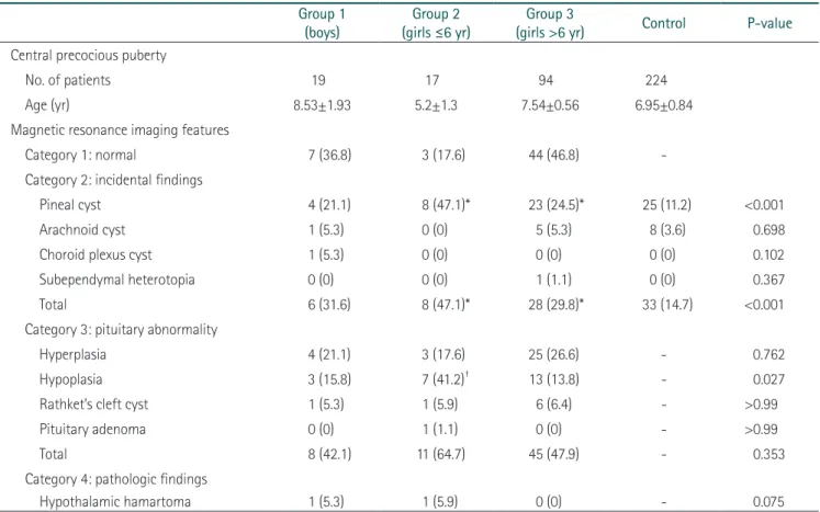
Table 1. Comparisons of prevalence of incidental findings, pituitary abnormalities and pathologic magnetic resonance imaging findings in central precocious puberty groups
Group 1
(boys) Group 2
(girls ≤6 yr) Group 3
(girls >6 yr) Control P-value Central precocious puberty
No. of patients 19 17 94 224
Age (yr) 8.53±1.93 5.2±1.3 7.54±0.56 6.95±0.84
Magnetic resonance imaging features
Category 1: normal 7 (36.8) 3 (17.6) 44 (46.8) -
Category 2: incidental findings
Pineal cyst 4 (21.1) 8 (47.1)* 23 (24.5)* 25 (11.2) <0.001
Arachnoid cyst 1 (5.3) 0 (0) 5 (5.3) 8 (3.6) 0.698
Choroid plexus cyst 1 (5.3) 0 (0) 0 (0) 0 (0) 0.102
Subependymal heterotopia 0 (0) 0 (0) 1 (1.1) 0 (0) 0.367
Total 6 (31.6) 8 (47.1)* 28 (29.8)* 33 (14.7) <0.001
Category 3: pituitary abnormality
Hyperplasia 4 (21.1) 3 (17.6) 25 (26.6) - 0.762
Hypoplasia 3 (15.8) 7 (41.2)† 13 (13.8) - 0.027
Rathket’s cleft cyst 1 (5.3) 1 (5.9) 6 (6.4) - >0.99
Pituitary adenoma 0 (0) 1 (1.1) 0 (0) - >0.99
Total 8 (42.1) 11 (64.7) 45 (47.9) - 0.353
Category 4: pathologic findings
Hypothalamic hamartoma 1 (5.3) 1 (5.9) 0 (0) - 0.075
Values are presented as mean±standard deviation or number (%).
*P<0.05 vs. control.
†P<0.05 vs. >6 years.
there was a statistically significant difference between the CPP groups. Statistical significance was defined as P<0.05.
All radiological diagnoses were diagnosed by a qualified ra- diologist. Among them, the criteria for diagnostic imaging of arachnoid cyst and pineal cyst were as follows. The arachnoid cyst was identified by imaging for extra-axial lesions, with similar signal intensity as that of the cerebrospinal fluid, in all sequences of the MRI. In the case of the pineal cyst, the diag- nosis was made when the signal intensity was shown on MRI T1-weighted image or in T2-weighted image or was slightly higher than cerebrospinal fluid [16].
Results
The study included a total of 354 participants. The CPP group comprised 130 children (19 boys [mean age, 8.53 years], 17 girls aged ≤6 years [mean age, 5.2 years], and 94 girls aged
>6 years [mean age, 7.54 years]). The control group included 224 children (mean age, 6.95 years) (Table 1).
The CPP group with the normal MRI brain findings com- prised seven boys, three girls aged ≤6 years, and 44 girls aged
>6 years. The incidence of normal brain findings in the CPP groups was 31.6% among the boys, 17.6% in the girls aged
≤6 years, and 46.8 % in the girls aged >6 years. The incidence of incidental brain findings in the CPP groups was 31.6%
among boys, 47.1% in girls aged ≤6 years, and 29.8% in girls aged >6 years, and no significant between-group differences were observed for the CPP groups. The incidence of incidental brain findings in the control group was 14.7%, which was sig- nificantly lower than in the CPP groups (P<0.001). Intergroup comparisons showed a significantly higher incidence among the
girls than in the control group (P<0.05 vs. control) (Table 2).
The incidence of pineal cyst in the control group was 11.2%
lower than in the CPP groups. Intergroup comparisons showed that the incidence of pineal cyst was 47.1% for the CPP group of girls aged ≤6 years and 24.5% in the CPP group with girls aged >6 years, and was significantly higher than in the control group (11.2%, P<0.001) (Table 2). However, there was no significant between-group difference in the incidence of arach- noid cysts, choroid plexus cysts, and subependymal heterotopia in the CPP groups, nor when compared with the control group (Table 2).
With regard to pituitary abnormalities, there was no sig- nificant intergroup difference (64.7% in girls aged ≤6 years, 47.9% in girls aged >6 years, and 42.1% in boys with CPP;
P<0.353) and the incidence of pituitary hyperplasia, pituitary hypoplasia, Rathke’s cleft cyst, and pituitary adenoma were compared among the three groups. The incidence of pituitary
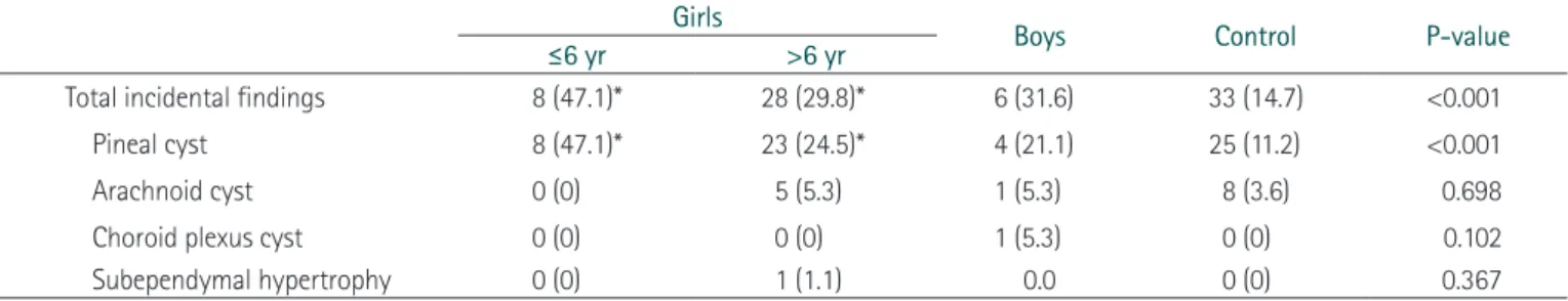
Table 2. Comparisons of prevalence of incidental brain magnetic resonance imaging findings in central precocious puberty groups vs. control group
Girls
Boys Control P-value
≤6 yr >6 yr
Total incidental findings 8 (47.1)* 28 (29.8)* 6 (31.6) 33 (14.7) <0.001
Pineal cyst 8 (47.1)* 23 (24.5)* 4 (21.1) 25 (11.2) <0.001
Arachnoid cyst 0 (0) 5 (5.3) 1 (5.3) 8 (3.6) 0.698
Choroid plexus cyst 0 (0) 0 (0) 1 (5.3) 0 (0) 0.102
Subependymal hypertrophy 0 (0) 1 (1.1) 0.0 0 (0) 0.367
Values are presented as number (%).
*P<0.05 vs. control.
Table 3. Comparisons of prevalence of pituitary abnormalities and pathologic findings of pituitary magnetic resonance imaging among central precocious puberty groups
Girls Boys P-value
≤6 yr >6 yr
Pituitary abnormalities 11 (64.7) 45 (47.9) 8 (42.1) 0.353 Hyperplasia 3 (17.6) 25 (26.6) 4 (21.1) 0.762 Hypoplasia 7 (41.2)* 13 (13.8) 3 (15.8) 0.027 Rathket’s cleft cyst 1 (5.9) 6 (6.4) 1 (5.3) >0.99 Pituitary adenoma 0 (0) 1 (1.1) 0 (0) >0.99 Pathologic findings
Hamartoma 1 (5.9) 0 (0) 1 (5.3) 0.075
Values are presented as number (%).
*P<0.05 vs. >6 years.
hypoplasia (41.2% in girls aged ≤6 years, 13.8% in girls aged
>6 years, and 15.8% in boys) differed significantly between the three CPP groups (P=0.027) (Table 3). Hypothalamic hamar- tomas were detected in one girl aged ≤6 years (5.9%) and in one boy (5.3%), but not in girls aged 6 years (P=0.075).
Discussion
Recent studies have reported an increase in the incidence of CPP, especially the incidence of idiopathic CPP [17,18]. Besides the development of specific tests for the diagnosis, history tak- ing, and physical examination, brain-imaging modalities have become an important diagnostic tool in CPP. However, brain MRI screening in patients with CPP remains controversial.
In a study by Mogensen et al. [19] in a CPP population in Denmark, most cases were idiopathic; 13 out of 208 girls (6.3%) with early or precocious puberty and no other CNS symptoms were found to have pathological CNS lesions on the MRI, which were connected with CPP. All 13 girls were
>6 years of age, and 6 girls were in the age range of 8–9 years.
A high proportion of girls in the of 6–8-year age range with early or precocious development had pathological findings on brain MRI. In the 6- to 8-year-old girls with early or preco- cious development, pathological causes were identified on the brain MRI that could not have been detected from a clinical investigation in this study. Thus, the findings of this study indi- cate that girls with precocious pubertal development of central origin before 8 years of age should be screened by using brain MRI [19]. However, another study conducted in Italy in 2014 in 182 girls showed 86% had no specific findings on the brain MRI; 11% had incidental findings unrelated with CPP, and 3%
reported hamartomas. In contrast to previous studies, routine screening with brain MRI is not recommended in all girls with CPP between 6 and 8 years of age [20]. Against this back- ground, we investigated the brain MRI findings of CPP patients and found pituitary abnormalities and incidental brain findings on the brain MRI examination.
In our study, the total incidences of incidental brain findings, including pineal cysts, arachnoid cysts, choroid plexus cysts, and subependymal heterotopia, were compared between CPP groups without any significant differences. However, com- parison with the control group showed significantly higher incidences in the CPP groups, with girls ≤6 years showing the
highest incidences.
Melatonin, which is released from the pineal gland, is con- sidered responsible for puberty, although the exact mechanism has not been defined [21]. Pineal cysts, which have a prevalence rate of 0.6% in the pediatric population, are well-known as- ymptomatic incidental findings, but have been associated with CPP [22-25]. Similarly, in the present study, the incidence of pineal cysts was significantly higher in the CPP group than in the control group, which showed normal MRI findings, espe- cially in girls aged ≤6 years with CPP. This suggests that pineal cysts may be considered an incidental brain lesion with regard to CPP pathogenesis. However, only the data on brain MRIs, excluding the hormone serum level, were obtained in the con- trol group of our study; therefore, it was difficult to clearly compare the prevalence of pituitary abnormalities between the CPP groups and the control group.
The proportion of diseases related to CNS diseases is higher in boys, but must also be excluded in girls. Approximately 95%
of girls with CPP have idiopathic CPP and only 5% have CPP arising from a secondary cause. However, more than 50% of boys with CPP have an identifiable etiology, and idiopathic CPP is a diagnosis of exclusion [26]. In the present study, we expected that incidental brain findings would be significantly higher than the control group in boys with CPP, but no statis- tical significance was found for higher incidental brain findings in boys with CPP. However, it should be considered that the number of boys included in this study was too small to gener- ate a significant difference with the control group. Hamartoma, also known as brain lesion, as the cause of CPP was identi- fied in only two in our study population (one in a girl aged ≤6 years, one in a boy), but there was no significant difference. In general, many articles have reported that hamartomas are a common brain lesion in patients with CPP. Faizah et al. [27]
reported that hypothalamic hamartoma was the commonest tumor causing CPP in their patients, accounting for 10 out of 34 (29%) cases with abnormal findings on brain imaging.
This data analysis has some limitations, which should be considered with regard to the clinical interpretation of the find- ings. First, the number of boys with CPP who were included in this study was insufficient for a proper analysis and did not result in significant intergroup results on comparative analysis.
Second, the CPP groups performed the pituitary MRI and the control group performed the brain MRI. In general, the meth-
ods of image acquisition of the pituitary MRI and the brain MRI are different. The pituitary MRI creates high resolution images that allow physicians to see the pituitary region better, and bias was included according to the inspection protocol.
In the control group, only the brain MRI was performed and the hormone tests not undertaken. Therefore, it was difficult to clearly compare the incidence of the pituitary gland abnormali- ties and the pineal cysts between the CPP and control groups.
Third, we did not define the control group as ‘healthy control’
which could have provided selection bias. In addition, there was no data of the control group for pituitary abnormalities.
Finally, as we examined only Korean children, it is possible that the regional prevalence of brain lesions in CPP patients is related to the study population, race, and healthcare and insur- ance systems.
In conclusion, this study suggests that pituitary MRIs are necessary as a diagnostic tool in CPPs, especially in young girls and boys. Children with CPP are more likely to have incidental findings on brain MRI. In particular, the relatively high inci- dence of MRI findings in the CPP group than in the control group suggests the possibility that pineal cysts, which were considered to be an incidental finding on brain MRI, may be related to the CPP pathogenesis. There is a need to further elu- cidate the brain MRI features of the CPP diagnosis in future large-sample research studies that include rigorous statistical analysis.
References
1. Partsch CJ, Heger S, Sippell WG. Management and outcome of central precocious puberty. Clin Endocrinol (Oxf) 2002;56:129- 148.
2. Nebesio TD, Eugster EA. Current concepts in normal and abnormal puberty. Curr Probl Pediatr Adolesc Health Care 2007;37:50-72.
3. Papadimitriou A, Beri D, Tsialla A, Fretzayas A, Psychou F, Nico- laidou P. Early growth acceleration in girls with idiopathic preco- cious puberty. J Pediatr 2006;149:43-46.
4. Cisternino M, Arrigo T, Pasquino AM, Tinelli C, Antoniazzi F, Be- duschi L, et al. Etiology and age incidence of precocious puberty in girls: a multicentric study. J Pediatr Endocrinol Metab 2000;13 Suppl 1:695-701.
5. Chalumeau M, Hadjiathanasiou CG, Ng SM, Cassio A, Mul D, Cisternino M, et al. Selecting girls with precocious puberty for brain imaging: validation of European evidence-based diagnosis rule. J Pediatr 2003;143:445-450.
6. Carel JC, Leger J. Clinical practice: precocious puberty. N Engl J Med 2008;358:2366-2377.
7. De Sanctis V, Corrias A, Rizzo V, Bertelloni S, Urso L, Galluzzi F, et al. Etiology of central precocious puberty in males: the results of the Italian Study Group for Physiopathology of Puberty. J Pe- diatr Endocrinol Metab 2000;13 Suppl 1:687-693.
8. Stephen MD, Zage PE, Waguespack SG. Gonadotropin-depen- dent precocious puberty: neoplastic causes and endocrine con- siderations. Int J Pediatr Endocrinol 2011;2011:184502.
9. Kaplowitz P, Bloch C; Section on Endocrinology, American Academy of Pediatrics. Evaluation and referral of children with signs of early puberty. Pediatrics 2016;137.
10. Kim YJ, Kwon A, Jung MK, Kim KE, Suh J, Chae HW, et al. Inci- dence and prevalence of central precocious puberty in Korea:
an epidemiologic study based on a national database. J Pediatr 2019;208:221-228.
11. Neely EK, Wilson DM, Lee PA, Stene M, Hintz RL. Spontaneous serum gonadotropin concentrations in the evaluation of preco- cious puberty. J Pediatr 1995;127:47-52.
12. Brito VN, Latronico AC, Arnhold IJ, Mendonca BB. Update on the etiology, diagnosis and therapeutic management of sexual precocity. Arq Bras Endocrinol Metabol 2008;52:18-31.
13. Carel JC, Eugster EA, Rogol A, Ghizzoni L, Palmert MR; ESPE- LWPES GnRH Analogs Consensus Conference Group, et al.
Consensus statement on the use of gonadotropin-releasing hor- mone analogs in children. Pediatrics 2009;123:e752-e762.
14. Denk CC, Onderoglu S, Ilgi S, Gurcan F. Height of normal pitu- itary gland on MRI: differences between age groups and sexes.
Okajimas Folia Anat Jpn 1999;76:81-87.
15. Keanninsiri C, Cheiwvit P, Tritrakarn S, Thepamongkhol K, Santiprabhop J. Size and shape of the pituitary gland with MR imaging from newborn to 30 years: a study at siriraj hospital. In:
Proceedings of the 6th Annual Scientific Meeting on Challenges of Quality Assurance in Radiation Medicine; 2012 Feb 23-26;
Phitsanulok, Thailand. Bangkok: Thai Medical Physicist Society;
2012. p.55-58.
16. Barkocich AJ, Raybaud C. Pediatric neuroimaging. Philadelphia:
Wolters Kluwer; 2019.
17. Alikasifoglu A, Vuralli D, Gonc EN, Ozon A, Kandemir N. Chang- ing etiological trends in male precocious puberty: evaluation of 100 cases with central precocious puberty over the last decade.
Horm Res Paediatr 2015;83:340-344.
18. Kim SH, Huh K, Won S, Lee KW, Park MJ. A significant increase in the incidence of central precocious puberty among Korean girls from 2004 to 2010. PLoS One 2015;10:e0141844.
19. Mogensen SS, Aksglaede L, Mouritsen A, Sorensen K, Main KM, Gideon P, et al. Pathological and incidental findings on brain MRI in a single-center study of 229 consecutive girls with early or precocious puberty. PLoS One 2012;7:e29829.
20. Pedicelli S, Alessio P, Scire G, Cappa M, Cianfarani S. Routine screening by brain magnetic resonance imaging is not indicated in every girl with onset of puberty between the ages of 6 and 8 years. J Clin Endocrinol Metab 2014;99:4455-4461.
21. Waldhauser F, Weiszenbacher G, Tatzer E, Gisinger B, Wald- hauser M, Schemper M, et al. Alterations in nocturnal serum melatonin levels in humans with growth and aging. J Clin Endo- crinol Metab 1988;66:648-652.
22. Aleandri V, Spina V, Morini A. The pineal gland and reproduc- tion. Hum Reprod Update 1996;2:225-235.
23. Jansen PR, Dremmen M, van den Berg A, Dekkers IA, Blanken LME, Muetzel RL, et al. Incidental findings on brain imaging in the general pediatric population. N Engl J Med 2017;377:1593- 1595.
24. Kumar KV, Verma A, Modi KD, Rayudu BR. Precocious puberty and pineal cyst: an uncommon association. Indian Pediatr
2010;47:193-194.
25. Benitez Fuentes R, Velazquez de Cuellar Paracchi M, Blanco Rodriguez M, Soriano Guillen L. Central precocious puberty and pineal gland cyst: an association or incidental finding? An Pediatr (Barc) 2008;68:72-73.
26. Kornreich L, Horev G, Blaser S, Daneman D, Kauli R, Grune- baum M. Central precocious puberty: evaluation by neuroimag- ing. Pediatr Radiol 1995;25:7-11.
27. Faizah M, Zuhanis A, Rahmah R, Raja A, Wu L, Dayang A, et al.
Precocious puberty in children: a review of imaging findings.
Biomed Imaging Interv J 2012;8:e6.