Vol. 19, No. 1, March, 2007
불안정 대퇴골 전자간 골절의 치료시 근위 대퇴정(Proximal femoral nail)과 압박 고 나사(Dynamic hip screw)의 후향적 비교
임 군 일
동국대학교 일산병원 정형외과
목적: 대퇴 전자간 불안정 골절을 근위 대퇴정과 압박 고나사 고정술로 치료한 결과를 후향적으로 분석하여 비교하고자 하였다.
대상 및 방법: 대퇴 전자간 골절의 AO 분류상 불안정성 골절(A22,23형)이 있는 환자들 중 근위 대퇴정과 압박 고 나사 고정술로 치료한 100명(근위 대퇴정 50명, 압박 고 나사 50명)을 대상으로 후향적 분석을 시행하였다. 나이, 성별, 수 술 전의 상태, 수술 중 자료(마취 종류, 수술 시간, 수혈량, 삽입물의 위치) 수술 후 경과와 합병증등을 비교하였다.
결과: 평균 수술시간은 근위대퇴정군에서 유의하게 짧았고(P=0.03) 수술중 수혈양은 큰 차이가 없었다. 수술중 합병증으 로 근위대퇴정군에서 대퇴간부골절 1례, 수술후 합병증으로 지연나사의 이탈과 골두천공으로 인공관절 치환술을 시행하였 던 경우가 근위 대퇴정과 압박고나사 군에서 각각 1례씩 있었으며 압박고나사군에서 고정의 소실로 근위 대퇴정으로 치환 한 경우가 1례 있었다. 환자의 기능 지표로 사용된 Parker and Palmer 운동 지수는 수술 전 큰 차이는 없었지만 수술 후 근위 대퇴정을 시행한 환자들에게 높았으며 통계적으로 유의한 차이가 있었다(P=0.02). 또 다른 지표로써 사용된 Jensen의 사회 기능 지수는 추시 기간 중 두 군 간에 큰 차이를 보이진 않았다.
결론: 근위 대퇴정의 사용은 압박 고 나사를 사용한 경우와 비교해 볼 때 대퇴 전자간 불안정 골절의 치료에 있어서 환자 의 전반적인 경과를 개선하지는 못하지만 수술시간의 단축과 술후 운동능력의 향상에 있어서는 장점을 가진 것으로 사료 된다.
색인단어: 대퇴골 전자간 불안정 골절, 압박 고 나사. 근위 대퇴정
Introduction
Intertrochanteric fractures of femora are common fractures occurring in elderly patients11). About half of intertrochanteric fractures are comminuted and unstable as these fractures occur in people with poor bone quality10,19,20). In unstable intertrochanteric fractures, excessive medialization of shaft and subsequent loss of contact between fragments can lead to fixation failures6,24). Even after the fracture ultimately unites, limb shortening and decreased length of abductor lever
arm disturb adequate hip function6,24). Therefore unstable intertrochanteric fractures are distinguished from their stable counterparts in regard to the treatment plan and prognosis.
As early mobilization is necessary to prevent complications of prolonged immobilization, a fixation method that enables immediate weight bearing must be employed in treating an unstable intertrochanteric fractures18). At the present time, the dynamic hip screw (DHS) which was devised to control the collapse of fracture is widely accepted as the standard treatment of intertrochateric fractures except in a reverse obliquity type8,15). However, this type of fixation is theoretically disadvantaged over an intramedullary nail because of greater length of lever arm14,21,26). While this mechanical drawback does not militate against the clinical results in
※ 통신저자 : 임 군 일
경기도 고양시 식사동 814 동국대학교 일산병원 정형외과 Tel: 82-31-961-7315 Fax: 82-31-961-9219 E-mail: [email protected]
stable fractures, relatively high complication rate have been reported with DHS in unstable intertrochanteric fractures8,9,20,30).
Proximal femoral nail (PFN) is a second- generation intramedullary nail which was designed to overcome the drawbacks of Gamma nail. It has two key differences from conventional nails29). First, an anti-rotation pin was added to the construct to reduce the rotation and collapse of head and neck fragment. Second, the nail tip has a smaller diameter and therefore the chance of fracture at the tip is reduced. In addition, smaller and tapering scale of lag screw enables placement of screw tip deeper into the subchondral bone. The purpose of this paper was to compare the short-term results of DHS and PFN in treating unstable intertrochanteric fractures.
Materials and Methods
From March 2000 to March 2003, a total of 232 patients were consecutively treated for intertrochanteric fractures in the author‘s institution. The figure did not include patients with pathologic fracture, polytrauma patients, patients with previous hip surgery, and fractures extending 5cm distal to inferior border of lesser trochanter.
Nor were patients with high-energy injuries or under 55 years included in this number. Of the patients, 108 patients were judged to have an A2 AO/OTA (ArbiergemeinshaftOsteosynthefragen/
Orthopaedic Trauma Association) unstable intertrochanteric fracture.
The fractures were classified as AO/OTA A22 and A23 types from agreements of three orthopaedic surgeons. On the period from March 2000 to March 2001, only DHS were used. From April 2001 to June 2002, either DHS (Synthes- Stratec, Oberdorf, Switzerland) or PFN (Synthes- Stratec, Oberdorf, Switzerland) was randomly used. From July 2002 to March 2003, only PFN was used for the treatment. Eight early cases of PFN group were excluded in order to diminish the bias caused by the inexperience with the implant.
So this study ultimately included fifty patients treated with DHS (Group I) and 50 patients
treated with PFN (Group II).
The operations were carried out under either epidural or general anesthesia with patient supine on the fracture table. Closed reduction was performed under fluoroscopic control in two planes. The operations were performed or supervised by the author. DHS was inserted according to the standard techniques8,15). Four-hole barrel plates with the angle of 135 degrees were used in all of the patients. After the operation, patients were allowed to get out of bed and sit on a chair on the third postoperative day. Patients usually started standing and walking with aid of walker by 5th to 8th postoperative day. Weight- bearing was allowed as much as tolerated by the patients. Proximal femoral nails were also used according to the manufacturer’s protocol. A 5 to 8 cm incision was made starting 2 cm proximal to the tip of greater trochanter and extending upward. A guide wire was inserted from the tip of greater trochanter. Then a 16.5 mm reamer was used to ream the area where proximal part of nail would be seated. A PFN with the diameter of 10mm was inserted by pushing inward without use of mallet. All the patients were given prophylactic antibiotics (Cefazoline) beginning on the day of operation and being continued until 48 hours after operation. Patients were usually discharged from hospital 2 to 3 weeks after surgery. They were followed every other week until complete healing of fracture, then every third month until 1 year after surgery (Fig. 1). Anteroposterior and lateral radiographs were taken at each time of visit. Tip-apex distance (TAD) from immediate postoperative radiographs was used to determine lag screw position within the femoral head. TAD of more than 25 mm was considered to indicate a poor position. Fractures were judged to be clinically healed when pain-free walking and range of motion of the hip were possible. Fractures were judged to be united when bridging callus was evident on three of four cortices as seen on two views.
Information on age, gender, ASA (American Society of Anesthesiologist) physical status score, the mobility score by Parker and Palmer23), the
social function score of Jensen16), living condition as well as preoperative morbidities was prospectively collected. Of the 100 patients, 5 patients of PFN group were lost to follow-up less than one year after surgery. Eight patients of DHS group and seven patients of PFN group died before a year had passed after surgery. It left 42 patients in DHS group and 38 patients in PFN group available for final assessment.
Preoperative data and intraoperative data including the type of anesthesia, the duration of operation, the amount of transfused blood and a need for open reduction was compared between the two groups. In addition, postoperative general morbidities, surgical complications, union time, living situation, the social function score and the mobility score at the final follow up were compared between the two groups. An independent biostatistician who was not directly involved with the study performed statistical analysis of the data. The parameters were compared between two groups. Student t-test was used for interval data. Chi square test was used for categorical data. Fisher two-sided exact test
was used for dichotomous data.
The level of significance was set at p<0.05.
Results
The mean age was 74 (range, 59~91) years in DHS group and 74 (range, 55~90) years in PFN group (p=0.58). There were 19 men and 31 women in DHS group while PFN group had 13 men and 37 women (p=0.28). ASA score, the mobility score, the social function score and living situation were similar between the two groups (Table 1). The preoperative morbidities also were comparable (Table 2). Most patients underwent regional anesthesia in both groups. The mean duration of operation was significantly shorter in PFN group (68 minutes: range 30~150) compared with DHS group (97 minutes; range 50~180) (p=0.03). The amount of transfusion was 2.1 ( range,0~6) unit for DHS group and 1.7 (range, 0~4) units for PFN group, which was not significantly different (p=0.28) (Table 3). There were poor implant positions in three and six patients respectively in the two groups (DHS v
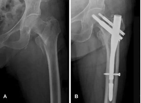
Fig. 1. A 72-year-old female sustained an A22 AO/OTA intertrochantertic fracture (A). She was treated with internal fixation with PFN, and the radiograph taken 12 months postoperatively shows complete healing of the fracture (B).
A B
PFN, p=0.49). TAD was 14.7 (S.D. 4.9) mm in the DHS group and 15.8 (S.D. 6.3) mm in the PFN group (p=0.29).
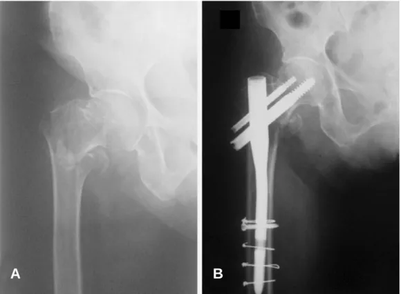
Surgical complications included a femoral shaft fracture in PFN group which occurred during the insertion of interlocking screw (Fig. 2). There were 2 cases of cut-through: 1 in DHS group and 1 in PFN group. These patients were managed with total hip arthroplasty. Gross loss of reduction without cut-through occurred in one hip in DHS group ,which was salvaged with PFN. The mean time to union was 21 weeks in DHS group and 20 weeks in PFN group (P=0.70). Postoperative morbidities included respiratory complication, urinary tract infection, pulmonary embolism, deep vein thrombosis and cardiovascular disorders
which affected each group alike (p=0.81) (Table 4). Three patients in DHS group and two patients in PFN group died within one month after surgery while staying in the hospital. The causes of death in those patients were pneumonia in two patients, pulmonary embolism in two patients and myocardial infarction in one patient. After one year of follow up, 8 patients (16%) of DHS group and 7 patients of PFN group died. The mobility score of Parker and Palmer was definitely higher in PFN group than in DHS group (p=0.02).
However, the social function score of Jensen failed to display significant difference between the two groups (p=0.09). Time to union was also similar between the two groups (Table 5).
Table 1. Preoperative data of the patients
DHS (n=50) PFN (n=50) p-value
Mean age (years) 74±8.8 74±7.1 0.58
Gender
Male 19 13
female 31 37
ASA physical-status score
Class I 04 03
Class II 34 29
Class III 12 17
Class IV 00 01
Social function score (Jensen)
Group I 11 15
Group II 24 22
Group III 13 10
Group IV 02 03
Mean 2.1±0.79 2.0±0.86 0.55
Mobility score (Parker & Palmer) 6.4±1.80 6.7±2.10 0.44
Living situation
Home 46 43 0.52
Nursing home or hospital 04 07
Table 2. Preoperative morbidities
DHS PFN
p-value (n=50) (n=50)
Cardiovascular disease 20 17 0.68
Diabetes 11 14 0.64
Pulmonary disease 05 03 0.71
Cancer 02 01 1.00
III or IV ASA risk 12 18 0.28
Table 3. Perioperative data of the patients
DHS PFN
p-value (n=50) (n=50)
Type of anesthesia
Regional 42 43 1.08*
spinal *8 *7
Operative time(minutes) 97±20 68±22 0.03*
Blood transfusion(units) 2.1±1.3 1.7±1.1 0.28*
Poor implant position *3 *6 0.49*
* p<0.05
Discussion
The purpose of this study was to find an answer to the question if PFN has any advantage over conventional DHS techniques in treating an A2 AO/OTA intertrochanteric fractures. While the use of PFN is advocated over 95-degree condylar plate for an A3 fracture27),there have
been controversies on the role intramedullary nails deserve in treating more common A2 fractures. First generation intramedullary nails such as Gamma nail were fraught with complication stemming from technical difficulties and design flaws. Therefore a plethora of studies failed to show advantage of intramedullary nail over DHS3,12,13,25)
. In a meta-analysis of unstable Fig. 2. Radiograph of a 90-year-old woman shows an unstable intertrochanteric fracture on her right hip (A). A femoral shaft fracture occurred while inserting an interlocking screw. Cerclage wiring was done and the fracture healed after 4 months (B).
A B
Table 4. Postoperative complications
DHS PFN
p-value
(n=50) (n=50)
General complications 0.81
UTI 1 2
Pneumonia 4 3
Myocardial Infarction 2 1
Pulmonary embolism 1 2
Deep vein thrombosis 2 1
Gastrointestinal bleeding 2 1
Death 3 2
Surgical complications
Femoral fracture 0 1
Cut out of lag screw 1 1
Loss of reduction 1 0
intertrochanteric fractures treated with dynamic screw versus intramedullary nail devices which investigated 17 trials, there was no significant difference in the frequency of implant related complications between the two types of implants3). Iatrogenic femoral fractures represent a rare but persistent risk. There was tendency for less frequent cut-through with intramedullary devices than with DHS3).
As PFN is a second generation intramedullary nail that corrected the rotational stability problems and stress concentration on the tip of a nail, different results may be expected in treating an unstable intertrochanteric fractures. Early studies on the results of PFN report on the persisting complications of cut-through of lag screws but few accidents of femoral shaft fractures2,4,5,29). Simmermacher et al published the first report on the results of PFN for unstable intertrochanteric fractures. They reported only one case of cut- through and overall technical failure rate of 4.6%
but there was no case of femoral shaft fracture29).
Three other series which investigated the results of unstable intertrochanteric fractures also reported cut-throughs in 4 to 7% of patients and occasional implant failures but no case of femoral shaft fracture2,4,5). However, there are few studies that illustrate the ultimate benefit of PFN as compared with DHS in A2 AO/OTA intertrochanteric fractures. A large series which compared the results of DHS versus PFN in A1 and A2 intertrochanteric fractures combined reported almost equal outcome in using either PFN or DHS28).
When the author began to use PFN, he was well aware of known complications of intramedullary nail. However, his focus in using a PFN, as original inventors of the system insisted, was first the mechanical advantage from shorter lever arm which would also promote early weight bearing, and secondly minimal dissection and shorter operative time which would facilitate patients’recovery. In addition, the author’s previous experience of uncontrollable collapse and subsequent loss of Table 5. Clinical data at the final follow-up
DHS PFN p-value
(n=50) (n=50)
Died 8 7 1.0
Lost to follow-up 0 5
Available for review 42 38
Final follow-up DHS PFN p-value
(n=42) (n=38)
Complications 01.0*
Nonunion 0 0
Infection 0 0
Loss of reduction 1 0
Cut-out of screw 1 1
Living situation 0.55*
Home 34 33
Hospital or Nursing home 8 5
Social functional score (Jensen)
Group I 10 16
Group II 14 12
Group III 12 6
Group IV 6 4
Mean 2.3±1.0 2.0±1.0 0.09*
Mobility score 4.4±1.5 5.2±1.4 0.02*
(Parker and Palmar)
Time to union (weeks) 21±7.2 20±7.6 00.7*
* p<0.05
fixation in treating unstable intertrochanteric fractures with DHS (data not given) led us to try PFN. In the author’s cases, these points were corroborated with shorter operative time and better mobility score in PFN group. The shorter operative time is attributed to lesser time taken for dissection and closure. This was confirmed by other studies which compared the results of PFN versus DHS17,22). However, it should be noted that the use of PFN did not reduced patients’postoperative morbidities or mortalities compared with DHS. This point was previously demonstrated by a study by Hardy et al.
which reported better mobility with intramedullary nail compared with DHS by 0.8 point12). The author agree with them in that the better mobility after intramedullary nailing may be due to fewer limbs shortening which can prevent older patients from recovering the ability to walk.
However, the similar social function index means that this advantage was insufficient to improve social independence. It should be noted that the author’s patients already had low social function score before the fracture. It is also interesting that there was not a critical difference in the amount of transfused blood despites smaller incision and shorter duration of operation.
It was similar to the results of Adams et al which prospectively compared Gamma nail with DHS1). In contrast, Hardy et al reported less blood loss with an intramedullary hip screw compared with dynamic hip screw12). It is surmised that in the authors’ cases the need for transfusion was determined by the fracture per rather than the fixation method.
In the author’s series, surgical complications such as cut-through of screws or reduction loss occurred in PFN groups as well as in DHS group.
A femoral shaft fracture took place in the early use of PFN while inserting an interlocking screw.
This devastating complication was due to technical error in the author’s incipient experience with PFN and may be eliminated with familiarity with the system.
This study has several limits. First, its retrospective nature does not permit a randomized experimental trial which would enable a more control of clinical variables although the
demographic data was comparable between the two groups. Second, the number of cases was not large enough to make possible the assessment of diverse surgical complications. Third, this series includes several learning curve cases in PFN groups though first 8 cases were excluded from the analysis. The strength lies in being a single surgeon series and including only A22 and A23 AO/OTA fractures.
In summary, the use of PFN has advantage in shorter operative time and better mobility of the patients while not altering overall course of patients’ recovery. In this regard, the PFN deserve a place in treating an A2 intertrochanteric fractures as a more effective tool for experienced hands. However, in view of the occurrence of surgical complications uncommon with DHS, the author would not recommend PFN as a standard form of treatment for surgeons who treat small number of patients.
REFERENCES
01) Adams CI, Robinson CM, Court-Brown CM and McQueen MM: Prospective randomized controlled trial of an intramedullary nail versus dynamic screw and plate for intertrochanteric fractures of the femur. J Orthop Trauma, 15: 394-400, 2001.
02) Al-yassari G, Langstaff RJ, Jones JW and Al-Lami M:
The AO/ASIF proximal femoral nail (PFN) for the treatment of unstable trochanteric femoral fracture.
Injury, 33(5): 395-399, 2002.
03) Audige L, Hanson B and Swiontkowski MF: Implant- related complications in the treatment of unstable intertrochanteric fractures: meta-analysis of dynamic screw-plate versus dynamic screw-intramedullary nail devices. Int Orthop, 27: 197-203, 2003.
04) Banan H, Al-Sabti A, Jimulia T and Hart AJ: The treatment of unstable, extracapsular hip fractures with the AO/ASIF proximal femoral nail (PFN)--our first 60 cases.
Injury 33: 401-405, 2002.
05) Boldin C, Seibert FJ, Fankhauser F, Peicha G, Grechenig W and Szyszkowitz: The proximal femoral nail (PFN)--a minimal invasive treatment of unstable proximal femoral fractures: a prospective study of 55 patients with a follow-up of 15 months. Acta Orthop Scand 74(1): 53-58, 2003.
06) Chan KS and Gill GS: Cemented hemiarthroplasties for elderly patients with intertrochanteric fractures. Clin Orthop, 371: 206-215, 2000.
07) Chevally F and Gamba D: Gamma nailing of
pertrochanteric and subtrochanteric fractures, clinical results of a series of 63 consecutive cases. J Orthop Trauma, 11: 412-415, 1997.
08) Clawson DK: Trochanteric fracture treated by the sliding screw plate fixation method. J Trauma, 4: 737-752, 1964.
09) Davis TR, Sher JL, Horsman A, Simpson M, Porter BB and Checketts RG: Intertrochanteric femoral fractures: mechanical failure after internal fixation. J Bone Joint Surg, 72-B: 26-31, 1990.
10) Dimon JH and Hughston JC: Unstable intertrochanteric fractures of the hip. J Bone Joint Surg, 49-A: 440-450, 1967.
11) Evans EM: The treatment of trochanteric fractures of the femur. J Bone Joint Surgery. 31-B: 190-203, 1949.
12) Hardy DC, Descamps PY, Krallis P, Fabeck L, Smets P, Bertens CL and Delince PE: Use of an intramedullary hip screw compared with a compression hip screw with a plate for intertrochanteric femoral fractures. J Bone Joint Surg, 80-A: 618-630, 1998.
13) Harrington P, Nihal A, Singhania AK and Howell FR:
Intramedullary hip screw versus sliding hip screw for unstable intertrochanteric femoral fractures in the elderly.
Injury, 33: 23-28, 2002.
14) Haynes RC, Poll RG, Miles AW and Weston RB: An experimental study of the failure modes of the Gamma Locking Nail and AO Dynamic Hip Screw under static loading: a cadaveric study. Med Eng Phys, 19: 446-453, 1997.
15) Heyse-Moore GH, MacEachern AG and Evans DC:
Treatment of intertrochanteric fractures of the femur. A comparison of the Richards screw-plate with the Jewett nail plate. J Bone Joint Surg, 65-B: 262-267, 1983.
16) Jensen JS: Determining factors for the morbility following hip fractures. Injury 15: 411-414, 1984.
17) Klinger HM, Baums MH, Eckert M and Neugebauer R.: A comparative study of unstable per- and intertrochanteric femoral fractures treated with dynamic hip screw (DHS) and trochanteric butt-press plate vs.
proximal femoral nail (PFN). Zentralbl Chir, 130: 301- 306, 2005.
18) Koval KJ, Fiend KD, Ahronohof GB and Zuckerman JD: Weight bearing after hip fracture. A prospective series of 596 geriatric hip fracture patients. J Orthop Trauma, 10: 526-530, 1996.
19) Koval KJ and Zuckerman JD: Intertrochanteric
fractures. In: Bucholz RW, Heckman JD(eds). Fractures in Adults. 5th ed. Philadelphia, Lippincott Williams and Wilkins: 1635-1663, 2001.
20) Kyle RF, Gustilo RB and Premier RF: Analysis of six hundred and twenty-two intertrochanteric fractures. J Bone Joint Surg, 61-A:216-221, 1976.
21) Leung KS, So WS, Shen WY and Hui PW: Gamma nails and dynamic hip screws for peritrochanteric fractures. A randomized prospective study in elderly patients. J Bone Joint Surg, 74-B: 345-351, 1992.
22) Nuber S, Schonweiss T, and Ruter A: Stabilisation of unstable trochanteric femoral fractures. Dynamic hip screw (DHS) with trochanteric stabilisation plate vs.
proximal femur nail (PFN). Unfallchirurg, 106: 39-47, 2003.
23) Parker MJ and Palmer CR: A new morbility score for predicting mortality after hip fracture. J Bone Joint Surg, 75-B: 797-798, 1993.
24) Rha JD, Kim YH, Yoon SI, Park TS and Lee MH:
Factors affecting sliding of the lag screw in intertrochanteric fractures. Int Orthop, 17: 320-324, 1993.
25) Radford PJ, Needoff M and Webb JK: A prospective randomized and comparison of the dynamic hip screw and Gamma locking nail, J Bone and Joint Surg, 75-B: 789- 793, 1993.
26) Rosenblum S F, Zuckerman JD, Kummer FJ and Tam BS: A biomechanical evaluation of the Gamma nail. J Bone and Joint Surg, 74-B: 352-357, 1992.
27) Sadowski C, Luebbeke A, Saudan M, et al: Treatment of reverse obliquity and transverse intertrochanteric fractures with use of an intramedullary nail of a 95o screwplate. J Bone Joint Surg, 84-A: 372-382, 2002.
28) Saudan M, Lubbeke A, Sadowski C, Riand N, Stern R and Hoffmeyer P: Pertrochanteric fractures: is there an advantage to an intramedullary nail?: A randomized, prospective study of 206 patients comparing the dynamic hip screw and proximal femoral nail. J Orthop Trauma, 16: 386-393, 2002.
29) Simmermacher RKJ, Bosch AM and van der Werken C: The AO/ASIF proximal femoral nail (PFN) a new device for the treatment of unstable proximal femoral fractures. Injury 30: 327-332, 1999.
30) Wolfgang GL, Bryant MH and Oneil JP: Treatment of intertrochanteric fractures of the femur using sliding screw plate fixation. Clin Orthop 163:148-158, 1982.
Treatment of Unstable Intertrochanteric Fractures :
A Comparison of Proximal Femoral Nail and Dynamic Hip Screw
Gun Il Im, M.D.
Department of Orthopaedics, Dongguk University International Hospital
Purpose: To retrospectively analyze and compare the results of proximal femoral nail (PFN) and dynamic hip screw (DHS) in the treatment of A22, 23 AO/OTA intertrochanteric fractures.
Materials and methods: Out of 100 patients who had an A22,23 unstable intertrochantericfractures, 50 patients were treated with DHS (Group I) and 50 patients were treated with PFN (Group II). The age, gender, preoperative morbidity, intraoperative data (type of anesthesia, duration of surgery, the amount of blood transfusion, the position of implant) and postoperative functional status and complications of both groups were compared.
Results: The mean surgical duration was shorter in the PFN group than in the DHS group (P=0.03) but the amount of transfusion was comparable. The intraoperative complications encountered were a femoral shaft fracture in the PFN group, cut-through of the lag screws in one patient from each group, and a loss of reduction in one hip in the DHS group. There were no significant differences in the union time, postoperative morbidity or mortality. The mobility score was higher in the PFN group than in the DHS group (P=0.02) even though the social function score was similar.
Conclusion: The use of PFN has the advantage of a shorter operative time and a better mobility of patients without altering the overall course of patients’ recovery.
Key Words: Intertrochanteric fracture, Proximal Femoral Nail, Dynamic Hip Screw A
ABBSSTTRRAACCTT