서 론
고령환자의 대퇴골 전자 주위 골절은 평균 수명의 연장에 따라 발생률이 증가하고 있으며, 장기간의 침상안정으로 유
발되는 여러 가지 합병증으로 인하여 높은 이환율과 사망률 을 보이는 골절이다1-3). 해부학적 정복 및 견고한 고정으로 술 후 조기 거동을 허용하는 것이 치료의 원칙이며4), 이를 위한 다양한 내고정물이 개발되고 있다. 과거 압박 고나사 (dynamic hip screw)가 개발되어 근위부 대퇴골 골절의 치료에 이용되어 만족할 만한 결과를 보였지만5,6), 불안정 골절 시 내반 전위되거나 금속판이 파손되는 등의 단점이 있다7-9). 이후 다양한 골수강 내 금속정이 개발되어 사용되 었지만, 감마정(gamma nail)의 경우 대퇴골 간부 골절, 근위 대퇴정(Proximal Femoral Nail, PFN)의 경우 나사 의 활강 및 Z-effect로 인한 대퇴 골두 천공 등의 합병증이 발생할 가능성이 있으며, 약 4~18%정도의 합병증 발생률 이 보고 되고 있다10-13). 이에 저자들은 최근 개발된 항회전 근위 대퇴 골수정(Proximal Femoral Nail Anti rotation, PFNA, AO Synthes, Paoli, Switzerland)을 사용하여 불 안정성 대퇴 전자 주위 골절을 치료하고 그 결과를 임상적 방사선학적으로 평가 하고자 하였다.
Submitted: October 29, 2010 1st revision: November 29, 2010 2nd revision: February 10, 2011 3rd revision: February 22, 2011 Final acceptance: February 23, 2011
�Address reprint request to Yerl-Bo Sung, MD
Department of Orthopedic Surgery, Sang-Gye Paik Hospital, College of Medicine, Inje University, 761-1 Sang-Gye 7-dong, Nowon-gu, Seoul 139-707, Korea
TEL: +82-2-950-1032 FAX: +82-2-934-6342 E-mail: [email protected]
�본 논문의 요지는 2010년도 대한정형외과학회 추계학술대회에서 발표되었음.
�본 논문은 2009년도 인제대학교 학술연구조성비 보조에 의한 것임 (This work was supported by the 2009 Inje University research grant).
Copyright ⓒⓒ 2011 by Korea Hip Society
Results of the Proximal Femoral Nail-Antirotation (PFNA) in Patients with an Unstable Pertrochanteric Fracture
Yerl-Bo Sung, MD, Sung-Il Jo, MD
Department of Orthopedic Surgery, Sang-Gye Paik Hospital, College of Medicine, Inje University, Seoul, Korea
Purpose: This study was performed to review the results of PFNA (Proximal Femoral Nail Antirotation) for treating unstable femoral intertrochanteric fractures.
Materials and Methods: Forty-seven out of 187 hips treated from September 2006 to March 2010 with PFNA for unstable femoral pertrochanteric fractures were enrolled in this study. The mean duration to radiologic bone union, the functional status and the complications were assessed. The Cleveland index, the tip apex distance, the sliding distance of the blade and the change in the neck-shaft angle were also measured.
Results: The mean duration to radiologic bone union was 15.8 weeks and 66% of the patients recovered to a premorbid functional status. The average amount of blade sliding was 5.5 mm and the mean change of the neck-shaft angle was varus 4.4。. There were 2 cases of penetration of the blade tip, 2 cases of impending penetration, one case of posttraumatic osteonecrosis of the femoral head and 2 cases of lateral wall fractures.
Conclusion: PFNA would be preferable for unstable femoral intertrochanteric fractures in terms of the short operation time, the rapid ambulatory recovery and the reduced complications. Yet careful handling is required to avoid a grave complication such as head penetration.
Key Words: Femur, Unstable pertrochanteric fracture, PFNA (Proximal Femoral Nail Antirotation)
대상 및 방법
1. 연구대상
2006년 9월부터 2010년 3월까지 대퇴골 전자 주위 골 절로 본원에 내원하여 항회전 근위 대퇴 골수정으로 치료 한 환자 187명중 AO group A2.2~A3.3에 해당하는 내측 지지가 부족한 불안정성 골절이며 6개월 이상의 추시가 가능하였던 47예를 대상으로 후향적으로 분석하였다. 추 시 기간은 최소 6개월에서 최장 44개월까지로 평균 10.6 개월이었다. 성별은 남자가 12명, 여자가 35명으로 여자 가 많았으며 평균 연령은 75.6세(64~94세)였다. 골절의 원인으로는 실족사고가 44예, 교통사고가 3예이었다. 골 절은 수술 전 방사선 사진을 이용한 AO/ASIF 분류에 따 라 구분하였으며 A2 45예, A3 2예이었다(Table 1). 동반 된 전신질환은 고혈압을 포함한 심혈관 질환이 가장 많았 고 치매 및 뇌졸중, 당뇨, 폐질환 그리고 파킨슨 병 등이 있었다.
2. 수술 방법
수술은 전신 마취 혹은 척추 마취 후 골절 수술대에 환 자를 양와위 자세로 위치한 후 견인 및 회전을 시행하여 영상 증폭장치(fluoroscopy)로 정복의 정확도를 확인하 였다. 근위 골편이 굴곡 변형되어 견인만으로 충분한 정복 이 이루어지지 않은 경우에서는 Homann retractor를 이 용하여 근위 골편을 전방에서 후방으로 밀어주면서 전방 피질고의 접촉을 유도하였다. 대전자부 외측 정점에서 근 위부로 약 5 cm 되는 위치에서 대퇴골 장축의 연장선을 따라 약 5 cm 정도 장골능 방향으로 절개하였다. 영상 증 폭기로 확인하면서 대전자부 정점에서 대퇴 골수강을 따 라 가이드 핀을 넣은 다음 확공기로 넓힌 후 골수정을 삽 입, 그리고 경부 회전을 막기 위한 가이드 핀을 삽입한 후
drilling을 통한 확공을 하였고, 타격에 의하여 나선형 날 (helical blade)을 삽입한 후 고정하고, 경부 회전을 막기 위한 가이드 핀을 제거 하였으며, 삽입 후 원위 고정나사 로 골수정의 원위부를 고정하였다. 술 후 다음날부터 앉는 것을 허용하였고, 골절 정도, 전신상태 및 통증 정도에 따 라 경사판 및 평행봉 보행을 시행하였고 이후 부분 체중 부하를 시작하였다. 추후에는 방사선학적 검사상 골유합 진행 정도 및 정복 상태에 따라서 전 체중부하 보행을 시 작하였다.
3. 연구 방법
수술 후 골절의 정복 정도를 판단함에 있어 해부학적 정 복을 시행하는 것을 목표로 하였으며, 근위 및 원위 두 골 편의 내측 및 후내측 피질골이 서로 맞닿도록 하는 것을 안정적인 정복으로 판단하였다. 수술 시간, 술 중 및 술 후 출혈량 및 수혈량, 재원기간을 측정, 수술 후 전후면 및 측 면 방사선 검사를 시행하여 Cleveland Index14)(Fig. 1), Tip Apex Distance (TAD)를 측정하였으며 추시 방사선 검사를 통해 골 유합 기간, PFNA blade 활강정도 및 대퇴 경간각의 차이를 측정하였다. 골 유합은 체중부하 시의 동 통 혹은 압통이 없으면서 전후면 및 측면 방사선 사진 상 피질골의 가골교(cortical callus bridge)가 3개 이상 보이 며 골절 선이 보이지 않는 경우로 하였다. Tip Apex distance는 전후방 및 측면 사진을 촬영하여 치환물의 첨부 와 대퇴 골두의 피질 사이의 거리를 측정하였다15). PFNA blade의 활강 정도는 전후면 방사선 사진에서 blade의 끝 부분과 대퇴골 외측 피질의 거리를 수술 직후와 최종 추시 시에 각각 측정하여 비교하였다. 대퇴 경간각은 수술 직후 와 마지막 경과 관찰에서 측정하였으며 금속 삽입물의 각
Table 1. Dermographics (N=47)
PFNA* (N=47)
Age 75.6 (64~94)
Gender
Male 12
Female 35
Type�of fracture
A2.2 32
A2.3 13
A3.1 -
A3.2 -
A3.3 02
*PFNA; Proximal Femoral Nail Antirotation,
�Type; AO classification Fig. 1. The Cleveland Index.
이 아닌 골 유합된 대퇴골의 실제 해부학적 경간각의 차이 를 측정하였다. 이는 수술 후 정복의 정도를 간접적으로 평 가할 수 있는 지표로서 통상 10。이상의 각 변형이 있는 경 우 불량으로 구분하였다. 이외에도 임상적인 평가를 위해 외래 방문과 전화를 통한 설문을 이용하여 Koval이 제안한 보행능력(walking ability) 측정방법을 이용하여, 수술 후 거동여부 및 정도를 측정하였다16)(Table 2).
결 과
피부 절개에서 봉합까지 걸린 수술시간은 평균 54분이 소요되었다. 수술 중 및 수술 후 수혈량의 합계치 평균은 305 ml였으며, 배액관(Hemovac)을 통해 배출된 평균 출 혈량은 55 ml이었다. 재원 기간은 평균 23일이었으며. 방 사선학적으로 골절부의 가골 혈성과 합께 골소주의 재형 성으로 판단한 골유합의 시기는 평균 15.8주였다(Fig. 2).
술 전 및 최종 관찰 시 측정한 보행능력의 차이는 31예 (66%)에서 변화가 없었으며, 16예(34%)에서 1단계이상
의 보행능력 감소를 보였다(Table 2). PFNA blade의 골 두 내 위치는 Cleveland Index상 5구역은 35예, 6구역은 2예, 4구역은 1예, 8구역은 9예였으며(Fig. 1), Tip Apex Distance는 평균 17.3 mm이었다. 수술 직후 및 최종 추 시 시 전후방 방사선 사진에서 측정한 대퇴경간각은 평균 4.4。의 내반 전위를 보였으며, PFNA blade의 활강 정도 는 평균 5.5 mm 이었고, 10 mm 이상은 9예에서 관찰되 었다. 하지만 돌출된 PFNA blade tip 으로 인해 증상을 호소한 경우는 3예 있었고, 그 중 증상이 심한 1예만 짧은 blade로 교체를 하였다. 수술 후 합병증은 수술 창상의 표 재 감염은 없었고, 외측벽 골절이 2예 있었지만, 2예 모두 보존적 치료 후 증상호전 되었다(Fig. 3). Blade의 골두 천공이 2예, 골두 천공의 우려가 있었던 경우가 2예 있어 서, 모두 짧은 blade로 교체하였다(Fig. 4). 1예에서 수술 후 대퇴골두 무혈성 괴사가 나타나 인공 고관절 반치환술로 전환하였다(Table 3).
Fig. 2. (A) Preoperative antero-posterior (AP) radiograph of a 81 year-old woman shows A.O classification 2.3 intertrochanteric fracture. (B) Four months after internal fixation with PFNA, AP radiograph shows solid fracture-union.
A B
Table 2. Walking Ability by Koval
Walking Ability by Koval Pre-Fracture (N=47) Last F/U (N=47)
Independent Community Ambulator 34 24
Community Ambulatory with Cane 08 09
Community Ambulatory with Walker 01 05
Independent Household Ambulatory 02 02
Household Ambulatory with Cane - -
Household Ambulatory with Walker 01 01
Nonfunctional Ambulator 03 04
고 찰
대부분의 대퇴 전자 주위 골절은 골다공증이 있는 노년 층에서 발생하므로 견고한 내고정을 통한 조기 활동 및 보 행을 허용함으로써 장기간의 침상 안정으로 인한 합병증 을 줄이고 사망률과 이환율을 감소시키는 것이 중요하다3). 하지만 골다공증과 골절의 분쇄로 인해 해부학적 정복 및 견고한 내고정이 쉽지 않으며 금속의 고정능력 상실과 내 반고, 하지 단축 등 휴유증의 발생빈도가 높으며 재활치료 에 많은 문제점이 있다. 대퇴 전자간 골절의 수술적 치료 는 크게 골수강 외 고정과 골수강 내 고정으로 나눌 수 있 으며, 전자의 경우, 관혈적 정복이 가능하며 술기가 상대 적으로 용이하고 안전한 것이 장점이고, 후자의 경우 최소 침습적으로 출혈량 및 술 후 감염률이 적으며 술 후 체중 부하가 가능하다는 장점이 있다. 이러한 골절의 치료에 있 어 압박고 나사는 골절 부위의 압박력을 얻을 수 있는 장 점으로 인해 전통적으로 선호되어 왔으나5,6) 지연나사의
골두내 천공, 고정 소실, 지연나사의 과도한 활강 등이 고 정 실패로 이어져 불유합, 하지 단축으로 인한 동통 등을 유발할 수 있으며, 특히 불안정성 골절 시에는 고정 실패 의 빈도가 10~16%까지 보고 되고 있다7-9). 이러한 문제점 을 극복하기 위하여 전자부 고정 금속판(TSP; Trochanteric Stabilizing Plate)의 부가적 사용이 소개되기도 하였다17). 이러한 단점들을 보완하기 위해 생체 역학적으로 보다 안 정적이고 덜 침습적인 골수강 내 금속정들이 개발되었는 데 1980년대 소개된 감마 골수정은 근위 골편을 활강나사 로 고정하여 대퇴골 간부의 골수강 내 고정과 맞물림 고정 으로 기존의 기구보다 견고한 고정을 얻게 하여 조기 체중 부하 운동이 가능하며 폐쇄적 방법으로 수술이 가능하고 이론적으로 지렛대 간격(lever arm)이 짧아 축성 토크 성 분(bending moment)이 적은 장점이 있는 반면18,19), 응력 차단 현상(stress shielding phenomenon)으로 하부 골 수정 주변으로 피로골절이 일어날 수 있다는 단점을 내포 하고 있었다20,21). 이후 개발된 대퇴 골수정(PFN; Proximal Femoral Nail)은 기존의 감마 골수정에 비하여 6。덜 외 반되어 있으며 원위부 직경이 작으며 확공이 필요하지 않 아 삽입도 간단하고, 원위부에 flute이 있도록 하여 피로 골절이 유발되지 않도록 하고, 출혈량 및 수술시간을 줄일 수 있는 등의 장점으로 인해 국내외 여러 저자들이 임상적 으로 좋은 결과를 보고 하였다13,22-25). 그러나 이러한 근위 대퇴 골수정도 지연나사나 반 회전 나사의 후방돌출로 인 한 피부자극22,26), 과도한 내반 변형이나 대퇴 경간각 소실 등의 문제점이 보고 되었고27), 특히 근위 대퇴 골수정에서 만 볼 수 있는 Z-effect 현상으로 인한 항회전핀의 관절 내
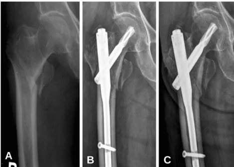
Fig. 3. (A) Preoperative antero-posterior (AP) radiograph of a 75 year-old female shows AO classification 2.2 intertrochanteric fracture. (B) Three weeks after internal fixation with PFNA, AP radiograph shows lateral wall fracture. (C) After conservative treatment, radiograph shows union of lateral wall fracture.
A B C
Table 3. Postoperative Complication
PFNA (N=47)
Lateral Wall Fracture 02
Lateral Sliding of the Blade (>10 mm) 09
Blade Penetration 02
Impending Penetration 02
AVN* 01
Total 16
*AVN; Avascular necrosis
돌출 및 대퇴 경부에 삽입된 지연나사의 후방돌출 등의 단 점이 12.5%까지 보고 되면서26), 새로운 디자인에 대한 요 구가 증가 되었다.
항회전 근위 대퇴 골수정(PFNA; Proximal Femoral Nail)은 기존 근위 대퇴 골수정과 비슷한 형태를 가지고 있으나 대퇴 경부에 각과 회전에 대한 안정성을 가지는 항 회전 나선 칼날(antirotation helical blade) 하나만을 삽 입하도록 고안되었다. 나선 칼날의 근위 1/2의 flange와 원위 1/2의 shaft는 잠금(locking) 전에는 자유로운 회전 이 가능하지만 잠금을 하게 되면 회전이 불가능하고 오직 활강(sliding)만 가능하게 되어 대퇴 골두가 회전하며 발 생하는 후방돌출을 예방할 수 있으며, 나선 칼날을 망치로
두드려서 삽입시키는 동안 flange가 회전하며 대퇴골두 내 의 해 면 골 (cancellolus bone)안 에 서 압 축 되 어 (compaction) 골조직과 고정물 사이의 접촉면을 증가시 켜 안정성을 나타내게 된다28). 본 연구에서도 평균 76.5세 로 대부분 골다공증이 있는 환자들이었으나, 4예에서 대 퇴골 골두 천공이 발생하긴 하였지만 전 예에서 골 유합을 얻을 수 있었다. 또한 유 등29)은 근위 대퇴 골수정과의 비 교 연구에서 수술시간이 두 군 간 통계적 유의성이 없었다 고 하였으나 이와 이2)는 근위 대퇴 골수정군에서 평균 73.0분, 항회전 근위 대퇴 골수정을 사용한 군에서 평균 56.7분으로 유의할만한 수술 시간의 감소가 있었다고 하 였다. 본 연구에서는 비교 연구가 아니나 평균 시간이 54
Fig. 4. (A) Preoperative antero-posterior (AP) radiograph of a 71 year-old female shows AO classification 2.2 intertrochanteric fracture. (B) Immediate postoperative AP radiograph shows internal fixation with PFNA. (C) AP radiograph 2 months after the operation shows blade penetration into the hip joint. (D) The blade was exchanged into shorter one. (E) Four months after the 2nd operation, AP radiograph shows union.
A B
D E
C
분으로 PFN 과 비교하여 상당한 수술 시간의 감소가 있었 다. 타 연구와 비교해 수술 전 보행 능력의 회복은 66% 로 타 연구에 비해 다소 낮게 측정되었다. 이 등30)은 80.9%
에서 술 전 보행 능력이 회복되었다고 보고하였고, Liu 등
31)은 74% 로 보고하였다. 그 이유로는 본 연구에서는 전 예가 불안정성 골절이었는데 반해, 이 등30)은 21예 중 10 예, Liu 등31)은 143예 중 19예가 안정성 골절이었다. 그리 고 Liu 등31)은 평균 연령이 67세로 본 연구(75.6세)보다 환자들의 나이가 다소 젊은 연령층이었다. 이와 같은 이유 로 타 연구에 비해 본 연구의 보행능력 회복률이 낮았던 것으로 보인다. 내고정물의 대퇴 골두 내 적절한 위치에 대해서는 의견이 많다. Davis 등7)과 Mulholland와 Gunn32)은 대퇴 골두 내 중심 위치를 권유하였고 Nunn33), Thomas34), Gundle 등35)은 상방 위치는 피할 것을 권유하 였으며 Kyle 등36)은 후방이나 후 중앙부에 위치시키는 것 이 좋다고 하였는데 저자들의 경우 Cleveland index 상 1, 2, 3등의 상방위치로 삽입된 경우가 없어 고정 실패와 의 상관 관계를 밝히기는 어려웠다. 그리고 술 후 정복의 적절성 여부에 대해 많은 저자들이7,37)해부학적 정복의 필 요성을 강조하였는데 특히 내측 및 후내측 피질골의 연속 성을 유지하는 것을 중요시 하였다.
적절한 나사의 활강은 골절부위의 감입을 유발시켜 moment arm을 감소시키며 골절 안정성을 높이고 골 유 합을 촉진시키는 것으로 알려져 있으나 나사 활강이 과도 할 경우, 충돌로 인한 술 후 통증 및 금속 고정물의 부전으 로 이어져 하지 단축이나 이로 인한 보행장애 등의 원인이 될 수 있다. 술 후 과도한 활강을 유발하는 이유로는 부정 확한 골절의 정복, 후내측 피질골의 분쇄 및 대전자 외벽 의 골절, 부적절한 골두 내 나사의 위치, 불량한 골질 등이 알려져 있다. 본 연구에서 blade의 활강은 평균 5.5 mm 로 10 mm이상은 15예가 관찰되었다. 위에서 말한 부적 절한 정복이 5예, BMD -2.5 이하의 골다공증은 9예에서 관찰되었다. 하지만 15명중 피부 자극 및 통증을 호소하 는 예는 단 4예로 이는 PFNA blade tip 부위가 둥근 모양 이기 때문에 피부나 근막에 자극이 심하지 않는 것으로 사 료 된 다38). blade의 내 측 이 동 에 의 한 골 두 천 공 은 Alexander 등39)도 보고한 바가 있다. PFNA는 blade가 해 면골을 압축시키며 삽입되기 때문에 내반(varus) 및 회전 (rotation) 안정성은 뛰어난 것으로 보인다. 그래서 내반 및 회전 변형에 의한 cutting-out이 적은 대신 내측 이동 혹은 과도한 활강에 의한 cut-through(penetration)가 이 기구의 문제점의 하나로 생각된다. 감마정의 경우 지연나 사(lag screw)가 골수정에서 완전히 분리되어 골반 내까 지 이동하여 모두 인공관절로 치환한 반면40), PFNA는 blade의 외측 끝(lateral end)이 골수정에서 더 이상 내측 으로 분리되지 않도록 잠금 장치가 되어있기 때문에 blade가 골수정과 분리되어 골반 내로 이동할 우려는 없
다. 본 연구에서도 골두의 천공 및 임박 천공이 4예 있었 지만, 연골의 손상이 적어 관절 치환술 대신에 짧은 blade 로 교체하여 해결할 수 있었다.
결 론
항회전 근위 대퇴 골수정은 골다공증을 동반한 고령의 불안정성 대퇴 전자 주위 골절 환자에서 짧은 수술 시간, 양호한 보행능력 및 적은 합병증으로 인해 유용한 치료 방 법의 하나로 생각된다. 하지만 기술적 어려움, 골두천공 및 blade의 과도한 활강 등의 합병증이 나타나는 문제점 을 가지고 있어 이를 줄이기 위한 노력이 지속적으로 필요 할 것으로 사료된다.
REFERENCES
01. Hwang DS, Rhee KJ, Choi JH. Recovery of walking ability after treatment of unstable intertrochanteric fractures in elderly patients: comparision of compression hip screw to primary hemiarthroplasty. J Korean Hip Soc. 1999;11:22-9.
02. Lee JY, Lee SY. Treatment of the proximal femoral extracapsular fracture with proximal femoral nail antirotation (PFNA): comparision with proximal femoral nail (PFN). J Korean Hip Soc. 2007;19:183-9.
03. Richmond J, Aharonoff GB, Zuckerman JD, Koval KJ.
Mortality risk after hip fracture. 2003. J Orthop Trauma.
2003;17 Suppl:S2-5.
04. Evans EM. Trochanteric fractures; a review of 110 cases treated by nail-plate fixation. J Bone Joint Surg Br. 1951;
33B:192-204.
05. Flores LA, Harrington IJ, Heller M. The stability of intertrochanteric fractures treated with a sliding screw- plate. J Bone Joint Surg Br. 1990;72:37-40.
06. Pugh WL. A self-adjusting nail-plate for fractures about the hip joint. J Bone Joint Surg Am. 1955;37-A:1085-93.
07. Davis TR, Sher JL, Horsman A, Simpson M, Proter BB, Checketts RG. Intertrochanteric femoral fractures.
Mechanical failure after internal fixation. J Bone Joint Surg Br. 1990;72:26-31.
08. Madsen JE, Naess L, Aune AK, Alho A, Ekeland A, strømsøe K. Dynamic hip screw with trochanteric stabilizing plate in the treatment of unstable proximal femoral fractures: a comparative study with the Gamma nail and compression hip screw. J Orthop Trauma. 1998;
12:241-8.
09. Rha JD, Kim YH, Yoon SI, Park TS, Lee MH. Factors affecting sliding of the lag screw in intertrochanteric fractures. Int Orthop. 1993;17:320-4.
10. Adams CI, Robinson CM, Courl-Brown CM, McQueen MM. Prospective randomized controlled trial of an intramedullary nail versus dynamic screw and plate for intertrochanteric fractures of the femur. J Orthop Trauma.
2001;15:394-400.
11. Albareda J, Laderiga A, Palanca D, Paniagua L, Seral F.
Complications and technical problems with the gamma nail. Int Orthop. 1996;20:47-50.
12. Friedl W, Colombo-Benkmann M, Dockter S, Machens HG, Mieck U. Gamma nail osteosynthesis of per- and subtrochanteric femoral fractures. 4 years experiences and their consequences for further implant development.
Chirurg. 1994;65:953-63.
13. Simmermacher RK, Bosch AM, Van der Werken C. The AO/ASIF-proximal femoral nail (PFN): a new device for the treatment of unstable proximal femoral fractures.
Injury. 1999;30:327-32.
14. Cleveland M, Bosworth DM, Thompson FR, Wilson HJ Jr, Ishizuka T. A ten-year analysis of intertrochanteric fractures of the femur. J Bone Joint Surg Am. 1959;41-A:
1399-408.
15. Baumgaertner MR, Curtin SL, Lindskog DM, Keggi JM.
The value of the tip-apex distance in predicting failure of fixation of peritrochanteric fractures of the hip. J Bone Joint Surg Am. 1995;77:1058-64.
16. Koval KJ, Skovron ML, Aharonoff GB, Meadows SE, Zuckerman JD. Ambulatory ability after hip fracture. A prospective study in geriatric patients. Clin Orthop Relat Res. 1995;310:150-9.
17. Choo SK, Oh HK, Kim YC, Lee DB. Excessive sliding of compression hip screw for the treatment of intertrochanteric fracture in elderly. J Korean Hip Soc.
2007;19:190-6.
18. Koval KJ, Zuckerman JD. Hip fractures: I. Overview and evaluation and treatment of femoral-neck fractures. J Am Acad Orthop Surg. 1994;2:141-9.
19. Koval KJ, Zuckerman JD. Hip fractures: II. Evaluation and treatment of intertrochanteric fractures. J Am Acad Orthop Surg. 1994;2:150-6.
20. Leung KS, Chen CM, So WS, et al. Multicenter trial of modified Gamma nail in East Asia. Clin Orthop Relat Res.
1996;323:146-54.
21. Radford PJ, Needoff M, Webb JK. A prospective randomised comparision of the dynamic hip screw and the gamma locking nail. J Bone Joint Surg Br. 1993;75:789-93.
22. Ahn SJ, Park JH. Proximal femoral nail (PFN) for the treatment of the femoral trochanteric fracture. J Korean Fract Soc. 2004;17:7-12.
23. Boldin C, Seibert FJ, Frankhauser F, Peicha G, Grechenig W, Szyszhowitz R. The proximal femoral nail (PFN)--a minimal invasive treatment of unstable proximal femoral fractures: a proxpective study of 55 patients with a follow- up of 15 months. Acta Orthop Scand. 2003;74:53-8.
24. Kim BS, Lew SW, Ko SH, Cho SD, Yang JH, Park MS.
Treatment of femoral intertrochanteric fracture with proximal femoral nail. J Korean Fract Soc. 2004;17:1-6.
25. Sung YB, Sohn YJ, Yum JK, et al. Proximal femoral nail (PFN) for intertrochanteric fracture: long-term follow up results. J Korean Hip Soc. 2005;17:141-8.
26. Papasimos S, Koutsojannis CM, Panagopoulos A, Megas P, Lambiris E. A randomised comparison of AMBI, TGN and PFN for treatment of unstable trochanteric fractures.
Arch Orthop Trauma Surg. 2005;125:462-8.
27. Moon YW, Suh DH, Kang ST, Kwon DJ, Ji YN, Lee KB.
The proximal femoral nail for intertrochanteric fracture of the femur. J Korean Soc Fract. 2003;16:29-36.
28. Steinberg GG, Desai SS, Kornwitz NA, Sullivan TJ. The intertrochanteric hip fracture. A retrospective analysis.
Orthopedics. 1988;11:265-73.
29. Yoo JH, Park JS, Noh KC, et al. The results of proximal femoral nail antirotation: a comparative study with proximal femoral nail. J Korean Hip Soc. 2008;20:286-92.
30. Lee KJ, Min BW, Cho CH, Song KS, Bae KC, Kim SG.
Results of treating senile osteoporotic peritrochanteric fracture with proximal femoral nail antirotation (PFNA). J Korean Hip Soc. 2009;21:162-8.
31. Liu Y, Tao R, Liu F, et al. Mid-term outcomes after intramedullary fixation of peritrochanteric femoral fractures using the new proximal femoral nail antirotation (PFNA). Injury. 2010;41:810-7.
32. Mulholland RC, Gunn DR. Sliding screw plate fixation of intertrochanteric femoral fractures. J Trauma. 1972;12:
581-91.
33. Nunn D. Sliding hip screws and medial displacement osteotomy. J R Soc Med. 1988;81:140-2.
34. Thomas AP. Dynamic hip screws that fail. Injury.
1991;22:45-6.
35. Gundle R, Gargan MF, Simpson AH. How to minimize failures of fixation of unstable intertrochanteric fractures.
Injury. 1995;26:611-4.
36. Kyle RF, Gustilo RB, Premer RF. Analysis of six hundred and twenty-two intertrochanteric hip fractures. J Bone Joint Surg Am. 1979;61:216-21.
37. Jensen JS. Classification of trochanteric fractures. Acta Orthop Scand. 1980;51:803-10.
38. Simmermacher RK, Ljungqvist J, Bail H, et al. The new proximal femoral nail antirotation (PFNA) in daily practice: results of a multicentre clinical study. Injury.
2008;39:932-9.
39. Brunner A, Jo¨ckel JA, Babst R. The PFNA proximal femur nail in treatment of unstable proximal femur fracture--3 cases of postoperative perforation of the helical blade into the hip joint. J Orthop Trauma. 2008;22:731-6.
40. Lucke M, Burghardt RD, Siebenlist S, Ganslmeier A, Sto¨ckle U. Medial migration of lag screw with intrapelvic dislocation in gamma nailing--a unique problem? A report of 2 cases. J Orthop Trauma. 2010;24:e6-e11.
국 국문문초초록록
불안정성 대퇴 전자 주위 골절을 가진 환자에서 항회전 근위 대퇴 골수정을 이용한 치료결과
성열보∙조성일
인제대학교 의과대학 상계백병원 정형외과학교실
목적: 불안정성 대퇴 전자 주위골절을 가진 환자의 치료에 있어서 항회전 근위 대퇴 골수정 (PFNA)을 이용한 치료결과에 대해 알아보고자 한다.
대상 및 방법: 2006년 9월부터 2010년 3월까지 PFNA를 이용하여 치료한 불안정성 대퇴 전자 주 위골절 환자 187명 중 47예를 대상으로 하였다. 평균 골 유합 기간, blade의 활강 정도 및 대퇴 경간각의 차이, 활동 정도 및 합병증 등을 점검하였다.
결과: 방사선학적 골 유합은 평균 15.8주에 얻을 수 있었으며 술 후 66% 에서 수술 전 보행을 회 복할 수 있었다. blade의 평균 활강은 5.5 mm 였으며 대퇴 경간각은 평균 4.4。의 내반 전위를 보 였다. 골두의 천공 및 임박 천공이 각각 2예씩 발생하였으며, 1예에서 외상 후 골두 무혈성 괴사가 발생하였다. 그리고 2예에서 술 후 추시 중 외측벽 골절(lateral wall fracture)이 발생하였다.
결론: 불안정성 대퇴전자주위골절에 있어서 항회전 근위 대퇴골수정을 이용한 수술은 수술 시간 이 짧고, 환자의 거동능력 회복이 양호하며, 합병증도 적어 유용한 치료법 중 하나라고 생각된다.
그러나 골두 천공 같은 심각한 합병증을 막기 위해서는 세심한 시술이 필요하다.
색인단어: 대퇴골, 불안정성 대퇴전자 주위 골절, 항회전 근위 대퇴 골수정