Introduction
Cardiac myxoma is the most common benign tumor of the heart. Complete surgical removal of the myxoma is usually curative. However, low incidence of recurrence and metastasis has been reported with the brain being the most common site of cardiac myxoma metastasis (1). We report a rare case of delayed
multiple cerebral metastases from the completely resected cardiac myxoma without evidence of local tumor recurrence.
Case Report
A 49-year-old female patient was admitted in the hospital due to sudden onset of left side weakness. She did not have any history of medical disease or recent
JKSMRM 15:165-169(2011)
1Department of Radiology, NHIC Ilsan Hospital
Delayed Cerebral Metastases from Completely Resected Cardiac Myxoma: Case Report and
Review of Literature
Ah Hyun Kim2, Jae-Wook Lee1, Mi-Kyung Lee3, Pyeong Ho Yoon1, Min Jung Kim2
Cardiac myxoma is the most common benign tumor of the heart. However, low incidence of recurrence and metastasis has been reported. A 49-year-old female patient was admitted in the hospital due to sudden onset of left side weakness.
Magnetic resonance imaging (MRI) of brain showed multifocal areas of diffusion restriction on diffusion weighted images. Echocardiography was performed to evaluate the cause of embolic brain infarction and cardiac myxoma was found in the left atrium. The patient underwent complete excision of the mass. One year later, the patient was readmitted with symptoms of dysarthria. Brain MRI showed newly developed multiple hemorrhagic metastatic lesions. The patient underwent radiotherapy of the metastatic lesions. Although rare, cardiac myxoma can cause delayed metastasis. We report a rare case of delayed multiple cerebral metastases from the completely resected cardiac myxoma.
Index words :Cardiac myxoma
Delayed cerebral metastases Magnetic resonance imaging
drug therapy. Brain magnetic resonance imaging (MRI) was performed and revealed multifocal areas of high signal intensity (SI) in bilateral cerebral hemispheres, cerebellar hemispheres, pons and right thalamus on T2- weighted images and diffusion-weighted image (DWI) which did not show enhancement on post contrast T1- weighted images. These lesions did not correlate with vascular territories, therefore, the likelihood of acute embolic infarction from cardiac origin was suggested.
Cardiac echocardiography was performed to further evaluate the cause of multifocal brain infarction. On echocardiography, a large round-shaped homogenous highly mobile mass was attached to the left atrium and excision of the mass was performed under cardiopul- monary bypass. Grossly, a loose gelatinous friable mass with a broad base on atrial septum was seen and microscopic findings showed spindled and stellite cells with eosinophilic cytoplasm without significant cytologic atypia (Fig. 1). The final histopathologic diagnosis was reported as cardiac myxoma. About one year later, the patient was readmitted to the hospital with symptoms of dysarthria. Brain MRI showed newly developed multiple hemorrhagic and enhancing lesions with perilesional edema and adjacent superficial siderosis in bilateral cerebral hemispheres (Fig. 2). The previously noted multifocal embolic areas showed complete encephalomalacia in this study. Evaluation of echocardiography did not show evidence of recurrence and full evaluation was performed to rule out the possibility of brain metastases from another origin.
However, no other focus of primary malignancy was found. Although histopathologic confirmation of the cerebral lesions was not performed, hemorrhagic cerebral metastases from the previously resected cardiac myxoma were suggested. The patient underwent whole brain radiation treatment 35 Gy/24 fractions for 3 weeks and follow up MRI revealed slight improvement of the multiple enhancing cerebral
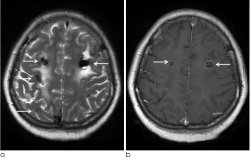
a b
Fig. 2. Patient unerwent MRI due to symptoms of sudden dysarthria 1- year after resection of cardiac myxo- ma.
(a) T2-weighted image shows multi- ple peripheral dark signal intensity rims in bilateral cerebral hemi- spheres, and adjacent superficial siderosis, suggestive of hemorrhagic metastases. (b) T1-post contrast im- age shows enhancement of the metastatic lesions.
Fig. 1. A 49-year-old female patient underwent resection of the cardiac myxoma that caused multiple cerebral embolic infarction. Photomicrograph of the histopathologic speci- men shows spindled and stellite cells with eosinophilic cy- toplasm and round to slender nucleus embedded in pale edematous myxoid matrix. No significant cytologic atypia including marked pleomorphism and abnormal mitotic fig- ures was seen (H-E, ×400).
lesions (Fig. 3).
Discussion
Cardiac myxoma is the most common benign heart tumor, most commonly found in the left atrium (1).
Cardiac myxoma is usually a sporadic lesion most commonly found in women over 30 years. However, it has been also related to the autosomal dominant syndrome called Carney complex characterized by spotty pigmentation (blue nevi and lentigines), myxomas (cardiac, cutaneous, and mammary), endocrine over-activity (Cushing’s syndrome and acromegaly), testicular tumors and schwannomas (2).
Patients with cardiac myxoma usually present with one of the classic triad; obstructive cardiac signs, embolic signs or systemic constitutional manifestations at the time of diagnosis (3). Because most cardiac myxomas are located in the left atrium, systemic emboli are frequent and occur in 10-45% of myxoma, with more than two-thirds of myxomatous emboli migrating to the central nervous system (4). In spite of the benign nature of cardiac myxoma, it infrequently recurs at the site of the original tumor or metastasizes to extracardiac sites with the brain known to be the most frequent sites for metastasis (5). Contrary to primary myxoma,
associated with Carney complex. Recurrence is reported to be associated with incomplete excision, multifocality and embolism of tumor fragments (6).
Metastasis of cardiac myxoma is rare and is known to be intravascular and presents with delayed occurrence after resection of the cardiac lesion (1). Metastatic lesions sometimes present earlier than the diagnosis of the primary lesion, however, has been diagnosed up to 8 years later than the primary lesion. These lesions are usually multiple and most commonly located in frontoparietal regions of the brain. Previous reports have shown interleukin-6 (IL-6) to be involved in the induction of intracellular adhesion molecule-1 (ICAM- 1) during the period of embolization and metastasis (7).
The mechanism of tumor metastases has not been fully established. However, it has been postulated that tumor tissues may grow into the walls of the vessels causing focal disruption of the internal elastic lamina, providing a nidus for cerebral hemorrhage and subsequent growth of metastatic tumor tissue (8). It is most likely, the intracerebral hemorrhagic metastases resulted from silent emboli of the cardiac myxoma occuring in the pre-operative period or due to operative manipulation. These myxomatous emboli survive in the vessels for varying unpredictable durations and later cause focal disruption of internal elastic lamina
a b
Fig. 3. Follow up MRI was per- formed 3-weeks after whole-brain radiotherapy.
(a) T2-weighted image shows resid- ual hemorrhagic metastatic lesions in the brain. (b) T1-post contrast im- age shows decrease in size of en- hancing portions suggestive of favor- able therapy response.
imaging findings of cerebral brain metastasis from cardiac myxoma correlate with the tumor metastatic mechanism. The typical angiographic features of myxoma-associated aneurysms are well described as fusiform outpouchings or saccular anerusyms (9). CT morphology of cerebral myxoma metastasis usually appears as a hyperdense lesion with some contrast enhancement due to extensive hemorrhage (10). MR images show peripheral dark signal intensity rim on T1 and T2-weighted images which can be explained by dense accumulation of iron and hemosiderin from chronic recurrent hemorrhage, and occasionally superfical siderosis. The multiplicity of the lesion, the typical imaging findings of hemorrhage and enhancement of the cerebral lesions in patients with completely resected cardiac myxomas such as our case can suggest a diagnosis of cerebral metastasis.
Treatment has not been well-established. Surgery is usually performed in cases having one or two metastatic lesions showing favorable prognosis. For multiple lesions, whole brain radiotherapy with 50 Gy doses has been preferred in reported literature.
However, there have been only a few reports on the outcome of whole brain radiotherapy and further study would be necessary to propose a treatment guideline for these patients.
In summary, cardiac myxomas show good prognosis when completely resected. However we report here a rare case of delayed cerebral metastases from completely resected cardiac myxoma. Periodic long- term follow-up evaluation including brain imaging would be helpful for early detection of both recurrence
and metastasis.
References
1.Amano J, Kono T, Wada Y, et al. Cardiac myxoma: its origin and tumor characteristics. Ann Thorac Cardiovasc Surg 2003;9:215-221
2.Stratakis CA, Kirschner LS, Carney JA. Carney complex:
diagnosis and management of the complex of spotty skin pigmentation, myxomas, endocrine overactivity, and schwannomas. Am J Med Genet 1998;80:183-185
3.Pinede L, Duhaut P, Loire R. Clinical presentation of left atrial cardiac myxoma. A series of 112 consecutive cases.
Medicine (Baltimore) 2001;80:159-172
4.Rodriguez FJ, Brown RD, Mohr JP, et al. Embolic atrial myxoma with neoplastic aneurysm formation and haemorrhage: a diagnostic challenge. Neuropathol Appl Neurobiol 2006;32:213-216
5.Altundag MB, Ertas G, Ucer AR, et al. Brain metastasis of cardiac myxoma: case report and review of the literature. J Neurooncol 2005;75:181-184
6.McCarthy PM, Piehler JM, Schaff HV, et al. The significance of multiple, recurrent, and “complex”cardiac myxomas. J Thorac Cardiovasc Surg 1986;91:389-396
7.Wada A, Kanda T, Hayashi R, Imai S, Suzuki T, Murata K.
Cardiac myxoma metastasized to the brain: potential role of endogenous interleukin-6. Cardiology 1993;83:208-211 8.Ng HK, Poon WS. Cardiac myxoma metastasizing to the
brain. Case report. J Neurosurg 1990;72:295-298
9.Hwang BJ, Connelly MM, Lev MH. Distinctive MR imaging appearance of hemorrhagic cerebral aneurysms associated with atrial myxoma. AJR Am J Roentgenol 2001;177:925-927 10.Hofmann E, Becker T, Romberg-Hahnloser R, Reichmann H,
Warmuth-Metz M, Nadjmi M. Cranial MRI and CT in patients with left atrial myxoma. Neuroradiology 1992;34:57- 61
통신저자 : 이재욱, (410-719) 경기도 고양시 일산동구 백석동 1232, 국민건강보험공단 일산병원 영상의학과 Tel. 82-31-900-0873 Fax. 82-31-900-0856 E-mail: [email protected]
완전히 절제된 심장 점액종의 지연된 뇌전이: 증례보고 및 문헌고찰
1국민건강보험공단 일산병원 영상의학과
2연세대학교 의과대학 세브란스병원 영상의학과
3국민건강보험공단 일산병원 병리과
김아현2∙이재욱1∙이미경3∙윤평호1∙김민정2
심장점액종은 심장의 가장 흔한 양성 종양이다. 하지만 국소전이와 원격전이의 증례들이 낮은 빈도로 보고되어 있 다. 뇌경색을 주소로 내원한 49세 여자 환자의 심장초음파에서 심장 점액종이 발견되었으며 수술로 절제 1년 후에 구 음장애가 발생하여 촬영한 자기공명영상에서 여러개의 조영증강이 되는 출혈성뇌전이 병변들이 관찰되었다. 방사선 치료 후 추적 자기공명영상에서 병변의 크기들이 작아지는 것을 확인할 수 있었다. 양성 종양이지만 심장 점액종은 드 물게 지연되어 전이를 보일 수 있다. 저자들은 심장 점액종을 수술로 완전히 절제 후 지연되어 나타난 뇌전이를 경험 하여 증례 및 문헌고찰을 하는 바이다.
대한자기공명의과학회지 15:165-169(2011)