D I A B E T E S & M E T A B O L I S M J O U R N A L D I A B E T E S & M E T A B O L I S M J O U R N A L
This is an Open Access article distributed under the terms of the Creative Commons Attribution Non-Commercial License (https://creativecommons.org/licenses/by-nc/4.0/) which permits unrestricted non-commercial use, distribution, and reproduction in any medium, provided the original work is properly cited.
Consequences of Obesity on the Sense of Taste: Taste Buds as Treatment Targets?
Kerstin Rohde1, Imke Schamarek2, Matthias Blüher1,2
1 Helmholtz Institute for Metabolic, Obesity and Vascular Research (HI-MAG) of the Helmholtz Center Munich at the University of Leipzig and University Hospital Leipzig, Leipzig,
2Medical Department III (Endocrinology, Nephrology and Rheumatology), University of Leipzig, Leipzig, Germany
Premature obesity-related mortality is caused by cardiovascular and pulmonary diseases, type 2 diabetes mellitus, physical dis- abilities, osteoarthritis, and certain types of cancer. Obesity is caused by a positive energy balance due to hyper-caloric nutrition, low physical activity, and energy expenditure. Overeating is partially driven by impaired homeostatic feedback of the peripheral energy status in obesity. However, food with its different qualities is a key driver for the reward driven hedonic feeding with tre- mendous consequences on calorie consumption. In addition to visual and olfactory cues, taste buds of the oral cavity process the earliest signals which affect the regulation of food intake, appetite and satiety. Therefore, taste buds may play a crucial role how food related signals are transmitted to the brain, particularly in priming the body for digestion during the cephalic phase. Indeed, obesity development is associated with a significant reduction in taste buds. Impaired taste bud sensitivity may play a causal role in the pathophysiology of obesity in children and adolescents. In addition, genetic variation in taste receptors has been linked to body weight regulation. This review discusses the importance of taste buds as contributing factors in the development of obesity and how obesity may affect the sense of taste, alterations in food preferences and eating behavior.
Keywords: Dysgeusia; Feeding behavior; Food preferences; Obesity; Taste; Taste buds; Taste perception
Corresponding authors: Kerstin Rohde https://orcid.org/0000-0001-5316-6870 Helmholtz Institute for Metabolic, Obesity and Vascular Research (HI-MAG) of the Helmholtz Center Munich at the University of Leipzig and University Hospital Leipzig, Philipp-Rosenthal-Straße 27, 04103 Leipzig, Germany
E-mail: [email protected] Matthias Blüher https://orcid.org/0000-0003-0208-2065
Helmholtz Institute for Metabolic, Obesity and Vascular Research (HI-MAG) of the Helmholtz Center Munich at the University of Leipzig and University Hospital Leipzig, Philipp-Rosenthal-Straße 27, 04103 Leipzig, Germany
INTRODUCTION
Obesity is defined as an excessive and abnormal storage of fat which can cause pathological conditions. Obesity increases the risk to develop type 2 diabetes mellitus, cardiovascular diseas- es, hypertension, fatty liver disease, sleep apnea, osteoarthritis, dyslipidemia, certain types of cancer, disruption of endocrine circuits, and low-grade systemic inflammation [1]. Although multiple factors contribute to the development of obesity, one crucial mechanism implicated is a profound mismatch be- tween calorie intake and expenditure. In the westernized
world, the need to engage in physical active behavior is vanish- ing whereas at the same time caloric dense food is constantly available. Food intake no longer fulfills the mere function of nourishment and sustaining physiological integrity and func- tion but serves to satisfy hedonic needs. Hedonic food intake, hence the consumption of food for its palatability and related
“pleasure” is fueled by flavor, which is the result of a complex interplay of sensory perception in which taste comprises an important role [2]. Therefore, the sense of taste plays a central role in the development of obesity as it contributes to food se- lection, caloric intake and consequently body weight regula- https://doi.org/10.4093/dmj.2020.0058
pISSN 2233-6079 · eISSN 2233-6087
tion (Fig. 1).
Indeed, accumulating evidence connects taste to food selec- tion and obesity [3-5]. People with obesity display decreased taste sensitivity [6,7]. Intriguingly, it appears that taste does not only contribute to obesity, but is also affected by obesity as ac- cumulating evidence suggests that the primary taste tissue, the tongue, is an obesity target organ [8,9]. For example, the adi- pokine leptin is involved in sweet sensation of sweet sensing taste cells [10,11]. Importantly, obesity has been linked to a about 25% reduction in taste bud abundancy [9]. Alterations in taste sensation and taste buds may become a novel target for
urgently needed obesity prevention and treatment strategies.
Therefore, this review discusses the current understanding of how the sense of taste is regulated on the level of taste buds and how this modulates food intake in obesity. We also highlight that obesity itself may affect the sense of taste by distinct mo- lecular mechanisms.
THE SENSE OF TASTE
The ingestion of food leads to a converging sensory perception which humans experience as a wide array of flavors, encoded
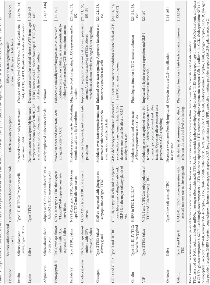
Fig. 1. Effects of obesity on the sense of taste and its relation to food intake. Food intake is driven by the interplay of hedonic and homeostatic feedback. During digestion, a variety of factors (meal quantity, nutrients, energy status, fermentation) feedback the nutrient load and energy status of the body towards the brain and control hunger and satiety circuits. However, the earliest signal priming the cephalic phase response is food quality reflected by smell and taste, which, moreover is the key driver for the reward driven hedonic eating. The earliest signals of food intake are processed in taste buds located on the tongue surface. Taste buds are very complex and consist of three functional taste cells (type I, type II, type III) and basal cells which can develop into either adult taste cell. Basal cells are post-mitotic cells which derive from proliferating progenitor cells clustering outside the taste bud and en- suring a lifelong cell turnover. Obesity is associated with alterations in taste sensation. This may be partially explained by the con- tribution of obesity on taste bud signaling, homeostasis and renewal. Thereby, the huge variety of factors potentially influencing the sense of taste on the level of taste buds (adipokines, cytokines, hormones etc.) may derive from the circulation, but also the sal- ivary glands, local fat cells or even endocrine taste cells. CCK, cholecystokinin; GLP-1, glucagon like peptide 1; PYY, peptide YY.
Type I Type II Type IIi Basal cell (Type IV) Progenitor cell Keratinocyte
Taste bud renewal/taste sensation
• Taste bud survival
• Proliferation of stem cells
• Cell fate
• Taste receptor abundancy
• Cell signalling
Meal quality Smell Taste
Meal quality Stretch osmotic load
CCK, GLP-1, GhrelinPYY, Nutrient status
Insulin, Glucose Amino acids,
oxidation Energy status
Insulin, Leptin Adiponectin Fermentation Hedonic feeding Homeostatic feeding
by the sense of taste. Although an uncountable range of flavors exists, it is generally accepted that only five taste qualities can be perceived by humans, namely sweet, sour, bitter, salty, and umami [12,13]. Nevertheless, the existence of “fatty” as a sixth taste quality is controversially discussed [14]. Taste perception commences when molecules from food (tastants) reach taste buds. Humans usually have 5,000 to 10,000 taste buds which are mainly, but not exclusively embedded in the epithelium of the tongues surface [15]. However, some taste buds are also present on the palate, epiglottis, pharynx, and esophagus [16- 18]. Lingual taste buds cluster into gustatory papillae which are categorized into papillae vallate, foliatae, and fungiformis ac- cording to their shape. They are located at the posterior, at the posterior-lateral sides and the anterior two-thirds of the tongue, respectively [19]. While papillae vallate accommodate hundreds of taste buds, papillae foliatae express around 50 and papillae fungiformis 3 to 5 taste buds [15]. Each taste bud is comprised of approximately 50 to 100 taste bud cells (TBCs) which are chemosensory cells of epithelial origin [20]. TBC can be categorized into three different cell types according to functional and structural features including patterns of protein expression (Fig. 2) [15].
All types of cells are present in each taste bud, with type I cells comprising roughly 50%, and type II and type III cells contribute 15% to 20% each [15]. In recent years, taste percep- tion has been very well characterized. Here, we focus on the major tasks of the diverse taste cells rather than describing de- tailed signaling mechanisms which have been recently re- viewed elsewhere [15].
Type I cells mainly serve for the maintenance of the support- ing structure of the taste bud. They display cytoplasmatic la- mellar processes which enfold the other cells comprising the taste bud [21]. This is believed to regulate molecules involved in cell signaling by reducing their dispersion such as clearing spare neurotransmitter level secreted from type II and type III cells or buffer K+ (potassium) by expressing cognate ion chan- nels [22-24].
Type II cells are responsible for the perception of sweet, bitter, and umami taste via binding of tastants to G-protein coupled receptors (GPCR) [25,26]. These receptors function as mono- mers or dimers. Sweet tastants bind to receptor type 1 member 2 (T1R2 or TAS1R2) and member 3 (T1R3 or TAS1R3), but be- side this well recognized mechanism, T1R-independent mech- anisms appear to be implicated in sweet taste recognition [27,28]. Thus, glucose transporter type 4 (GLUT4) and sodium/
glucose transporter 1 (SGLT1) have been identified for trans- porting glucose into type II cells which results in the depolar- ization of the taste cell. This may be primarily of relevance for the cephalic phase insulin response, preparing for incoming food rather than signaling taste per se [27]. Heterodimers of T1R1 and T1R3 serve as receptor for the detection of L-gluta- mate (rodents) and L-aspartate (humans) and are recognized as umami [29,30]. The presence of glutamate receptors has been demonstrated to be important for the response to ingested glu- tamate in mice [31]. Bitter taste is sensed by T2Rs (or TAS2Rs) which comprises 25 receptor encoding genes in humans giving a hint for the various compounds stimulating bitter taste [32].
In contrast to T1Rs, only one type of bitter taste receptor is ex- pressed per taste cell pointing towards the enormous role to distinguish diverse bitter compounds in contrast to just detect nutritious, sweet and savory food [33]. A single type II cell mainly responds to one specific taste quality forming separate populations of type II cells. However, this does not imply that the taste of a specific quality is restricted to a certain location on the tongue, as each taste bud located anywhere on the tongue harbors sweet, umami and bitter type II cells [13]. Despite their diversity, T1Rs and T2Rs both activate gustducin subunits when being stimulated by a tastant which further stimulate phospholipase Cβ2 and raise the intracellular Ca2+ level [34].
This results in opening transient receptor potential cation chan- nel subfamily M member 5 (TRPM5) followed by a subsequent depolarization of taste cells. The latter process occurs by atypi- cal and non-vesicular secretion of adenosine triphosphate (ATP) through calcium homeostasis modulator protein 1 (CALHM1) activating afferent nerve fibers [34-36]. Secreted ATP feeds back to type II cells in order to push further signaling or to stimulate type III cells [37]. Additionally, acetylcholine (Ach) is secreted by type II cells and serves as autocrine media- tor for the secretion of ATP [38]. A degradation of extracellular ATP to adenosine diphosphate (ADP) is mediated enzymati- cally by ectonucleoside triphosphate diphosphohydrolase 2 (NTPDase2) in type I cells, which in turn increases the re- sponse to sweet stimuli by binding of ADP to adenosine 2B (A2B) receptors in type II cells [39].
Type III cells, are referred to as presynaptic, neuron-like cells, as they form ordinary, neuronal synapses with afferent nerve fi- bers at the basal side of a taste bud. These cells release common neurotransmitters such as gamma aminobutyric acid (GABA), serotonin (5-HT) and noradrenaline upon depolarization via voltage gated Ca2+ channels [25,40]. In addition to secreted ATP
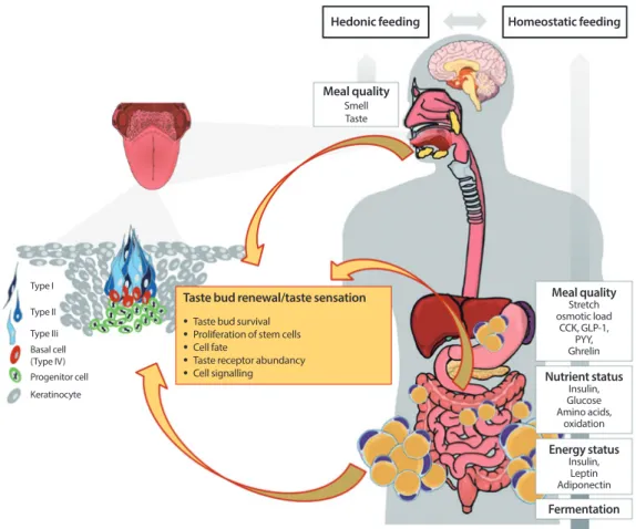
Fig. 2. Schematic presentation of cell signaling within taste bud cells. Type II cells express G-protein coupled receptors (GPRs) for bitter (taste receptors type 2 [T2Rs]), sweet (taste receptor type 1 member 1 [T1R1], T1R2, T1R3) and umami (T1R1+T1R3), but also GPR40 and 120 and glycoprotein 4 (also named cluster of differentiation 36, CD36) transducing the taste quality “fatty” [15].
In addition, metabotropic glutamate receptors (mGluRs) and glucose- and sodium/glucose transporters (GLUTs, SGLT1) are thought to transduce umami and sweet, respectively [27,31]. Binding of tastants to their cognate receptors increase intracellular calcium level ([Ca2+]i) which activates transient receptor potential cation channel subfamily M member 5 (TRPM5), a Ca2+/Na+ cotransporter [34]. This leads finally to an activation of the calcium homeostasis modulator protein 1 (CALHM1) which is meant to release adenosine triphosphate (ATP) [35,36]. ATP signals to afferent nerve fibers via binding to P2X receptors but also feeds back in an autocrine fashion via binding to P2X and P2Y receptors on type II cells [36]. In addition, type II cells secrete Acetylcho- line (Ach) which further stimulates ATP secretion [38]. Moreover, ATP activates type III cells by binding to P2Y receptors [37].
This in turn initiates the release of neurotransmitters gamma aminobutyric acid (GABA), serotonin (5-HT) and noradrenaline into the presynaptic space as consequence of raised [Ca2+]i [25,40]. In addition, this release is mediated as a result of changes in pH through the uptake of H+ by ion channels such as polycystic kidney disease proteins 1 like 3 and 2 like 1 (PKD1L3, PKD2L1), in- ward rectifying K+ channel (KIR2.1) and the epithelial Na+ channel (ENaC) [41-45]. GABA and 5-HT activate afferent nerve fibers but feedback to type II cells in order to decrease further ATP secretion [15]. Glutamate is released by activated nerve fibers and tune the release of GABA and 5-HT, finally shutting down ATP secretion from type II cells [15]. Type I cells seem to have glia like function as they express several ion channels (inward rectifying K+ channel [ROMK], glutamate-aspartate transporter [GLAST], ENaC) which are supposed to clear ion currents [22,23,35]. Moreover, as ENaC has been identified in type I cells and this is thought to be the main receptor for the detection of low NaCl-salts, these cells may transduce salty [46]. Further channels involved in transducing salty are mucolipin 3 (TRPML3) and transient receptor potential cation channel subfamily V member 1 (TRPMV) [47]. However, their cellular localization has yet to be elucidated. In addition, the enzyme nucleoside triphosphate diphosphohy- drolase-2 (NTPDase2) located on the surface of type I cells, is responsible for the degradation of ATP to adenosine diphosphate (ADP) [39]. In turn, sweet receptor expressing type II cells bind ADP by adenosine 2B receptors (A2B) which further increases sweet sensation [39]. TRPV1, transient receptor potential cation channel subfamily V member 1; PLCβ2, Phospholipase C beta 2.
Type I Type II Type III
Sweet Fatty
Sweet ?
Sour Umami
Bitter Salty
TRPML3 TRPV1 ENaC
Glutamat GLAST
K+ROMK
K+ Na+
Ca2+
A2B
ATP
ATP Ach ADP
TRPM5 NTPDase2
Na+ CALHM1 GABA
5HTNoradrenalin (?)
P2X; P2Y TRPM5
Glutamat P2Y
SGLT1 GLUTs T1R2+T1R3
T1R1+T1R3 T2Rs
CD36 GPR40 GPR120
mGluRs (?)
PKD1L3 PKD2L1 KIR2.1
ENaC H+
Na+
P2X
P2X
from type II cells, type III cells get depolarized in response to acids [41]. Various ion channels are implicated in the transduc- tion of sour taste such as the epithelial Na+ channel (ENaC), the polycystic kidney disease protein 1 like 3 (PKD1L3) and PK- D2L1 or the inwardly rectifying K+ channel (KIR2.1) [42-45].
ENaC is thought to be the main receptor for the detection of NaCl or the taste salty [46]. Moreover, other receptors have been discussed to play a role in sensing salt such as transient receptor potential cation channel subfamily V member 1 (TRPV1) or TRPML3 (mucolipin 3) [47], but clear evidence for how salty is sensed is still missing.
The existence of a sixth taste quality “fat” is discussed to be located in type II cells employing glycoprotein 4 (also named cluster of differentiation 36 [CD36]), and the free fatty acid transporters GPR40 and GPR120 (GPCRs 40 and 120) [48].
These receptors recognize fatty acids rather than triglycerides derived from foods. However, fatty acids activate GPR40, GPR120, and CD36 in the same manner as substances bound to T1Rs and T2Rs by increased intracellular Ca2+ level [48-50].
There are further taste qualities discussed, namely a taste of water [51], a taste of Ca2+ salts potentially by a calcium sensing receptor (CaSR) [52] or sensations including pungency [15].
Information about taste acquired in the TBC is finally trans- mitted to the central nervous system via the so called “labeled line principle.” This means, that for example, a single sweet- taste cell is connected to a specific sweet-neuron within the ge- niculate, trigeminal (fungiform taste buds) or petrosal gangli- on (circumvallate and filiform papillae) [53]. From there, higher order neurons follow this labeling over the brainstem to the primary gustatory cortex which seems to be located in the insula [54,55].
Taste bud renewal
Although the sense of taste is already developed very early in life, the tongue is incomparably affected by external and inter- nal stimuli leading to a need for permanent taste cell renewal throughout life. Taste cells have an average life span of 10 to 14 days [56]. This means that approximately 10% of TBC are re- newed in each taste bud every day. A huge variety of factors may contribute to taste bud formation and taste cell fate during adulthood. As early as during pregnancy (10 to 13 weeks), the fetus develops differentiated and innervated taste buds sug- gesting functionality [56]. It has therefore been proposed that tastants present in the amniotic fluid, which are influenced by the maternal diet, may already prime early taste preferences in
the developing fetus and thereby contribute to the develop- ment of obesity in later life [57]. The initial taste cells derive from the epithelial placodes and differentiate into functional taste cells after birth [56,58]. Wnt/β-catenin signaling is thought to be the main initiating driver of taste placode devel- opment. Sonic hedgehog (Shh) is co-expressed and involved in placode formation and taste cell differentiation at least in fun- giform papillae [59,60]. In contrast, circumvallate papillae for- mation seems to be initiated by the fibroblast growth factor (FGF) pathway [61]. Interestingly, early taste cells derived from embryonic Shh+ precursors are lost 4 months after birth and do not contribute to the life-long taste cell turn over [62]. In- stead, renewal of taste cells during adulthood is sustained by a stem cell pool surrounding and/or laying within the basal niche of a taste bud [63]. Thereby, taste buds on the anterior and posterior tongue harbor distinct types of K14+ stem/pro- genitor cells (K14+/Lgr6+ and K14+/Lgr5+, respectively) [56,63]. The Wnt/β-catenin pathway seems to prompt these progenitor cells to either become Shh expressing cells or non- taste keratinocytes. Shh+ cells are a post-mitotic precursor cell population (also named type IV or basal cells) entering the taste bud and developing into either type of taste cells [64].
Further downstream, specific transcription factors such as Skn1a (or POU class 2 homeobox 3 [POU2F3]) or achaete- scute family BHLH transcription factor 1 (Ascl1) are thought to control which exact cell type is finally developing [65,66].
Shh positive basal cells signal towards the epithelial progenitor pool which express the Shh target genes patched 1 (Ptch1) and GLI family zinc finger 1 (Gli1) in order to promote cell turn- over [56,58,59]. In addition, the transcription factor SRY-box transcription factor 2 (SOX2) has been shown to interact with Shh and regulate progenitor differentiation [67]. GLI family zinc finger 3 (Gli3), another target of Shh, is as highly ex- pressed in Lgr5+ stem cells and contribute to their prolifera- tion. Gli3 increases the number of Tas1r3 expressing cells indi- cating that cell fate decisions already take place in progenitor cells rather than only in mature type IV basal cells [68]. Collec- tively, specific factors trigger a proper taste cell formation and differentiation at all stages and contribute to cell fate decisions.
The level of β-catenin is thought to mediate type IV cell forma- tion and differentiation into type I, II, or III cells [62,69].
The complex taste bud structures and distinct differentiation patterns may be relevant for taste (and food) preferences, eat- ing habits and calorie consumption. Together with this impor- tant function in the regulation of food intake, the rapid TBC
turn-over suggest their manipulation as a novel strategy in the prevention and treatment of obesity. Indeed, the loss of taste is often seen in relation to weight loss due to decreased appetite [70,71]. Therefore, the following sections summarize current knowledge on the sense of taste in relation to obesity and how obesity may affect adult taste buds.
THE ALTERED SENSE OF TASTE IN OBESITY
Association of taste perception with body weight
Over the past decade, the association between body mass in- dex (BMI) and taste perception has been studied extensively with heterogeneous results. It has been repeatedly reported that increased BMI is associated with reduced perceived inten- sity of different taste qualities and weakened sense of taste [4,6,72,73]. For example, an increasing salty taste threshold (reduced salty sensitivity) was reported to be associated with higher BMI [6]. Other studies reported that adults with obesity perceive sweet and salty taste as less intensive [72] or that women with obesity have an increased mono-sodium-gluta- mate detection threshold (less sensitive) [73]. However, in the latter study no difference was detected between obese and nor- mal weight women regarding sucrose threshold [73]. Con- trasting these data, Hardikar et al. [4] observed a lower sucrose and sodium chloride threshold in obese compared to lean par- ticipants and concluded that obese individuals show a higher sensitivity for sweet and salty taste. Further evidence connect- ing taste sensitivity with obesity is derived from weight loss studies investigating changes in taste after bariatric surgery in rodents and humans. In rodents, preferential behavior is com- monly used as a proxy for taste [74]. It is assumed that brief ac- cess exposure (10 to 20 seconds) better reflects sensory re- sponse to stimuli minimizing the impact of possible post-in- gestive effects of direct gut-brain interaction [74]. Numerous studies found a decrease in (high concentrated) sweet taste preference after Roux-en-Y gastric bypass (RYGB) [7,75,76], while no change was observed in response to other stimuli (salt, sour, bitter) [75]. An observed decreased preference for highest concentration of sucrose, but not for the lowest con- centration of sucrose was interpreted as postsurgical lower sensitivity to sucrose compared to sham treated controls [7].
Moreover, rats exhibited a strong and persistent aversion to sweet tasting solutions after jejuno-ileal bypass compared to sham-operated rats. This effect could be attenuated by pre-sur- gery exposure to sucrose [77]. Importantly, observed sweet
aversion diminished after reversal of the surgical procedure which was interpreted as an inducible change in taste percep- tion. RYGB and vertical sleeve gastrectomy led to a similar high-sucrose aversion suggesting that sweet taste alterations are not regulated by alterations of the upper gastrointestinal tract [78]. However, opposite effects of RYGB on appetite and responsiveness to sucrose have also been reported [79,80]. Per- ceived changes in taste after bariatric surgery have also been investigated in humans, using subjective questionnaires as well as more objective controlled and standardized taste studies.
Most studies report a perceived increase in sweet taste post- surgically, while some also found patients reporting an in- creased sensitivity to other taste qualities including salt, sour and bitter taste [81]. Increased sensitivity to sweet, sour and salty taste in some participants was reported after RYGB as well as vertical sleeve gastrectomy, although these appear to be more common in RYGB [82,83]. Beyond that, multiple studies demonstrate an association between the amount of weight loss and taste changes outlined above and even conclude that taste changes post-surgically may be exacerbated by weight loss sug- gesting a causal relationship between weight and taste percep- tion [83,84]. Studies employing standardized measures to de- tect taste threshold largely support a post-surgical increase in sweet taste sensitivity, hence a reduced threshold for sweet taste [85-87]. Such taste desensitization may subsequently con- tribute to reduced intake of sweet and energy dense foods.
Data reporting lower sweet taste sensitivity after bariatric sur- gery further suggest large intra-individual differences how weight loss and the bariatric surgery intervention may affect taste and food preferences [5,73]. Similar to the observations for sweet taste, a number of studies point towards a decreased threshold for bitter taste [86,88] while others were not able to demonstrate any change of this taste quality upon weight loss [85]. Few studies reported a decreased threshold for sour taste [87,88], increased sensitivity to umami taste [87], increased sensitivity to salty taste [87] and a decrease in fat taste sensitiv- ity [89]. In line with the aforementioned, sweet taste threshold was shown to decrease (increasing sensitivity) with gradual weight loss in the context of a conservative weight loss pro- gram in obese women [90].
Taken together, associations between body weight regulation and changes in taste seem to exist and may underlie the het- erogeneous response to weight loss interventions. However, it cannot be ruled out, that other factors than weight loss per se, such as reward value and gut-brain-interaction, drive the ob-
served changes in taste perception. Beyond that, the nature of these described changes in taste perception as well as their as- sociation with food intake remains somewhat ambiguous. Al- though most studies point towards reduced taste sensitivity in obesity, which however is reversible with weight loss, some studies report opposing results [4,6,72,73]. It appears that changes in taste perception effect food selection and food in- take [89]. In this sense it is interesting to mention that people with obesity often exhibit an eating behavior characterized by bigger meal sizes, increased snacking behavior and score high- er on disinhibition as well as restraint scores which comprise major factors of eating behavior [91,92]. Interestingly, several studies focusing on the genetic background of obesity, eating behavior and food preferences identified genes which play a role in taste sensation. Therefore, the following section will briefly highlight what is known about genetics of eating behav- ior and food preferences in the context of taste sensation.
Genetics and gene-environment interaction of food preferences and eating behavior in obesity
In twin studies, the heritability for liking/disliking as well as the preference for a certain food over others has been reported in both, children and adolescents [93]. For instance, mothers of 4 to 5-year-old twins were given a food-questionnaire of 95 foods which were finally grouped into four groups of vegeta- bles, fruits, desserts and meat and fish. For these food groups, lowest heritability estimates were observed for desserts (0.20), moderate for vegetables (0.37) and fruits (0.51) and highest for meat and fish (0.78) [94]. Another study shows that genetic ef- fects on the liking of foods in children may be more related to foods with lower nutrient density such as vegetables (54%), fruits (53%), or proteins (48%) than for snacks (29%), starches (32%), or diary (27%). Interestingly, the opposite was found for environmental factors which seem to have a greater effect on liking of energy dense foods (snacks 60%, starches 57%, and diary 54%) [95]. One study showed high genetic heritability scores for the liking of a sucrose solution with 41% [96]. In ad- dition, the frequency of consumption for high caloric sweets (for example, ice cream, sweets, sweet pastry, chocolate) as well as craving for sweets display high heritability scores (40% and 31%, respectively) suggesting that especially the preference for sweets is strongly genetically determined [96]. This may not be surprising as it ensures sufficient nutrient load and therefore represent an evolutionary survival advantage. Mechanistically, genetic variability of food preferences may be explained by dif-
ferences in taste perception. Indeed, several studies show ge- netic variations, such as single nucleotide polymorphisms (SNPs) to be related to the sensation of taste or smell [97-99].
For example, SNPs within T1R1 and T1R3, which encode for sweet and umami receptors, have been associated with the loss of monosodium glutamate taste [100]. Most prominently, a common haplotype of the TAS2R38 is critical for the sensitivity to taste 6-n-propylthiouracil (PROP), a component for the taste of bitterness. In own studies, we could show that subjects carrying the PROP-sensitive haplotype consume less coffee and cigarettes compared to those with a different genotype [101]. Women carrying the PROP-insensitive haplotype dis- play higher BMI and waist circumference and have been cate- gorized by a lower restraint level [102]. In another cohort, this
“non-taster haplotype” has been associated with higher disin- hibition scores in women [99]. Beside these studies connecting sensitivity for tasting bitter components to eating behavior, further evidence shows specific food preferences being related to the genotype of several bitter taste receptors [103]. Interest- ingly, the sensation of caffeine bitterness was directly associat- ed with messenger ribonucleic acid (mRNA) levels of several T2Rs in human taste papillae and caffeine intake in humans [104]. Therefore, gene-environment interactions seem to be an important mediator for food preference, consumption and ca- loric intake. Food preferences are mediated very early in life by diverse nutrients challenging the taste system [105]. CD36, a membrane transporter is an important gustatory lipid sensor which also facilitates digestion and further feeds back satiety to the brain [49]. Importantly, a SNP within CD36 is associated with the preferential consumption of oily and lipid-rich foods along with changes in gene expression level of CD36 [49]. De- spite CD36, also GPR120 has been identified as lipid receptor in circumvallate taste papillae [48]. Martin and colleagues showed that mRNA expression of CD36, but not GPR120, is directly mediated by dietary fat intake and translated into spontaneous fat preference [48]. Another study could show that linoleic acid induced intracellular Ca2+ level of TBC by ac- tivating CD36 and GPR120 whereby the latter was only acti- vated by high concentrations of this fatty acid [106]. Moreover, this activation seems to feedback resulting in downregulation of CD36 and upregulation of GPR120. Indeed, TBC from obese mice showed lower Ca2+ response to fatty acids [106].
Numerous other SNPs within taste receptor genes have been correlated to taste perception and food preferences and are summarized by Dioszegi et al. [103]. Collectively, these studies
demonstrate that nutrients seem to partially drive the tran- scriptional machinery in taste cells with consequences on food intake. In addition, epigenetic mechanisms may mediate inter- actions between genetic variations and taste receptor expres- sion. However, mechanistic studies on how genetic variants in- teract with environmental factors and food intake are scarce.
Despite SNPs in genes functionally related to taste buds, also genetic variations in other genes have been shown to affect the taste perception and are likewise correlated with parameters of obesity. This includes genes which have long been known as obesity candidate markers identified by genome wide associa- tion studies (GWASs) including fat mass and obesity (FTO) associated gene or brain derived neurotrophic factor (BDNF) gene. For instance, the variant rs9939609 in FTO is related to dietary fat intake and variants in BDNF are related to increased BMI [107-109]. Interestingly, BDNF is also a very important factor in taste bud maintenance as it is crucial to connect new taste cells with afferent nerve fibers [110]. Thereby, BDNF was shown to derive directly from taste cells rather than from the circulation [110]. In this context, it is interesting that variants in BDNF have been linked to altered eating behavior and di- etary intake [111,112]. Furthermore, the common missense variant rs6265 previously associated with obesity, was also found in a GWAS for habitual coffee consumption, suggesting a relevance in taste perception [113]. Genetic variations in sema- phorin 3 (SEMA3) are associated with obesity as this guidance molecule is relevant in building hypothalamic melanocortin circuits being important for energy homeostasis [114]. In taste cells, SEMA3 is expressed in newly generated bitter taste cells and crucial for mediating a labeled-line connection to appro- priate afferent bitter neurons [54]. These findings highlight the importance of oral taste tissue in explaining parts of the miss- ing heritability of obesity. Genetic variations within FGF21 gene, a hormone primarily secreted by the liver, and its co-re- ceptor β-klotho (KLB) are associated with food intake and al- cohol consumption [115,116]. Moreover, FGF21 serum con- centrations are strongly mediating effects on the consumption of sugar and ethanol [116,117]. Mice with high Fgf21 levels display reduced sugar intake when given the free choice be- tween a regular diet and a diet enriched in sucrose compared to those with lower circulating Fgf21 [116]. These data suggest that already the taste of these diets at the level of lingual papil- lae might be influenced by serum FGF21 level. Indeed, several studies point out this kind of scenario where homeostatic modulators such as leptin, ghrelin or tumor necrosis factor α
(TNF-α) affect the taste sensation by directly targeting the taste tissue [10,118,119]. Therefore, the following paragraph attempts to integrate findings regarding hormonal/inflamma- tory and epigenetic factors influencing taste bud homeostasis and how obesity-specific changes may impact food selection, further pointing out possible associations with eating behavior.
HOW DOES OBESITY AFFECT TASTE BUD PHYSIOLOGY
Obesity is linked to alterations in taste sensitivity accompanied by changes in food intake. In addition to genetic factors, nutri- ents such as long chain fatty acids (LCFAs) [120] or caffeine [104], have been related to the mRNA expression of taste re- ceptor genes. Moreover, hormones, cytokines and their recep- tors have been found to be involved in mediating taste signal- ing in taste cells or being relevant for the early cephalic phase response [121]. Here we will focus on integrating this knowl- edge to understand mechanisms in taste bud biology, regula- tion of taste perception and eating behavior under obese con- ditions and point out the effect of metabolic disturbances on taste bud biology.
Hormonal modulation of the sense of taste in obesity Hormones can modulate the sense of taste. Strongest support for this notion has been obtained for the adipokine leptin.
Leptin acts via binding to its receptor obese receptor (Ob-R) in type II TBC and interferes with local KATP channels [10,122].
The activation of KATP channel results in reduced sweet re- sponse signaling to the afferent nerve fiber in the taste cell and dampens sweet perception [10]. Circadian variations in leptin concentrations are thereby overlay with variations for the sense of sweet [123]. Subjects needed higher concentrations of su- crose and saccharin when they were tested in the evening, when leptin levels peak, compared to the morning, when leptin levels decrease [123]. This diurnal variation was only ev- ident for sweet taste and was not observed in thresholds for other taste stimuli (salty, bitter, sour). Moreover, a phase shift of the diurnal variation of leptin was performed by varying the number of meals leading to significant time-dependent chang- es in leptin levels which was associated with a parallel phase shift in sweet taste threshold [123]. Circulating leptin levels di- rectly correlate with BMI [124]. As leptin dampens sweet taste sensitivity, it may account for an increased sweet taste thresh- old (decreased taste sensitivity) often observed in people with
obesity [125-127]. On the other hand, increased sensitivity to sweet taste in obesity has also been found [128]. This raises the question whether leptin resistance may be of relevance in TBC regulation. In neurons signaling hunger and satiety, leptin re- sistance can occur as a consequence of obesity [129].
It is of note, that taste receptors have been found in the brain and play a fundamental role in regulating energy metabolism [130]. Highest expression levels have been demonstrated for T1R3, T1R2, T2R116. More interestingly, a decreased expres- sion in obese ob/ob mice and diet induced obese mice com- pared to a lean control group as well as significant changes af- ter fasting has been reported for these factors. Moreover, leptin but also glucose had an effect on gene expression level of sweet taste receptors in murine hypothalamic neuron derived cells.
These results indicate that taste receptors in the brain are in- volved in recognizing and sensing energy status and might be implicated in the control of energy homeostasis. It was further concluded that these extra-oral taste receptors may contribute to obesity and are modulated by endocrine factors [130].
More recently, adiponectin, has been supposed to be rele- vant in TBC which express all types of adiponectin receptors [131]. Although adiponectin knock-out mice show no differ- ences in licking behavior by presenting a lipid emulsion, over- expression of salivary adiponectin in these mice resulted in in- creased responses [131]. This suggests that local adiponectin secretion is more relevant than systemic levels. Local adipo- nectin might derive from the salivary glands [121]. However, obese people often show elevated tongue fat which, might be a local source of adipokines affecting taste signaling [132].
However, TBC themselves have an endocrine function. Like it is known in extra-oral tissues, taste receptors can be under- stood as a sort of sensor for environmental factors and are in- volved in regulating hormone production or secretion. For ex- ample, the thyroid hormones triiodothyronine and thyroxine are released through thyroid stimulating hormone dependent Ca2+ signaling [133]. This was influenced by bitter compounds which are recognized by Tas2Rs in the thyroid gland [133].
Tas1Rs in β-cells are implicated in insulin secretion and there- fore also mediating the homeostatic response to food intake. In entero-endocrine cells they regulate the release of glucagon like peptide-1 (GLP-1). Therefore, hormonal influence on taste cells may regulate taste signaling, but taste receptors seem to regulate local hormone release which indicates a fine-tuning mechanism for dietary feedback.
In mouse taste buds, GLP-1 enhances the response to su-
crose and reduces the taste of sour [134]. Moreover, GLP-1 is released into the blood in response to lingual sugar supply dur- ing the cephalic phase response [135]. The release of GLP-1 from taste cells was also shown to be mediated by the binding of LCFAs to the lipid receptor GPR120 which is a mechanism for the secretion of GLP-1 in L-cells [134]. On the other hand, LCFAs are known to downregulate mRNA level of CD36 in taste cells; however, GLP-1 secretion is independent of this re- ceptor [20]. Overall, GLP-1 signaling in TBC does not only af- fect taste sensation, but triggers the activity of this hormone during the cephalic phase and therefore mediates satiety sig- nals.
Ghrelin, an appetite stimulating hormone derived from the stomach, has been found to be expressed in all types of TBC together with its receptor growth hormone secretagogue recep- tor (GSHR) [119] and the ghrelin O-acetyltransferase (GOAT) necessary for post-transcriptionally active ghrelin. Specific knock-out of these components in mice resulted in altered re- sponses to salty and sour food stimuli [119,136]. Moreover, GOAT knock-out animals exhibit lower consumption of sweet solutions (sucrose and maltodextrin), reduced weight gain and improved glucose and insulin homeostasis [136]. Hence, the acetylation of ghrelin in taste cells might also relate to sweet sensation. At this point, it is interesting to mention that also type 2 diabetes mellitus has been related to dysgeusia and al- tered taste thresholds [137].
It may therefore not be surprising that important modula- tors of the glucose homeostasis, including glucagon and insu- lin, have been proven to be functionally relevant in TBC. Brief- ly, glucagon together with its receptor is expressed in TBC with highest abundance in T1R3 cells [138]. Not surprisingly, the authors found that glucagon increases the response to sweet stimuli in mice [138]. Insulin mRNA has been found in type II and III cells, and insulin secretion from taste cells has now been confirmed [139]. Like in the gut, insulin is thought to up- regulate the expression of ENaC receptors in taste cells. Indeed, lingual administration of insulin increases the response to salty foods. [140]. Recently, Takai et al. [141] treated taste bud or- ganoids in vitro with insulin which resulted in a dose-depen- dent downregulation of several TBC genes. Moreover, they found a loss of taste cell number in these organoids related to the mechanistic target of rapamycin pathway suggesting that insulin is relevant for taste cell proliferation. With respect to the fact that obesity is linked to the development of insulin re- sistance and type 2 diabetes mellitus, alterations in gustatory
taste perception seems to be a consequence of metabolic dis- turbances caused by extreme overweight. Due to the high flex- ible nature of taste cell turnover in adulthood, especially nutri- tional and hormonal mediators, might be used to alter food preferences and eating behavior by directly targeting the taste cells rather than whole body metabolism.
The relevance of further hormones and signaling peptides in- volved in energy homeostasis in taste cells has been elegantly summarized by recent reviews [18,20,121,142]. In brief, endocan- nabinoids, oxytocin, neuropeptide Y (NPY), peptide YY (PYY), cholecystokinin (CCK), vasoactive intestinal peptide (VIP), and galanin may play a role in TBC regulation (Table 1) [10,20, 75,119,121,131,134,138-141,143-164].
Consequences of inflammation on taste buds
Extra-oral expression of taste receptors are implicated in in- nate immunity, inflammatory and autoimmune diseases are associated with altered taste perception, highlighting the po- tential relevance of taste cell-immune system-crosstalk [142, 165]. For example, intranasal taste receptors as well as those found in the respiratory tract, urethra and intestine have been shown to be protective against pathogens [142].
Accumulating evidence also identifies TBC on the tongue as a direct target for inflammatory agents and demonstrates an effect of acute inflammation on taste bud renewal and life span as well as structural integrity [118,166-169]. Feng et al. [168]
demonstrated that TBC express the TNF receptors TNFR1 and TNFR2 and that the level of TNF in taste cells can be induced by inflammatory stimuli. Beyond that, behavioral tests showed that TNF-deficient mice are significantly less sensitive to the bitter compound quinine while their responses to sweet, uma- mi, salty, and sour components remained comparable to those of wild-type mice [118]. A potential underlying mechanism was described by Cohn et al. [169] (2010) who demonstrated that acute inflammation following intraperitoneal lipopolysac- charide injection inhibited proliferation of taste progenitor cells as indicated by reduced expression of Ki67 (antigen Ki67), a cell proliferation marker, resulting in a decreased number of new taste cells. Beyond that a shorter average life span of taste buds became evident. In a different study, toll-like receptors (TLRs) and type I and II interferon (IFN) receptors as well as their downstream signaling components were identified in taste tissue [166] The IFN signaling pathway was activated by administration of TLR ligands (lipopolysaccharide and dou- ble-stranded RNA polyinosinic:polycytidylic acid) and upreg-
ulates the expression of IFN-inducible genes in taste buds.
Moreover, systemic administration of IFNs increased apopto- sis of TBC in mice [166]. From these results, a potential effect of inflammation on TBC turnover was postulated. However, obesity comprises a state of low-grade systemic inflammation [170,171]. More recently an implication of low grade inflam- mation in taste bud homeostasis in obesity has been shown.
Kaufman et al. [9] (2018) demonstrated, that taste bud renewal is affected by a specific rather than overall immune response in taste epithelium which was linked to obesity. Mice fed a high fat diet showed an increased expression of TNF-α in taste epi- thelium compared to lean controls [9]. Morphologically, sig- nificant reduced taste bud abundance was observed in obese mice while no differences in size or a change in balance be- tween type I, II, and III cells was observed. TNF-α knockout mice were protected against these effects. Obesity-resistant transgenic mice did neither show an increased TNF-α expres- sion nor changes in markers of taste bud renewal [9]. Taken together, these results strongly suggest, that low grade inflam- mation and TNF-α affect taste bud homeostasis and renewal.
Taste bud renewal in obesity
Obesity seems to interfere with taste bud renewal through dif- ferent mechanisms. Shh plays a crucial role in taste bud devel- opment during embryogenesis but also in TBC renewal throughout life. A study from 2019 shows a differential ge- nome wide gene expression pattern of human fungiform taste buds between lean and obese which includes Shh [3]. How obesity might affect this differential gene expression pattern was not elucidated. However, effects on the transcriptional ma- chinery in taste cells by nutrients, hormones or inflammatory factors via potential epigenetic mechanisms might be of high relevance in taste cell turnover during obesity. Indeed, some studies support epigenetic regulation of key genes involved in taste cell turnover. Just recently, Bar and colleagues found, the polycomp repressive complex 1 (PRC1), a chromatin remodel- ing complex, to prevent ectopic Shh expression in non-taste lingual epithelium in the developing tongue [172]. Moreover, enrichment analyses of genes being downregulated in Prc1 null mice revealed pathways such as epithelial development and cell differentiation as well as tissue development pathways [172]. Nevertheless, this mechanism has not been shown in adult taste tissue. To date, it can only be hypothesized that DNA methylation changes of key genes such as Shh may be a relevant mechanism to control renewal, fate decision and
Table 1. Expression of hormones and their receptors in taste buds are associated with taste perception and signaling and/or structural changes in taste buds HormoneSources within the oral cavityHormone-receptor location in taste budsEffects on taste perceptionEffects on taste signaling and/ or structural changes in taste budsReference InsulinSalivary gland and saliva; Type II TBCs Type I, II, III TBCs; Progenitor cellsIncreased response to salty tastants and avoidance to NaClChanges in mRNA level of NTPDase2; T1R3; Gustducin; CA4; GLUT8; SGLT1; Regulation of taste cell generation[121,139-141] LeptinSalivaType II TBCDampens sweet taste (possibly increased temperature obscures leptin’s effects); No effects on salty, sour, bitter, umami taste Decreases sweet-stimulus evoked release of ATP from type II TBC; Increases 5-HT release from type III TBC (may not directly involve leptin binding)
[10,20,143- 145] AdiponectinSaliva/salivary gland ductile cellsAdipor1 and Cdh13 in a subset of TBCs; AdipoR2 in TBC surrounding cellsPossibly implicated in the taste of lipidsUnknown[121,131,146] Neuropeptide Y TBC (overlaps almost entirely with CCK expression); Saliva
NPY1-R on type 11 TBC that express T1Rs; NPY4-R expressed on taste nerve fibers Involved in bitter and sweet taste; Acts antagonistically to CCK Enhances an inwardly rectifying potassium current and modulates taste cell excitability; Acts antagonistically to inhibitory effect exerted by CCK on potassium current
[147-150] Peptide YY Type II TBC; SalivaNPY1-R, on type II TBC; NPY4-R on type II TBC and afferent nervesModulates responsiveness to bitter-tasting stimuli, as well as to lipid emulsionsMight be involved in regulating CD36 expression and/or function[20,149,151, 152] Cholecystokinin TBC (overlaps almost entirely with NPY expression); Saliva
CCK-AR on type II TBC and afferent nervesInvolved in bitter, sweet, and umami taste perceptionImplicated in regulation of inward and outward potassium current; Alter electrical activity of TBCs; Altered intracellular calcium levels; Prolonged bitter signaling
[75,121,150, 153,154] GlucagonType II TBC; Saliva/ salivary glandGluR co-expressed with glucagon on subpopulation of type II TBCModulates sweet taste responsiveness and maintains/ enhances sweet taste; No effect on sour, salty bitter taste
Enhanced sweet taste signaling; Appears to function as an autocrine signal for taste cells[20,121,138, 151] GLP-1 and GLP-2Type II and III TBCGLP-1R, on type III cells and on adjacent nerve fibers innervating the taste bud; GLP-1R in the major salivary glands of rats
GLP-1 deficiency contributes to decreased sweet and increased umami taste; GLP-1 decreases sour taste; No effect of GLP-1 on salty, bitter taste
GLP-1 acts as a paracrine modulator of taste; Role of GLP- 2 in TBC remains unknown[20,134,151, 155-157] GhrelinType I, II, III, IV TBC; Saliva/salivary glandGHRP in TBC I, II, III, IVModulates salty, sour, and sweet taste; Affects responsiveness to LCFAsPhysiological function in TBC remains unknown[20,119,158, 159] VIP Type II TBC; SalivaVIPR-1 and VIPR-2 in subpopulation of T1R3 and T2R expressing TBC Modulating taste perception of sweet, bit- ter, sour; VIP-deficiency associated with enhanced perception of sweet and bitter tastants; Opposing effects on sweet taste perception as GLP-1 signaling
Implicated in leptin receptor expression and GLP-1 expression in taste cells [20,160] OxytocinSalivaType I (low salt sensing) TBCDecreases sweet taste; Possibly implicated in salty tasteOxytocin acts via Ca2+ mobilization[161-163] GalaninType II and Type II TBCGAL2-R in TBC (its co-expression with taste cell markers was not investigated) Implicated in fat consumption but direct effects at the taste bud level are lacking.Physiological function in taste buds remains unknown[121,164] Table 1 summarizes current knowledge about hormone secretion and/or action as well as hormone receptor expression within taste cells and their contribution to taste sensation. TBC, taste bud cell; NaCl, sodium chloride; mRNA, messenger ribonucleic acid; NTPDase2, nucleoside triphosphate diphosphohydrolase-2; T1R3, taste receptor type 1 member 3; CA4, carbonic anhydrase 4; GLUT8, glucose transporter 8; SGLT1, sodium/glucose transporter 1; ATP, adenosine triphosphate; 5-HT, serotonin; Adipor, adiponectin receptor; Cdh13, cadherin 13; CCK, cholecystokinin; NPY1-R, neuropeptide 1-receptor; NPY4-R, neuropeptide 4-receptor; CD36, cluster of differentiation 36 ; NPY, neuropeptide Y; CCK-AR, cholecystokinin-A receptor; GluR, glucagon receptor; GLP-1R, glucagon like peptide 1 receptor; GHRP, G protein-coupled growth hormone secretagogue receptor; LCFA, long chain fatty acid; VIPR, vasoactive intestinal peptide receptor; GAL2-R, galanin receptor 2.
![Fig. 2. Schematic presentation of cell signaling within taste bud cells. Type II cells express G-protein coupled receptors (GPRs) for bitter (taste receptors type 2 [T2Rs]), sweet (taste receptor type 1 member 1 [T1R1], T1R2, T1R3) and umami (T1R1+T1R3),](https://thumb-ap.123doks.com/thumbv2/123dokinfo/5220783.123385/4.892.186.725.143.614/schematic-presentation-signaling-express-protein-receptors-receptors-receptor.webp)