THE KOREAN JOURNAL OF HEMATOLOGY O R I G I N A L A R T I C L E
Characterization of antiphospholipid antibodies in chronic hepatitis B infection
Ji Young Huh
1, Dae Young Yi
1, Seong Gyu Hwang
2, Jin Jung Choi
2, Myung Seo Kang
3Departments of 1Laboratory Medicine, 2Internal Medicine, CHA Bundang Medical Center, Seongnam, 3Department of Laboratory Medicine, CHA Kangnam Medical Center, Seoul, Korea
p-ISSN 1738-7949 / e-ISSN 2092-9129 DOI: 10.5045/kjh.2011.46.1.36 Korean J Hematol 2011;46:36-40.
Received on November 5, 2010 Revised on February 13, 2011 Accepted on February 15, 2011
Background
Many infections are associated with antiphospholipid antibodies (aPLs). The purpose of this study was to investigate the prevalence, persistence, clinical significance, and charac- teristics of aPLs in hepatitis B virus (HBV)-infected patients.
Methods
This study included 143 patients with HBV infection and 32 healthy individuals as controls. The presence of anticardiolipin antibodies (aCL Ab), anti-β2-glycoprotein I anti- bodies (β2GPI Ab), and lupus anticoagulant (LA) was assessed.
Results
The total prevalence of aPLs in HBV-infected patients was 12.6% (18 of 143). Of these 18 patients, 15 had low to medium titers of aCL Ab (10 with IgM, 4 with IgG, and 1 with both isotypes). β2GPI Ab and LA were detected in 3 (2.1%) and 2 (1.4%) patients with HBV infection, respectively. In follow-up specimens from 14 patients with elevated levels of aCL Ab or β2GPI Ab, 10 (71.4%) showed the persistent presence of aPLs. No clinical manifestations related to aPLs were identified.
Conclusion
In HBV-infected patients, the most frequently detected antiphospholipid antibodies were IgM aCL Ab, which have a weak association with the clinical manifestations of APS. Unlike the transient presence reported for other infection-associated aPLs, most aPLs were per- sistently detected over a 12-week period in patients with HBV infection
Key Words Anticardiolipin antibodies, Anti-β2-glycoprotein I antibodies, Lupus coagulation inhibitor, Hepatitis B virus
*This work was supported by a Korea Research Foundation Grant funded by the Korean Government (MOEHRD, Basic Research Promotion Fund)
(KRF-2008-331-E00313).
Correspondence to Ji Young Huh, M.D., Ph.D.
Department of Laboratory Medicine, CHA Bundang Medical Center, CHA University, Yatap-dong, Bundang-gu, Sungnam 463-712, Korea
Tel: +82-31-780-5451 Fax: +82-31-780-5476 E-mail: jiyoungh@cha.ac.kr
Ⓒ 2011 Korean Society of Hematology
INTRODUCTION
Antiphospholipid antibodies (aPLs) are a heterogeneous group of autoantibodies or alloantibodies with an affinity for anionic phospholipids [1]. aPLs occur in patients with antiphospholipid syndrome (APS), systemic lupus eryth- ematosus (SLE), and various other rheumatic diseases. Fur- thermore, an elevated level of aPLs is recognized as a risk factor for thrombosis [2]. Several studies have suggested that the pathogenic mechanisms of aPLs are related to platelet activation, endothelial cell activation, and activation of the complement cascade [3, 4]; however, their pathogenic func- tions in patients with autoimmune diseases like SLE and primary APS are not fully understood.
aPLs have also been detected in numerous infectious dis-
eases, including those caused by parvovirus B19, cytomegalo- virus, varicella-zoster virus, human immunodeficiency virus (HIV), hepatitis B virus (HBV), hepatitis C virus (HCV), Helicobacter pylori, streptococci, and staphylococci [5-7].
The reported prevalence of aPLs in infected patients varies across studies [5, 8]. The variation in prevalence between studies may be partly due to differences in infection types, antibody types, and aPL measurement methods. For example, in HIV patients, a high prevalence of anticardiolipin anti- bodies (aCL Ab) (46.5%) [8] and lupus anticoagulant (LA) (43%) [9] was reported. In contrast, anti-β2-glycoprotein I antibodies (β2GPI Ab) were rarely detected in HIV patients [5].
The clinical significance of aPLs associated with various infections is controversial. In many studies, the presence of aPLs associated with infections has been regarded as
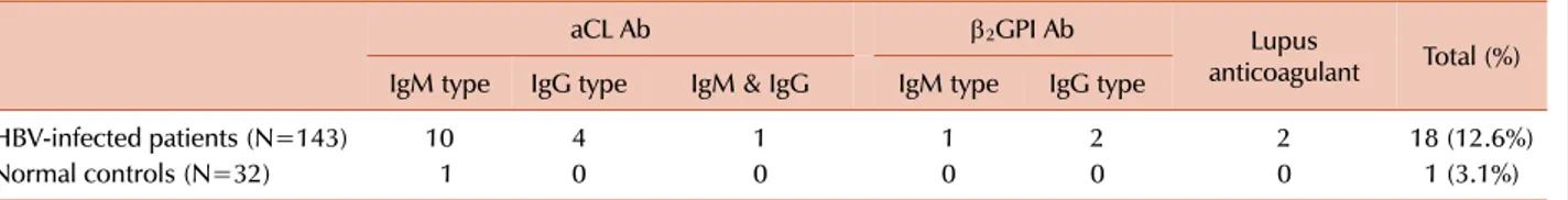
Table 1. Prevalence of antiphospholipid antibodies (aCL Ab, β2GPI Ab, and lupus anticoagulant) in HBV-infected patients.
aCL Ab β2GPI Ab Lupus
anticoagulant Total (%) IgM type IgG type IgM & IgG IgM type IgG type
HBV-infected patients (N=143) 10 4 1 1 2 2 18 (12.6%)
Normal controls (N=32) 1 0 0 0 0 0 1 (3.1%)
Abbreviations: HBV, hepatitis B virus; aCL Ab, anticardiolipin antibodies; β2GPI Ab, anti-β2-glycoprotein I antibodies.
non-pathogenic [5, 8]; however, in patients with various infections, thrombotic manifestations such as portal vein thrombosis and pulmonary embolism have been reported [10-13].
Recently, several studies showed a higher prevalence of aCL Ab in chronic viral hepatitis patients than in control individuals [14-17]. In the present study, we investigated the prevalence, persistence, clinical significance, and charac- teristics of aPLs in HBV-infected patients.
MATERIALS AND METHODS
1. Patient selection
The prevalence of aPLs was prospectively determined in HBV-infected patients and healthy controls who visited the Gastroenterology Department of the Bundang CHA hospital between 2008 and 2009. This study included 143 HBV-in- fected patients, irrespective of their treatment (59 women and 84 men; age range, 16-71 years; mean, 42.7 years), and 32 healthy individuals as controls (13 women and 19 men;
age range, 27-65 years; mean, 40 years). All patients with HBV infection tested positive for HBV surface antigen (HBsAg) or HBV DNA and negative for anti-HCV antibody.
Informed consent was obtained from all patients. The patients were divided into 2 groups on the basis of the hepatitis B e antigen and antibody (HBeAg, HBeAb) status and serum HBV DNA level: chronic hepatitis B patients (N=97) and patients with inactive HBsAg carrier state (N=46) [18].
Patients with positive HBeAg or high levels of HBV DNA (≥104 copies/mL) were considered chronic hepatitis B pa- tients, and patients with negative HBeAg and low HBV DNA level (<104 copies/mL) were considered inactive HBsAg carriers. All normal controls were negative for HBsAg, anti- HCV antibody, anti-HIV antibody, and antinuclear antibody.
Alanine aminotransferase (ALT) levels were normal (5-40 IU/L) in all healthy controls. Thromboembolic complications associated with aPLs were identified through medical record review. The local ethics committee of CHA Bundang Medical Center approved this study.
2. aCL Ab
aCL Ab (IgG and IgM isotype) were measured using a commercial enzyme-linked immunosorbent assay (ELISA) (Zeus Scientific Inc., Raritan, NJ). According to the manu- facturer’s instructions, the cut-off values for positivity were
20 GPL or MPL (IgG or IgM phospholipid units). Specimens with less than 20 GPL or MPL in the initial ELISA were considered negative. Specimens with greater than 20 GPL or MPL in the initial ELISA were retested in duplicate.
The specimens were considered positive, when the value was repeatedly greater than 20 GPL or MPL.
3. β2GPI Ab
β2GPI Ab (IgG and IgM isotype) were measured using a commercial ELISA (INOVA Diagnostics, Inc., San Diego, CA, USA). According to the manufacturer’s instructions, the cut-off values for positivity are 20 SGU or SMU (standard IgG or standard IgM units). Specimens with less than 20 SGU or SMU in the initial ELISA were considered negative.
Specimens with greater than 20 SGU or SMU in the initial ELISA result were retested in duplicate. The specimens were considered positive when the value was repeatedly greater than 20 SGU or SMU.
4. LA
All plasma samples from patients and normal individuals were evaluated for the presence of LA according to the criteria defined by the Subcommittee on LA/antiphospho- lipid antibody of the International Society of Thrombosis and Haemostasis [19]. LA was measured by performing the dilute Russell’s viper venom test using the HemosIL Kit (Instrumentation Laboratory, Milano, Italy).
5. Statistical analysis
Statistical analyses were performed using SAS Statistical Analysis Software Version 9.1 (SAS Institute Inc., Cary, NC, USA). A chi-square test was used to determine the difference in the presence of aPLs between the chronic viral hepatitis patient group and the control group.
RESULTS
The total prevalence of aPLs (aCL Ab, β2GPI Ab, or LA) in HBV-infected patients was 12.6% (18 of 143) (Table 1).
Among these 18 patients, aPLs were detected in 15 of 97 chronic hepatitis B patients and 3 of 46 inactive HBsAg carrier patients. No clinical manifestations related to aPLs were identified. Among the 143 HBV-infected patients, 15 patients (10.5%) had a low to moderate level of aCL Ab (10 with IgM, 4 with IgG, and 1 with both isotypes) compared
Table 2. Characteristics of HBV-infected patients with antiphospholipid antibodies and the types of antiphospholipid antibodies.
Patient ID Gender Age Status of HBV infectiona) Type of antiphospholipid antibodies 021058
062082 098099 104 112114 122130 134 137140 144149 150 153
MF MF FM F FF FF F MM MF M F
4339 3461 4234 46 2639 4037 70 5245 4764 47 28
Inactive HBsAg carrier Chronic hepatitis B Chronic hepatitis B Chronic hepatitis B Chronic hepatitis B Chronic hepatitis B Chronic hepatitis B Chronic hepatitis B Chronic hepatitis B Chronic hepatitis B Chronic hepatitis B Inactive HBsAg carrier Chronic hepatitis B Chronic hepatitis B Chronic hepatitis B Inactive HBsAg carrier Chronic hepatitis B Chronic hepatitis B
IgG and IgM aCL IgM β2GPI and LA IgG β2GPI IgM aCL IgG aCL IgM aCL IgM aCL IgG aCL IgG aCL IgM aCL
IgM aCL and IgG β2GPI IgM aCL
LAIgG aCL IgM aCL IgM aCL IgM aCL IgM aCL
a)Patients were divided into 2 groups on the basis of the hepatitis B e antigen and antibody status and the HBV DNA level: chronic hepatitis B patients and patients with inactive HBsAg carrier state.
Abbreviations: HBV, hepatitis B virus; M, male; F, female; aCL, anticardiolipin antibodies; β2GPI, anti-β2-glycoprotein I antibodies; LA, lupus anticoagulant.
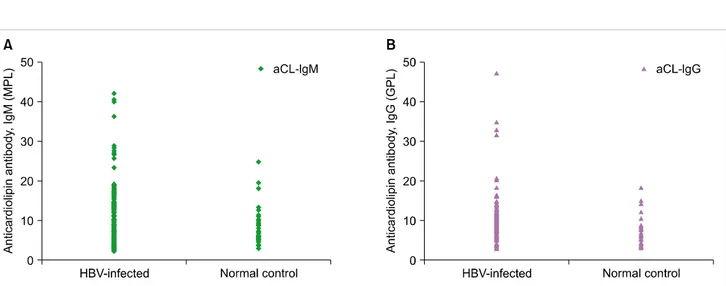
Fig. 1. Distribution of anticardiolipin antibody titers in hepatitis B virus (HBV)-infected patients and normal controls, IgM isotype (A) and IgG isotype (B).
to only 1 of the 32 control subjects (3.1%). The difference between the groups was not significant (P=0.19). The median values for aCL Ab IgM and IgG isotypes were 28.1 MPL (range, 23.4-42.2) and 32.9 GPL (range, 20.5-47.2), respec- tively (Table 2 and Fig. 1).
The prevalence of β2GPI Ab and the LA activity in the HBV-infected patient group was 2.1% (3 of 143) and 1.4%
(2 of 143), respectively. In contrast, none of the healthy controls had elevated levels of β2GPI Ab or LA; however, the difference between the groups was not significant (P= 0.40). The isotype distribution for β2GPI Ab in HBV-infected patients was 1 with IgM and 2 with IgG. Two HBV-infected patients simultaneously had 2 types of aPLs; 1 patient had IgM aCL Ab with cofactor dependency (β2GPI Ab, IgG type),
and the other had β2GPI Ab (IgM type) and LA.
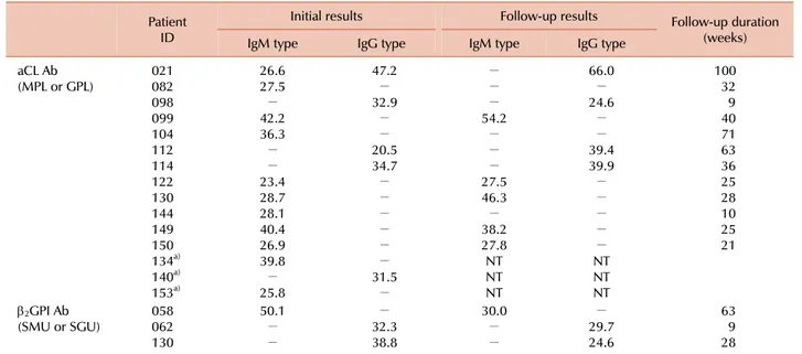
Follow-up specimens were obtained from 14 of the 17 patients with elevated levels of aCL Ab or β2GPI Ab (11 with aCL Ab, 2 with anti-β2GPI Ab, and 1 with both aCL Ab and β2GPI Ab). The median follow-up duration was 30 weeks (range, 9-100 weeks). The aPLs persisted in 10 of the 14 patients (71.4%), and β2GPI Ab persisted in all 3 patients with β2GPI Ab (Table 3).
DISCUSSION
aPLs have been found not only in patients with auto- immune diseases like SLE, but also in patients with various
Table 3. Follow-up duration and results of aCL Ab and β2GPI Ab testing in antiphospholipid antibody-positive HBV-infected patients.
Patient ID
Initial results Follow-up results Follow-up duration
(weeks)
IgM type IgG type IgM type IgG type
aCL Ab
(MPL or GPL) 021
082 098099 104 112114 122130 144 149150 134a) 140a) 153a)
26.627.5
− 42.236.3
−
− 23.428.7 28.1 40.426.9 39.8
− 25.8
47.2
− 32.9
−
− 20.534.7
−
−
−
−
−
− 31.5
−
−
−
− 54.2
−
−
− 27.546.3
− 38.227.8
NTNT NT
66.0
− 24.6
−
− 39.439.9
−
−
−
−
− NTNT NT
10032 409 71 6336 2528 10 2521
β2GPI Ab
(SMU or SGU) 058
062130
50.1
−
−
− 32.338.8
30.0
−
−
− 29.724.6
639 28
a)Follow-up specimens of 3 patients (134, 140, and 153) could not be obtained.
Abbreviations: HBV, hepatitis B virus; aCL Ab, anticardiolipin antibodies; β2GPI Ab, anti-2-glycoprotein I antibodies; MPL, IgM phospholipid unit; GPL, IgG phospholipid unit; SMU, standard IgM unit; SGU, standard IgG unit;-, negative; NT, not tested.
infections [5, 8]. Molecular mimicry is the proposed mecha- nism for the development of aPLs in infections. Previous reports showed that antigenic determinants shared between the antigens of infectious agents and host tissue might trigger the immune response [20, 21]. However, the mechanisms that lead to the development of aPLs and their possible pathophysiological implications in patients with infections have not been well established.
The reported prevalence of aPLs in infectious patients is variable, in part because of a methodological problem.
Currently, the ideal approach for standardized measurement for aCL Ab is under debate [22]. A discrepancy is observed among the different assay kits and methods, particularly in the lower range of antibody levels. Furthermore, one of the critical problems in the standardization process is the absence of a defined cut-off value for positivity. In pre- vious reports, the prevalence of aCL Ab in patients with HBV infection was between 14% and 42% [14, 17, 23], and that of β2GPI Ab was 2% [17] and 7.5% [14]. In the present study, the prevalence of aCL Ab and β2GPI Ab was 10.5%
and 2.1%, respectively, which was lower than that in pre- vious reports. The lower prevalence of aCL Ab in the present study may be partly because of a higher cut-off value for positivity than that of previous reports. We used 20 GPL or MPL as the cut-off values, whereas 10 GPL or MPL were used as the cut-off values in the other reports [14, 23]. The prevalence of β2GPI Ab in the present study was similar to those in previous reports.
In most cases, infection-associated aPLs appear tempora- rily and disappear within 2 or 3 months [24, 25], and except in rare cases, they are unrelated to thrombotic complications.
Contrary to previous reports, in the present study, most aCL Ab and β2GPI Ab showed persistence and were present over a 12-week period. We speculated that continuous anti- genic stimulation might be associated with persistent aPL production, because in the present study, the prevalence of aCL Ab in chronic hepatitis B patients was higher than that in inactive HBsAg carrier patients (15% vs. 6.5%).
Furthermore, it was shown that in a patient with HCV in- fection and aPLs, elimination of the virus was accompanied by the disappearance of aPLs, and when HCV infection re- lapsed, aPLs also reappeared [26]. Therefore, aPLs associated with other chronic infections caused by HIV or HCV may also show persistent positivity.
According to a previous report [27], IgM aPLs are asso- ciated with the clinical manifestations of APS less often than the IgG isotype is, and the diagnostic criteria for APS include only a medium or high titer of aCL Ab (>40 GPL or MPL) [28]. In the present study, IgM aCL Ab were found more frequently (11 of 15) than the IgG isotype (5 of 15), and the titers of aCL Ab were mostly low. These findings support that infection-associated aPLs rarely manifest with the clinical features of APS. Because the rates of aCL Ab according to isotype were not provided in previous reports of aPLs in patients with HBV infection, comparison with our results was not possible.
In conclusion, the most frequently detected aPLs in HBV-infected patients were IgM aCL Ab, which has a weak association with the clinical manifestations of APS. Unlike the transient presence of other infection-associated aPLs, most aPLs were persistently detected over a 12-week period in patients with HBV infection
REFERENCES
1. Levine JS, Branch DW, Rauch J. The antiphospholipid synd- rome. N Engl J Med 2002;346:752-63.
2. Tarr T, Lakos G, Bhattoa HP, et al. Clinical thrombotic manifes- tations in SLE patients with and without antiphospholipid anti- bodies: a 5-year follow-up. Clin Rev Allergy Immunol 2007;
32:131-7.
3. Giannakopoulos B, Passam F, Rahgozar S, Krilis SA. Current con- cepts on the pathogenesis of the antiphospholipid syndrome.
Blood 2007;109:422-30.
4. Vega-Ostertag ME, Pierangeli SS. Mechanisms of aPL-mediated thrombosis: effects of aPL on endothelium and platelets. Curr Rheumatol Rep 2007;9:190-7.
5. Cervera R, Asherson RA. Antiphospholipid syndrome asso- ciated with infections: clinical and microbiological characte- ristics. Immunobiology 2005;210:735-41.
6. Avcin T, Toplak N. Antiphospholipid antibodies in response to infection. Curr Rheumatol Rep 2007;9:212-8.
7. Sorice M, Pittoni V, Griggi T, et al. Specificity of anti-phospholi- pid antibodies in infectious mononucleosis: a role for anti-co- factor protein antibodies. Clin Exp Immunol 2000;120:301-6.
8. Sène D, Piette JC, Cacoub P. Antiphospholipid antibodies, anti- phospholipid syndrome and infections. Autoimmun Rev 2008;7:272-7.
9. Bloom EJ, Abrams DI, Rodgers G. Lupus anticoagulant in the ac- quired immunodeficiency syndrome. JAMA 1986;256:491-3.
10. Blank M, Shoenfeld Y. Beta-2-glycoprotein-I, infections, anti- phospholipid syndrome and therapeutic considerations. Clin Immunol 2004;112:190-9.
11. Gómez RM, García ES, Lacomba DL, Marchante I, Grande L, Fernandez MC. Antiphospholipid antibodies are related to por- tal vein thrombosis in patients with liver cirrhosis. J Clin Gastroenterol 2000;31:237-40.
12. Kida Y, Maeshima E, Yamada Y. Portal vein thrombosis in a pa- tient with hepatitis C virus-related cirrhosis complicated with antiphospholipid syndrome. Rheumatol Int 2009;29:1495-8.
13. Ramos-Casals M, Cervera R, Lagrutta M, et al. Clinical features related to antiphospholipid syndrome in patients with chronic viral infections (hepatitis C virus/HIV infection): description of 82 cases. Clin Infect Dis 2004;38:1009-16.
14. Guglielmone H, Vitozzi S, Elbarcha O, Fernandez E. Cofactor de- pendence and isotype distribution of anticardiolipin antibodies in viral infections. Ann Rheum Dis 2001;60:500-4.
15. Ordi-Ros J, Villarreal J, Monegal F, Sauleda S, Esteban I, Vilardell
M. Anticardiolipin antibodies in patients with chronic hepatitis C virus infection: characterization in relation to antipho- spholipid syndrome. Clin Diagn Lab Immunol 2000;7:241-4.
16. Harada M, Fujisawa Y, Sakisaka S, et al. High prevalence of anti- cardiolipin antibodies in hepatitis C virus infection: lack of ef- fects on thrombocytopenia and thrombotic complications. J Gastroenterol 2000;35:272-7.
17. Zachou K, Liaskos C, Christodoulou DK, et al. Anti-cardiolipin antibodies in patients with chronic viral hepatitis are in- dependent of beta2-glycoprotein I cofactor or features of anti- phospholipid syndrome. Eur J Clin Invest 2003;33:161-8.
18. Lee KS, Kim DJ. Management of Chronic Hepatitis B. Korean J Hepatol 2007;13:447-88.
19. Brandt JT, Triplett DA, Alving B, Scharrer I. Criteria for the diag- nosis of lupus anticoagulants: an update. On behalf of the Subcommittee on Lupus Anticoagulant/Antiphospholipid Anti- body of the Scientific and Standardisation Committee of the ISTH. Thromb Haemost 1995;74:1185-90.
20. Harel M, Aron-Maor A, Sherer Y, Blank M, Shoenfeld Y. The in- fectious etiology of the antiphospholipid syndrome: links be- tween infection and autoimmunity. Immunobiology 2005;210:
743-7.
21. Amin NM. Antiphospholipid syndromes in infectious diseases.
Hematol Oncol Clin North Am 2008;22:131-43.
22. Devreese K, Hoylaerts MF. Laboratory diagnosis of the anti- phospholipid syndrome: a plethora of obstacles to overcome. Eur J Haematol 2009;83:1-16.
23. Elefsiniotis IS, Diamantis ID, Dourakis SP, Kafiri G, Pantazis K, Mavrogiannis C. Anticardiolipin antibodies in chronic hepatitis B and chronic hepatitis D infection, and hepatitis B-related hep- atocellular carcinoma. Relationship with portal vein throm- bosis. Eur J Gastroenterol Hepatol 2003;15:721-6.
24. Uthman IW, Gharavi AE. Viral infections and antiphospholipid antibodies. Semin Arthritis Rheum 2002;31:256-63.
25. Dalekos GN, Zachou K, Liaskos C. The antiphospholipid syn- drome and infection. Curr Rheumatol Rep 2001;3:277-85.
26. Alric L, Oskman F, Sanmarco M, et al. Association of anti- phospholipid syndrome and chronic hepatitis C. Br J Rheumatol 1998;37:589-90.
27. Galli M, Luciani D, Bertolini G, Barbui T. Lupus anticoagulants are stronger risk factors for thrombosis than anticardiolipin anti- bodies in the antiphospholipid syndrome: a systematic review of the literature. Blood 2003;101:1827-32.
28. Devreese K, Hoylaerts MF. Challenges in the diagnosis of the an- tiphospholipid syndrome. Clin Chem 2010;56:930-40.