Prediction of Axillary Lymph Node
Metastasis in Early Breast Cancer Using Dynamic Contrast-Enhanced Magnetic Resonance Imaging and Diffusion-
Weighted Imaging
This is an Open Access article distributed under the terms of the Creative Commons Attribution Non-Commercial License (http://creativecommons.org/licenses/
by-nc/4.0/) which permits unrestricted non-commercial use, distribution, and reproduction in any medium, provided the original work is properly cited.
Received: March 27, 2019 Revised: May 8, 2019 Accepted: May 17, 2019 Correspondence to:
Eun Jung Choi, M.D., Ph.D.
Department of Radiology and Research Institute of Clinical Medicine of Chonbuk National University-Biomedical Research Institute of Chonbuk National University Hospital, Chonbuk National University Medical School, 20 Geonji-ro, Deokjin-gu, Jeonju, Jeollabuk-do 54907, Korea.
Tel. +82-63-250-1150 Fax. +82-63-272-0481 E-mail: [email protected]
Copyright © 2019 Korean Society of Magnetic Resonance in Medicine (KSMRM)
Original Article
Purpose: The purpose of this study was to evaluate dynamic contrast-enhanced breast magnetic resonance imaging (DCE-MRI), and diffusion-weighted imaging (DWI) variables, for axillary lymph node (ALN) metastasis in the early stage of breast cancer.
Materials and Methods: January 2011-April 2015, 787 patients with early stage of breast cancer were retrospectively reviewed. Only cases of invasive ductal carcinoma, were included in the patient population. Among them, 240 patients who underwent 3.0- T DCE-MRI, including DWI with b value 0 and 800 s/mm2 were enrolled. MRI variables (adjacent vessel sign, whole-breast vascularity, initial enhancement pattern, quantitative kinetic parameters, signal enhancement ratio (SER), tumor apparent diffusion coefficient (ADC), peritumoral ADC, and peritumor-tumor ADC ratio) clinico-pathologic variables (age, T stage, multifocality, extensive intraductal carcinoma component (EIC), estrogen receptor, progesterone receptor, HER-2 status, Ki-67, molecular subtype, histologic grade, and nuclear grade) were compared between patients with axillary lymph node metastasis and those with no lymph node metastasis. Multivariate regression analysis was performed, to determine independent variables associated with ALN metastasis, and the area under the receiver operating characteristic curve (AUC), for predicting ALN metastasis was analyzed, for those variables.
Results: On breast MRI, moderate or prominent ipsilateral whole-breast vascularity (moderate, odds ratio [OR] 3.45, 95% confidence interval [CI] 1.28-9.51 vs.
prominent, OR = 15.59, 95% CI 2.52-96.46), SER (OR = 1.68, 95% CI 1.09-2.59), and peritumor-tumor ADC ratio (OR = 6.77, 95% CI 2.41-18.99), were independently associated with ALN metastasis. Among clinico-pathologic variables, HER-2 positivity was independently associated, with ALN metastasis (OR = 23.71, 95% CI 10.50- 53.54). The AUC for combining selected MRI variables and clinico-pathologic variables, was higher than that of clinico-pathologic variables (P < 0.05).
Conclusion: SER, moderate or prominent increased whole breast vascularity, and peritumor-tumor ADC ratio on breast MRI, are valuable in predicting ALN metastasis, in patients with early stage of breast cancer.
Keywords: Breast neoplasm; Lymph node; Magnetic resonance imaging;
Diffusion magnetic resonance imaging
Eun Ha Jeong1, Eun Jung Choi1, Hyemi Choi2, Eun Hae Park1, Ji Soo Song1
1Department of Radiology and Research Institute of Clinical Medicine of Chonbuk National University-Biomedical Research Institute of Chonbuk National University Hospital, Chonbuk National University Medical School, Jeonju, Korea
2Department of Statistics, Chonbuk National University, Research Institute of Applied Statistics, Jeonju, Korea
INTRODUCTION
Breast cancer is the most common cancer, in women worldwide. There are approximately 1.7 million cases of breast cancer annually, representing 25.2% of female cancer patients (1). Axillary lymph node (ALN) status is one of the most critical factors to consider in breast cancer stage, treatment plan, and prognosis. Approximately 70%
of patients with early stage of breast cancer, have no ALN metastasis. During the past 15 years, sentinel lymph node biopsy (SLNB) has replaced axillary lymph node dissection (ALND), for nodal staging in patients with clinically node- negative breast cancer (2, 3). SLNB involves complications, including pain, paresthesia, and lymphedema (4).
A randomized study by the American College of Surgeon’s Oncology Group (Z0011 trial) demonstrated that ALND is not essential, in breast cancer patients with T stage 1 or 2, with up to 2 positive SLNs (5). Recent studies have reported significant benefits because of implementation of the Z0011 guideline, such as reduced morbidity, decreased operative time, and reduced length of hospital stay (6). These data show that SLNB, can be omitted in select cases. So, it would be clinically valuable to predict ALN metastasis in patients with early stage of breast cancer, using a non-invasive method.
Among non-invasive methods, dynamic contrast enhanced magnetic resonance imaging, (DCE-MRI) and diffusion weighted imaging (DWI), have been used to evaluate ALN metastasis. A previous study reported, that DCE-MRI and DWI exhibit high sensitivity and negative predictive value (2, 7, 8). DCE-MRI is based on estimation of velocity, of contrast enhancement in a lesion. Breast cancers show a faster and stronger signal intensity, increase after intravenous injection of gadolinium-containing contrast agents, compared to benign lesions (7). DCE-MRI can visualize tumor angiogenesis. Breast cancer with a high degree of angiogenesis, is much more likely to have lymph node metastasis, and may be valuable in predicting lymph node metastasis (9). DWI, a technology based on mobility of water molecules, is very sensitive to tissue properties such as cell density, membrane integrity, and microstructure (10). Malignant tumors have significantly lower apparent diffusion coefficients (ADC), than benign breast lesions and normal tissues. Also, DWI and ADC values have been used to differentiate benign and malignant lesions, or to detect metastatic lymph nodes (11, 12).
Association between primary tumor MRI variables and ALN metastasis, have been reported (13). Luo et al. (14)
reported significantly lower ADC ratios in metastatic malignant breast tumors, in terms of ADC value and ADC ratio, of metastatic lymph node and primary tumor.
Razek et al. (15) reported that ADC value is a promising prognostic parameter, in relation to ADC value of the primary tumor, histologic grade, tumor size, and axillary lymph node presence. In our study, we added peritumor ADC and peritumor-tumor ratio as well, as ADC value of the primary tumor, and we hypothesize that use of DCE-MRI and DWI in patients with early stage of breast cancer, will prevent unnecessary invasive procedure for prediction of ALN metastasis, and improve local care if preoperative ALN metastasis is predicted.
So, the purpose of our study was to evaluate DCE-MRI and DWI variables, for prediction of ALN metastasis in patients with early stage of breast cancer.
MATERIALS AND METHODS
Study SubjectsFollowing Institutional Review Board approval, a retrospective review of 787 patients with early breast cancer at our hospital January 2011-April 2015, was performed.
Among them, 475 women underwent preoperative 3.0 Tesla (T)-breast MRI examination including DCE-MRI and DWI, followed by curative surgery for breast cancer. Among these patients, 240 women, were enrolled in this study. Inclusion criteria were as follow: (a) Clinical TNM stage according to the 8th edition of the American Joint Committee on Cancer (AJCC): T1-2 and N0-1; (b) Underwent SLNB at our hospital;
(c) No history of cancer in the breast, or at any other site;
(d) Preoperative breast MR imaging including DCE-MRI and DWI.
Additional exclusion criteria were as follow: (a) Underwent neoadjuvant chemotherapy or endocrine therapy (n = 31); (b) Contralateral breast cancer (n = 8) ; (c) Had not undergone ALND (n = 13); (d) Region of interest (ROI) too small to be drawn in DCE-MRI and DWI (n = 71);
(e) Vacuum-assisted breast biopsy or excisional biopsy was performed, before preoperative MRI (n = 63); and (f) evaluation limited by low image quality (n = 49).
Breast MRI Examination Technique
All images were acquired with a 3.0 T system (Verio or Skyra; Siemens Healthcare, Erlangen, Germany), using a dedicated receive-only double breast coil. This coil extends into the axillary region, and allows coverage of the breast
and axilla. Patients were examined in the prone position, and breasts were gently cushioned, to reduce patient motion. MR imaging examination consisted of turbo spin-echo T1-and T2-weighted sequences, and a three- dimensional dynamic contrast-enhanced sequence. The following DCE-MRI parameters were used with a 3.0- T Skyra scanner: repetition time msec/echo time msec, 4.5/1.6; matrix, 448 × 381; field of view, 360 × 360 mm;
flip angle, 10°; section thickness, 1 mm without gaps. MR imaging parameters used with the 3.0-T Verio scanner were:
repetition time msec/echo time msec, 4.3/1.6; matrix, 448
× 354; field of view, 340 × 340 mm; flip angle, 6°; section thickness, 1 mm without gaps.
DCE-MRI was performed with one precontrast and five postcontrast dynamic series, obtained immediately after intravenous administration of a bolus injection of 0.1 mmol/
kg gadobutrol (Gadovist; Schering AG, Berlin, Germany), followed by a 20 ml saline flush at an injection rate of 3 ml/sec, using an automatic injector. Standard subtraction images were obtained by subtracting the precontrast images, from the second dynamic series (or early peak) of postcontrast images, on a pixel-by-pixel basis. Additionally, maximum-intensity-projection reconstructions were applied, to subtraction images. Before contrast agent injection, DWI with readout segment echo planar imaging at TR/TE 5200/53 ms, FOV 340 × 205 mm, matrix size 192 × 116, slice thickness of 4 mm, acquisition time of 2 minutes and 31 seconds, and five readout segments, were performed with b values of 0 and 800 s/mm2.
Breast MRI Analysis
Two radiologists independently analyzed images.
Radiologists knew that patients had malignant breast tumors, but did not have knowledge about histopathology results, or patient outcome. For adjacent vessel signs on subtracted images, presence of vessels either entering the enhancing lesion, or in contact with the lesion edge, was accepted as a positive adjacent vessel sign (16).
Whole breast vascularity of the ipsilateral breast was compared with that of the contralateral breast, at each maximum-intensity-projection image, based on number of vessels 3 cm or longer and 2 mm or larger, in maximal transverse diameter (17). Degree of vascularity difference was classified as “prominent” if the number of vessels in the ipsilateral breast was three or more than that in the contralateral breast; “moderate” if higher by two; “mild” if higher by one; and “not increased” if the number of vessels in the ipsilateral breast was the same as or lower, than that
in the contralateral breast (18).
Quantitative kinetic parameters were derived, from time- intensity curve images. For each curve, initial enhancement percentage (E1), peak enhancement percentage (Epeak), and time to peak enhancement (TTP), were measured as follow: E1 = 100 × (S1-S0)/S0, Epeak = 100 × (Speak-0)/S0, wherein E1 is initial percentage enhancement, Epeak is peak percentage enhancement, S1 is signal intensity in the ROI at the first contrast-enhanced point, Speak is peak signal intensity, and S0 is unenhanced signal intensity in the ROI.
Time to peak enhancement is time in seconds between injection of contrast material, and peak of signal intensity- time curve (16). Signal enhancement ratio (SER) was calculated, as a measure of washout as follow: SER = (S1- S0)/(Slast-S0), wherein Slast is signal intensity in the ROI, at the last point of contrast enhancement (19).
A T2-weighted image (T2WI) was used for visual evaluation of edema, and degree of signal intensity surrounding the tumor was classified (0-2) as “grade 0,”
absence of high signal intensity; “grade 1,” signal intensity surrounding the tumor was moderate but less than that of water; “grade 2,” signal intensity surrounding the tumor as high as that of water (20).
ADC values were obtained from ADC maps as follow:
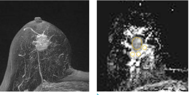
largest tumor cross section was selected on the ADC map, and an oval or round ROI as large as possible, was placed inside the tumor. Three ROIs > 20 mm2 were placed where ADCs visually appeared to be most increased on breast parenchymal tissue, adjacent to tumor contour, to measure mean ADC values of individual ROIs: the maximum of these was designated the peritumor ADC (Fig. 1). Peritumor- tumor ADC ratio was evaluated according to the following formula: peritumor-tumor ADC ratio = peritumoral ADC/
tumor ADC (20).
Clinico-Pathologic Variables
The following factors were examined, regarding potential to predict axillary lymph node involvement: patient age, T stage, multifocality, extensive intraductal carcinoma component (EIC), estrogen receptor (ER), progesterone receptor (PR), human epidermal growth factor receptor 2 (HER-2) status, proliferation related gene (Ki-67), molecular subtype, histologic grade, and nuclear grade. Patients were subdivided by age as follow: < 50 years and ≥ 50 years. Additionally, patients were divided into two groups, according to ALN status.
All specimens obtained from breast resections and axillary lymph node dissections, were paraffin-embedded for
Hematoxylin and Eosin (H&E) staining, molecular subtype, and fluorescence in situ hybridization (FISH) staining. Lymph node tissue was histologically dissected, countered, and analyzed. Immunohistochemistry (IHC) staining was used to analyze expression of ER, PR, and HER-2, and determine presence of micrometastasis (0.2-2 mm cancer foci). ER and PR were positive if immunostaining was present, in more than 10% of neoplastic cells. HER-2 positive status was defined as a score of 3+, on IHC or amplification on FISH.
When an IHC score of 2+ was obtained, FISH was conducted to defined positivity. IHC score of 1+ was negative for HER- 2 expression. Molecular subtypes are as follow: luminal A (ER + and/or PR + and HER-2 -), luminal B (ER+ and/or PR+/HER2+ or ER +/PR -/Ki-67 +), HER-2 enriched (ER -/
PR -/HER-2 +), and triple negative (ER -/PR -/HER-2 -). Ki- 67 was considered positive if nuclear staining of tumor cells was ≥ 14%, and negative, if nuclear staining of tumor cells was < 14%.
Statistical Analysis
DCE-MRI, DWI, and clinico-pathologic variables were compared between no ALN metastasis and ALN metastasis,
using Student’s t test for continuous variables, and the χ2 test for categorical variables. Univariate and multivariate logistic regressions were performed, to determine features independently associated with ALN metastasis. Sensitivity, specificity, positive predictive value, negative predictive value, and accuracy were reported. The receiver operating characteristic (ROC) curve was drawn, and the area under the curve (AUC), was used to assess predictive accuracy of the model. Statistical analyses were performed, using SAS (version 9.3; SAS Institute, Cary, NC, USA). P values less than < 0.05 were significant.
RESULTS
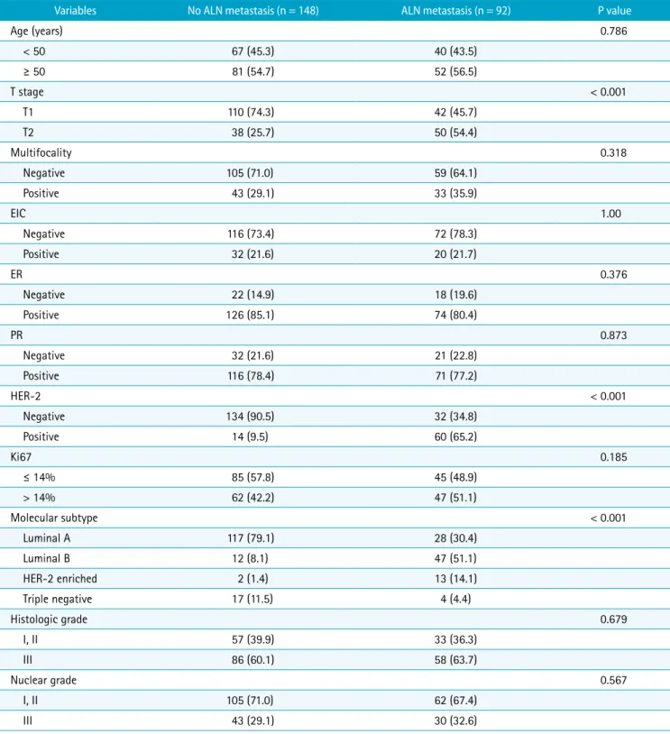
A total of 240 patients were enrolled; 148 patients (61.7%) had no ALN metastasis, and 92 patients (38.3%) had ALN metastasis. Histologic diagnoses consisted of 213 invasive ductal carcinomas, 22 invasive lobular carcinomas, one medullary carcinoma, two tubular carcinomas, and two mucinous carcinomas. For clinico-pathologic variables (Table 1), the ALN metastasis group showed significantly
Fig. 1. Maximum-intensity-projection image (a) and ADC map (b) in a 70-year-old woman with invasive ductal carcinoma, show the method used for placing ROIs to obtain the tumor ADC, peritumor ADC. Regarding the tumor ADC, the slice with the largest tumor cross section is selected, and the largest oval ROI is placed inside the tumor (b) with reference to the MIP image (a). Mean value of the ROI 1193 × 10-6 mm2/s, is recorded as tumor ADC. For the peritumor ADC, three ROIs are placed where the ADC visually appears to be most increased on breast parenchymal tissue, adjacent to tumor contour on the ADC map (b): the three ROIs are 2328, 2062, and 2646 x 10-6 mm2/s, respectively. Maximum value 2646 x 10-6 mm2/s is recorded as peritumor ADC. ADC=apparent diffusion coefficient; ROI=region of interest
a b
higher rates of T2 stage (54.4% vs. 25.7%, P < 0.001), HER- 2 positivity (65.2% vs. 9.5%, P < 0.001), luminal B (51.1%
vs. 8.1) type, and HER-2 enriched type (14.1% vs. 1.4%, P <
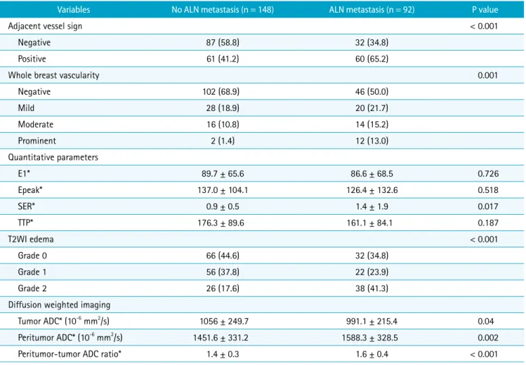
0.001). For DCE-MRI and DWI variables (Table 2), the ALN metastasis group showed higher rates of grade 2 edema (41.3% vs. 17.6%, P < 0.001), adjacent vessel sign (65.2%
Table 1. Comparison of Clinico-Pathologic Variables between No Axillary Lymph Node Metastasis and Axillary Lymph Node Metastasis
Variables No ALN metastasis (n = 148) ALN metastasis (n = 92) P value
Age (years) 0.786
< 50 67 (45.3) 40 (43.5)
≥ 50 81 (54.7) 52 (56.5)
T stage < 0.001
T1 110 (74.3) 42 (45.7)
T2 38 (25.7) 50 (54.4)
Multifocality 0.318
Negative 105 (71.0) 59 (64.1)
Positive 43 (29.1) 33 (35.9)
EIC 1.00
Negative 116 (73.4) 72 (78.3)
Positive 32 (21.6) 20 (21.7)
ER 0.376
Negative 22 (14.9) 18 (19.6)
Positive 126 (85.1) 74 (80.4)
PR 0.873
Negative 32 (21.6) 21 (22.8)
Positive 116 (78.4) 71 (77.2)
HER-2 < 0.001
Negative 134 (90.5) 32 (34.8)
Positive 14 (9.5) 60 (65.2)
Ki67 0.185
≤ 14% 85 (57.8) 45 (48.9)
> 14% 62 (42.2) 47 (51.1)
Molecular subtype < 0.001
Luminal A 117 (79.1) 28 (30.4)
Luminal B 12 (8.1) 47 (51.1)
HER-2 enriched 2 (1.4) 13 (14.1)
Triple negative 17 (11.5) 4 (4.4)
Histologic grade 0.679
I, II 57 (39.9) 33 (36.3)
III 86 (60.1) 58 (63.7)
Nuclear grade 0.567
I, II 105 (71.0) 62 (67.4)
III 43 (29.1) 30 (32.6)
Numbers in parenthese represent percentages (%).
ALN = axillary lymph node; EIC = extensive intraductal carcinoma component; ER = estrogen receptor; HER-2 = human epidermal growth factor receptor-2; PR = progesterone receptor
vs. 41.2, P < 0.001), prominent increased whole breast vascularity (13% vs. 1.4%, P < 0.001), lower mean tumor ADC (991.1 × 10⁻⁶ mm2/s vs. 1056.0 × 10-6 mm2/s, P = 0.04), higher mean peritumor ADC (1588.3 × 10-6 mm2/s vs. 1451.6
× 10-6 mm2/s, P = 0.002), and higher mean peritumor-tumor ADC ratio (1.65 vs. 1.40, P < 0.001).
Univariate and Multivariate Analyses with ALN Metastasis
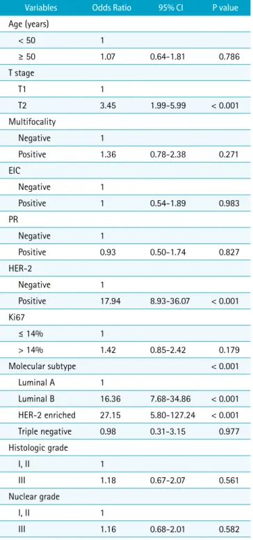
In the univariate logistic analysis, T2 stage (odds ratio [OR], 3.45; 95% confidence interval [CI], 1.99-5.99; P <
0.001), HER-2 positivity (OR, 17.95; 95% CI, 8.93-36.07;
P < 0.001), luminal B molecular subtype (OR, 16.37; 95%
CI, 7.69-34.86; P < 0.001), and HER-2 enriched molecular subtype (OR, 27.15; 95% CI, 5.80-127.24; P < 0.001) were significantly associated with ALN metastasis (Table 3).
Among MRI variables, SER (OR, 1.60; 95% CI, 1.10-2.30; P =
0.013), tumor ADC (OR, 1.00; 95% CI, 0.99-1.00; P = 0.043), peritumor ADC (OR, 1.00; 95% CI, 1.00-1.00; P = 0.003), peritumor-tumor ADC ratio (OR, 8.70; 95% CI, 3.73-20.30;
P < 0.001), grade 2 edema (OR, 3.01; 95% CI, 1.57-5.79;
P = 0.001), positive adjacent vessel sign (OR, 2.67; 95%
CI, 1.56-4.59; P < 0.001), and prominent increased whole breast vascularity (OR, 13.30; 95% CI, 2.86-61.85; P = 0.001) were significantly associated with ALN metastasis (Table 4).
In the multivariate logistic analysis, HER-2 positivity (OR, 23.72; 95% CI, 10.50-53.53; P < 0.001), SER (OR, 1.67; 95%
CI, 1.08-2.59; P = 0.02), peritumor-tumor ADC ratio (OR, 6.77; 95% CI, 2.41-19.00; P < 0.001), moderate increased whole breast vascularity (OR, 3.44; 95% CI, 1.24-9.51;
P = 0.017), and increased whole breast vascularity (OR, 15.59; 95% CI, 2.52-96.45; P = 0.003) were significantly associated with ALN metastasis (Table 5) (Fig. 2).
Regarding prediction of ALN metastasis, the area
Table 2. Comparison of DCE-MRI and DWI Variables between No Axillary Lymph Node Metastasis and Axillary Lymph Node Metastasis
Variables No ALN metastasis (n = 148) ALN metastasis (n = 92) P value
Adjacent vessel sign < 0.001
Negative 87 (58.8) 32 (34.8)
Positive 61 (41.2) 60 (65.2)
Whole breast vascularity 0.001
Negative 102 (68.9) 46 (50.0)
Mild 28 (18.9) 20 (21.7)
Moderate 16 (10.8) 14 (15.2)
Prominent 2 (1.4) 12 (13.0)
Quantitative parameters
E1* 89.7 ± 65.6 86.6 ± 68.5 0.726
Epeak* 137.0 ± 104.1 126.4 ± 132.6 0.518
SER* 0.9 ± 0.5 1.4 ± 1.9 0.017
TTP* 176.3 ± 89.6 161.1 ± 84.1 0.187
T2WI edema < 0.001
Grade 0 66 (44.6) 32 (34.8)
Grade 1 56 (37.8) 22 (23.9)
Grade 2 26 (17.6) 38 (41.3)
Diffusion weighted imaging
Tumor ADC* (10-6 mm2/s) 1056 ± 249.7 991.1 ± 215.4 0.04
Peritumor ADC* (10-6 mm2/s) 1451.6 ± 331.2 1588.3 ± 328.5 0.002
Peritumor-tumor ADC ratio* 1.4 ± 0.3 1.6 ± 0.4 < 0.001
ADC= apparent diffusion coefficient; ALN = axillary lymph node; DCE-MRI = dynamic contrast-enhanced magnetic resonance imaging; E1 = the initial enhancement percentage; Epeak = peak enhancement percentage; SER = signal enhancement ratio; T2WI = T2 weighted image; TTP = time to peak
* Data are mean ± standard deviation.
under the receiver operating characteristic curves (AUC) of combined clinico-pathologic and MRI variables (AUC
= 0.879) was significantly higher, than that of clinico-
pathologic variables alone (AUC = 0.835) (P < 0.05) (Fig. 3).
DISCUSSION
This study attempted to non-invasively predict ALN metastasis, using DCE-MRI and DWI in patients with early stage of breast cancer. SER, moderate or prominent increased ipsilateral whole breast vascularity, and peritumor-tumor ADC ratio, were shown to be valuable.
This study provides non-invasive methods to assess ALN status to guide clinical treatment of patients with breast cancer, by eliminating unnecessary SLNB and its associated complications.
Moderately or prominently increased ipsilateral whole Table 3. Univariate Analysis for Clinico-Pathologic Variables
Associated with Axillary Lymph Node Metastasis
Variables Odds Ratio 95% CI P value
Age (years)
< 50 1
≥ 50 1.07 0.64-1.81 0.786
T stage
T1 1
T2 3.45 1.99-5.99 < 0.001
Multifocality
Negative 1
Positive 1.36 0.78-2.38 0.271
EIC
Negative 1
Positive 1 0.54-1.89 0.983
PR
Negative 1
Positive 0.93 0.50-1.74 0.827
HER-2
Negative 1
Positive 17.94 8.93-36.07 < 0.001
Ki67
≤ 14% 1
> 14% 1.42 0.85-2.42 0.179
Molecular subtype < 0.001
Luminal A 1
Luminal B 16.36 7.68-34.86 < 0.001 HER-2 enriched 27.15 5.80-127.24 < 0.001
Triple negative 0.98 0.31-3.15 0.977
Histologic grade
I, II 1
III 1.18 0.67-2.07 0.561
Nuclear grade
I, II 1
III 1.16 0.68-2.01 0.582
CI = confidence interval; EIC = extensive intraductal carcinoma component; HER-2=human epidermal growth factor receptor-2; PR
= progesterone receptor
Table 4. Univariate Analysis for MRI Variables Associated with Axillary Lymph Node Metastasis
Variables Odds Ratio 95% CI P value
Adjacent vessel sign
Negative 1
Positive 2.67 1.56-4.59 < 0.001
Whole breast vascularity 0.004
Negative 1
Mild 1.58 0.81-3.10 0.179
Moderate 1.94 0.87-4.31 0.103
Prominent 13.3 2.86-61.85 0.001
Quantitative parameters
E1 1 0.99-1.00 0.725
Epeak 1 0.99-1.00 0.494
SER 1.59 1.10-2.30 0.013
TTP 1 0.99-1.00 0.194
T2WI Edema < 0.001
Grade 0 1
Grade 1 0.81 0.42-1.55 0.526
Grade 2 3.01 1.57-5.79 0.001
Diffusion weighted imaging
Tumor ADC (10-6 mm2/s) 1 0.99-1.00 0.043 Peritumor ADC (10-6 mm2/s) 1 1.00-1.00 0.003 Peritumor-tumor ADC ratio 8.69 3.73-20.30 < 0.001 ADC = apparent diffusion coefficient; CI = confidence interval; DCE-MRI = dynamic contrast-enhanced magnetic resonance imaging; E1 = the initial enhancement percentage; Epeak = peak enhancement percentage; SER = signal enhancement ratio; T2WI = T2 weighted image; TTP = time to peak
breast vascularity, was a significant DCE-MRI variable predictive of ALN metastasis. On DCE-MRI, increased whole breast vascularity indicated that there are more blood vessels in the breast, than in the contralateral normal breast (18, 21, 22). Although angiogenesis is a necessary factor for tumor cells to escape into the bloodstream (23), the
effects of large-vessel angiogenesis, including whole breast vascularity, remain largely unknown. However, previous studies have reported that whole breast vascularity in DCE- MRI, is associated with ipsilateral invasive cancer, well expressed in multifocal disease, and has high frequency of distant metastasis (21, 22). We found that whole breast
a
b c
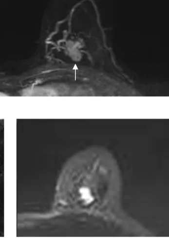
d
Fig. 2. A 66-year-old woman with palpable mass (arrows) on the left breast, had pathologically confirmed invasive ductal carcinoma, with axillary lymph node metastasis after surgery. (a) Maximum-intensity-projection images show that the adjacent vessel sign is positive, and increased breast vascularity is prominent. (b) T2-weighted image shows that degree of edema around the tumor, corresponds to grade 1. Peritumor-tumor ADC ratio was calculated, using DWI (c) and ADC (d). On an ADC map, mean value of the tumor ADC, and mean value of three ROIs for peritumor ADC, were 782, 654, and 1615 × 10-6 mm2/s, respectively.
So, peritumor-tumor ratio was calculated as 2.0. ADC = apparent diffusion coefficient; DWI = diffusion weighted image; ROI = region of interest
vascularity was associated with ALN metastasis, consistent with previous results that increased blood flow in cancer reflects growth of breast primary tumors, and potential for seeding tumor cells into peripheral blood. So, to accurately and reliably quantify breast vascularity in DCE-MRI may be a valuable indicator (24).
SER is one of the conventional kinetic statistic features.
DCE-MRI kinetic features, which characterize contrast absorption by tissue, are used to characterize tumors (20).
SER reflects vascular endothelial growth factor (VEGF) expression in breast tumors. Quantification of SER has also been used to measure rate constants, for transport from extravascular space to plasma (25). A previous study reported that SER of a primary lesion had potential for use as a surrogate marker, for ALN extracapsular extension (26).
Another study has shown that normal tissue surrounding a tumor or cancer-related stroma, plays an integral role in progression of breast cancer (27). SER around the tumor on DCE-MRI has been shown to help characterize vascular structures of breast tumors, and is associated with local recurrence, response to chemotherapy, and disease-free survival in patients with breast cancer (25). This indicates that SER, is associated with poor prognosis and ALN metastasis.
In this study, peritumor-tumor ADC ratio was found to predict ALN metastasis, in DWI variables. A previous study reported that breast tumor edema was associated with lymphovascular invasion, and that presence or absence of
edema, is associated with tumor size, extent of invasion into adjacent breast stroma, and ALN metastasis (28). One study reported that, if quantitative peritumoral lymphedema could be quantified in DCE-MRI, LVI status could be predicted; DWI and ADC were used to visually measure the size of extracellular space (20). Another study reported that, because signal intensity of DWI visualizes water molecule diffusion, DWI and ADC values are closely related to tumor cell density. So, higher values suggest lower cellularity and a benign lesion, while lower values suggest higher cellularity, and a malignant lesion (29). Peritumor-tumor ADC ratio is a relative value of peritumor ADC and tumor ADC. So, low tumor ADC and high peritumor ADC result in higher peritumor-tumor ADC ratio, and are associated with lymph edema and ALN metastasis due to LVI (20).
Among clinico-pathologic variables, only HER-2 status significantly differed by ALN metastasis. The epidermal growth factor receptor known as HER-2, encodes a transmembrane receptor with tyrosine kinase activity, involved in cell proliferation, differentiation, migration, and apoptosis (30). Too, a previous study reported that overexpression and/or gene amplification of the HER-2 gene, is highly correlated with breast cancer (31).
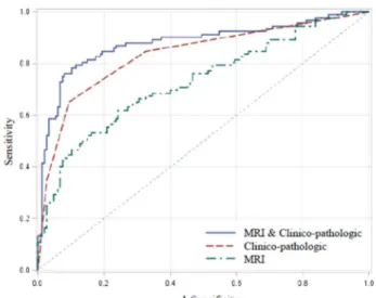
There was significant difference in AUC for combined selected DCE-MRI and clinico-pathologic variables, Fig. 3. Receiver-operating characteristic (ROC) curve of predictive variables in MRI, and clinico-pathologic validation, clinico-pathologic validation and MRI validation.
The area under the receiver operating characteristics curve (AUC) of MRI and clinico-pathologic, clinico-pathologic, and MRI variables, were 0.879, 0.835, and 0.729, respectively (P
< 0.05).
Table 5. Multivariate Analysis for Factors Associated with Axillary Lymph Node Metastasis
Variables Odds Ratio (95% CI) P value MRI variables Whole breast
vascularity
Negative 1
Mild 1.72 (0.69-4.28) 0.240 Moderate 3.44 (1.24-9.51) 0.017 Prominent 15.59 (2.52-96.45) 0.003
SER 1.67 (1.08-2.59) 0.020
Peritumor-tumor
ADC ratio 6.77 (2.41-18.99) < 0.001 Clinico-pathologic
variables HER-2
Negative 1
Positive 23.71 (10.50-53.53) 0.020 ADC = apparent diffusion coefficient; CI = confidence interval; HER-2 = human epidermal growth factor receptor-2; SER = signal enhancement ratio
compared to AUC of clinico-pathologic or DCE-MRI variables alone (P < 0.05). Clinico-pathologic variables associated with ALN metastasis are only useful after core biopsy, or surgical resection, and are not useful for predicting preoperative breast cancer. However, DCE- MRI is a non-invasive imaging technique that evaluates characteristics of preoperative tumors, and determines stage through tumor vessels. So, DCE-MRI variables may be beneficial in predicting preoperative ALN metastasis.
This study has several limitations. First, a small number of patients was included from one institution, offering limited representation of the population. Second, only cases of invasive ductal carcinoma, were included in the patient population. Other malignancies such as mucinous carcinoma, or invasive lobular carcinoma, were not included. If other malignancies were included, results may have differed. Third, micrometastatic nodes defined as metastatic foci less than 2 mm were excluded. Fourth, MRI was performed without considering the menstrual cycle, which could affect BPE. MRI must be obtained in the second week of the menstrual cycle, to have minimal effect on BPE. However, at many institutions, MRI is not considered for breast cancer staging. Last, ROI values were calculated using manual methods, which may be less reproducible between radiologists, than more automated volumetric techniques. However, manual methods are practical, because such methods can be used by all radiologists, and are commonly used for multiple imaging of lesions.
In conclusion, SER, moderate or prominent increased whole breast vascularity, and peritumor-tumor ADC ratio on DCE-MRI and DWI, may be valuable in predicting ALN metastasis in patients with early breast cancer.
REFERENCES
1. Park EH, Min SY, Kim Z, et al. Basic facts of breast cancer in Korea in 2014: The 10-year overall survival progress. J Breast Cancer 2017;20:1-11
2. Kuijs VJ, Moossdorff M, Schipper RJ, et al. The role of MRI in axillary lymph node imaging in breast cancer patients: a systematic review. Insights Imaging 2015;6:203-215 3. Qiu SQ, Zeng HC, Zhang F, et al. A nomogram to predict
the probability of axillary lymph node metastasis in early breast cancer patients with positive axillary ultrasound. Sci Rep 2016;6:21196
4. Roaten JB, Pearlman N, Gonzalez R, Gonzalez R, McCarter MD. Identifying risk factors for complications following sentinel lymph node biopsy for melanoma. Arch Surg
2005;140:85-89
5. Giuliano AE, Hunt KK, Ballman KV, et al. Axillary dissection vs no axillary dissection in women with invasive breast cancer and sentinel node metastasis: a randomized clinical trial. JAMA 2011;305:569-575
6. Caudle AS, Hunt KK, Tucker SL, et al. American College of Surgeons Oncology Group (ACOSOG) Z0011: impact on surgeon practice patterns. Ann Surg Oncol 2012;19:3144- 3151
7. Kvistad KA, Rydland J, Smethurst HB, Lundgren S, Fjosne HE, Haraldseth O. Axillary lymph node metastases in breast cancer: preoperative detection with dynamic contrast- enhanced MRI. Eur Radiol 2000;10:1464-1471
8. Valente SA, Levine GM, Silverstein MJ, et al. Accuracy of predicting axillary lymph node positivity by physical examination, mammography, ultrasonography, and magnetic resonance imaging. Ann Surg Oncol 2012;19:
1825-1830
9. Bahri S, Chen JH, Yu HJ, Kuzucan A, Nalcioglu O, Su MY.
Can dynamic contrast-enhanced MRI (DCE-MRI) predict tumor recurrence and lymph node status in patients with breast cancer? Ann Oncol 2008;19:822-824
10. Zaiton F, Shehata SM, Abo Warda MH, Alekrashy MA.
Diagnostic value of MRI for predicting axillarylymph nodes metastasis in newly diagnosed breastcancer patients:
Diffusion-weighted MRI. Egypt J Radiol Nucl Med 2016;47:659-667
11. Hatakenaka M, Soeda H, Yabuuchi H, et al. Apparent diffusion coefficients of breast tumors: clinical application.
Magn Reson Med Sci 2008;7:23-29
12. Marini C, Iacconi C, Giannelli M, Cilotti A, Moretti M, Bartolozzi C. Quantitative diffusion-weighted MR imaging in the differential diagnosis of breast lesion. Eur Radiol 2007;17:2646-2655
13. Dietzel M, Baltzer PA, Vag T, et al. Application of breast MRI for prediction of lymph node metastases - systematic approach using 17 individual descriptors and a dedicated decision tree. Acta Radiol 2010;51:885-894
14. Luo N, Su D, Jin G, et al. Apparent diffusion coefficient ratio between axillary lymph node with primary tumor to detect nodal metastasis in breast cancer patients. J Magn Reson Imaging 2013;38:824-828
15. Razek AA, Gaballa G, Denewer A, Nada N. Invasive ductal carcinoma: correlation of apparent diffusion coefficient value with pathological prognostic factors. NMR Biomed 2010;23:619-623
16. Dietzel M, Baltzer PA, Vag T, et al. The adjacent vessel sign on breast MRI: new data and a subgroup analysis for 1,084 histologically verified cases. Korean J Radiol 2010;11:178- 186
17. Knopp MV, Bourne MW, Sardanelli F, et al. Gadobenate dimeglumine-enhanced MRI of the breast: analysis of dose response and comparison with gadopentetate dimeglumine.
AJR Am J Roentgenol 2003;181:663-676
18. Kang DK, Kim EJ, Kim HS, Sun JS, Jung YS. Correlation of whole-breast vascularity with ipsilateral breast cancers using contrast-enhanced MDCT. AJR Am J Roentgenol 2008;190:496-504
19. Esserman L, Hylton N, George T, Weidner N. Contrast- enhanced magnetic resonance imaging to assess tumor histopathology and angiogenesis in breast carcinoma.
Breast J 1999;5:13-21
20. Mori N, Mugikura S, Takasawa C, et al. Peritumoral apparent diffusion coefficients for prediction of lymphovascular invasion in clinically node-negative invasive breast cancer.
Eur Radiol 2016;26:331-339
21. Sardanelli F, Iozzelli A, Fausto A, Carriero A, Kirchin MA.
Gadobenate dimeglumine-enhanced MR imaging breast vascular maps: association between invasive cancer and ipsilateral increased vascularity. Radiology 2005;235:791- 797
22. Wright H, Listinsky J, Quinn C, Rim A, Crowe J, Kim J.
Increased ipsilateral whole breast vascularity as measured by contrast-enhanced magnetic resonance imaging in patients with breast cancer. Am J Surg 2005;190:576-579 23. Han M, Kim TH, Kang DK, Kim KS, Yim H. Prognostic role
of MRI enhancement features in patients with breast cancer: value of adjacent vessel sign and increased ipsilateral whole-breast vascularity. AJR Am J Roentgenol 2012;199:921-928
24. Bielenberg DR, Zetter BR. The contribution of angiogenesis
to the process of metastasis. Cancer J 2015;21:267-273 25. Li KL, Henry RG, Wilmes LJ, et al. Kinetic assessment
of breast tumors using high spatial resolution signal enhancement ratio (SER) imaging. Magn Reson Med 2007;58:572-581
26. Loiselle CR, Eby PR, Peacock S, Kim JN, Lehman CD.
Dynamic contrast-enhanced magnetic resonance imaging and invasive breast cancer: primary lesion kinetics correlated with axillary lymph node extracapsular extension. J Magn Reson Imaging 2011;33:96-101
27. Cichon MA, Degnim AC, Visscher DW, Radisky DC.
Microenvironmental influences that drive progression from benign breast disease to invasive breast cancer. J Mammary Gland Biol Neoplasia 2010;15:389-397
28. Uematsu T, Kasami M, Watanabe J. Is evaluation of the presence of prepectoral edema on T2-weighted with fat- suppression 3 T breast MRI a simple and readily available noninvasive technique for estimation of prognosis in patients with breast cancer? Breast Cancer 2014;21:684- 692
29. Durur-Subasi I, Durur-Karakaya A, Karaman A, Seker M, Demirci E, Alper F. Is the necrosis/wall ADC ratio useful for the differentiation of benign and malignant breast lesions?
Br J Radiol 2017;90:20160803
30. Ahmed AR. HER2 expression is a strong independent predictor of nodal metastasis in breast cancer. J Egypt Natl Canc Inst 2016;28:219-227
31. Dittrich A, Gautrey H, Browell D, Tyson-Capper A. The HER2 signaling network in breast cancer--like a spider in its web.
J Mammary Gland Biol Neoplasia 2014;19:253-270