• 검색 결과가 없습니다.

A Comparative Study on Recovery of Motor Function in Stroke Patients with Corona Radiata Infarcts and Intracerebral Hemorrhage

N/A
N/A
Protected

Academic year: 2021

Share "A Comparative Study on Recovery of Motor Function in Stroke Patients with Corona Radiata Infarcts and Intracerebral Hemorrhage"

Copied!
6
0
0

로드 중.... (전체 텍스트 보기)

전체 글

(1)

Chung-Sun Kim, PT, PhD; Sang-Young Park, PT, MS

1

; Jung-Won Kwon, PT

1

Department of Physical Therapy, College of Rehabilitation Science, Daegu University;

1

Department of Rehabilitation Science, Graduate School, Daegu University

Purpose: Our goal was to determine the difference in motor recovery between two stroke types: the corona radiata (CR)  infarct type and the intracerebral hemorrhage (ICH) type, by using assessment methods for motor functions. 

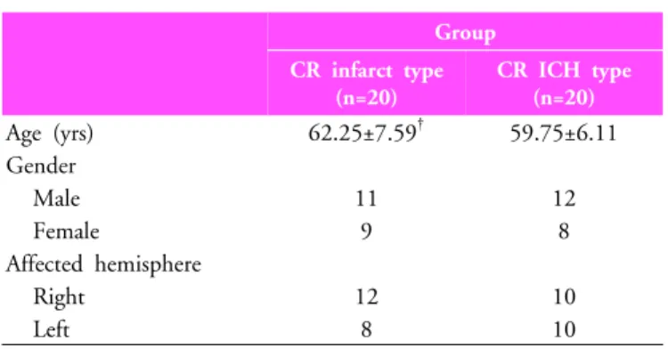
Methods:  Forty  subjects  who  were  diagnosed  as  having  had  a  stroke  with  an  infarct  (men:  11,  women:  9,  mean  age: 

62.25±7.59)  or  a  stroke  with  an  ICH  (men:  12,  women:  8,  mean  age:  59.75±6.11)  were  recruited.  In  all  subjects,  motor  functions of the affected extremities were measured 2 times: at stroke onset (initial) and 6 months after the onset (final) by  the  motricity  index  (MI),  the  modified  Brunnstrom  classification  (MBC),  and  functional  ambulatory  category  (FAC).  We  compared the final assessment with the initial one.

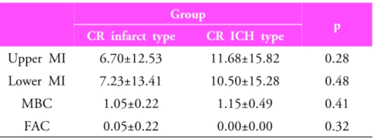
Results: Motor functions of all patients improved with the passing of time. All scores of motor function assessment in the  ICH type were higher than in the infarct type. Comparing the initial assessment with the final one, upper MI and MBC scores  of the upper extremities were significantly different between the two stroke types (p<0.05), but lower MI and FAC scores of  the lower extremities were not (p>0.05). 

Conclusion: These findings imply that patterns of motor recovery in patients with either the infarct type or the ICH type of  stroke change for the better over time. The degree of motor recovery in the ICH type was better than in the infarct type. 

Therefore, one can introduce clinical interventions by the aspect of progress in functional motor recovery.

Keywords: Corona radiata infarct, Intracerebral hemorrhage, Motor function, Motor recovery, Stroke  Received: October 4, 2010

Revised: November 15, 2010 Accepted: December 8, 2010

Corresponding author: Jung-Won Kwon, [email protected]

Patients with Corona Radiata Infarcts and Intracerebral Hemorrhage

The Journal Korean Society of Physical Therapy

I. Introduction

Stroke is a major neurological syndrome that is characterized by chronic inability to perform a variety of activities due to a hemiplegic deficit of motor and sensory function, impaired cognition and perception, and psychosocial problems.

1,2

Among individuals who survive a stroke, motor dysfunction is one of the most serious sequelae, with over 50% of stroke patients experiencing a physical limitation on functional activities.

3

Motor dysfunctions such as muscle weakness and atrophy, abnormal muscle tone and abnormal movement patterns of the extremities, impaired motor control, and dysregulation of neuromuscular systems are found inover 80% of these patients.

4

The corona radiata (CR), which passes the corticospinal

tract (CST), is the most important region because it is

commonly affected by infarct or intracerebral hemorrhage

(ICH).

5,6

The patients who experience a stroke caused by a CR

infarct or intracerebral hemorrhage are part of a varied clinical

spectrum including incomplete motor and sensory distribution

patterns, and additional neuropsychological deficits.

7

Among

the many studies that have been done, the clinical picture and

recovery patterns of infarct and ICH have been reported.

8-11

Since the CST is located in the posterior portion of the corona

radiata, preservation or restoration of the CST is mandatory for

good recovery of impaired motor function, and it seems to be

significant for predicting motor outcomes on the affected

side.

12,13

Accordingly, elucidation of the motor recovery mech-

anism in infarct and ICH types is really important because such

(2)

Group CR infarct type

(n=20)

CR ICH type (n=20)

Age (yrs) 62.25±7.59

59.75±6.11

Gender

Male 11 12

Female 9 8

Affected hemisphere

Right 12 10

Left 8 10

Mean±Standard deviation

CR: Corona radiata, ICH: Intracerebral hemorrhage

Table 1. General characteristic of subjects (N=40) information could provide a scientific basis for stroke reha-

bilitation.

According to advanced neuroimaging methods such as functional magnetic resonance imaging (fMRI) and diffusion tensor tractography (DTT), we can visualize and localize neuropathological lesions. Many investigators have become focused on motor recovery mechanisms.

5,6,14-17

Jang et al.

18

reported that the affected motor function of a stroke patient with ICH has recovered through the healing process of the damaged CST of the affected hemisphere. Kwon et al.

19

reported that motor recovery in patients with a corona radiata infarct reflects functional reorganization of motor pathways, depending on damage to the corticospinal tract. Therefore, the assessment of motor recovery with time may be used to predict motor outcomes of the affected extremities in stroke patients with infarct and ICH.

In previous studies of motor recovery, impaired motor function in stroke patients has generally recovered through a process of brain plasticity, and it is well known that they will recover by themselves within 3 months (although the time to appearance of motor recovery is variable).

20,21

However, many previous studies have also revealed that there are many differences among stroke patients in terms of their extent of motor recovery as well as with regard to recovery mechanisms.

Furthermore, differences in recovery mechanisms have not yet been clearly elucidated.

22-24

Therefore, we now report differences in motor recovery between the two stroke types: corona radiata infarct type and intracerebral hemorrhage type. We also present fundamental details about recovery mechanisms.

II. Methods

1. Subjects

Forty subjects who were diagnosed as having a stroke with a corona radiata infarct (men: 11, women: 9, mean age:

62.25±7.59) and intracerebral hemorrhage (men: 12, women: 8, mean age: 59.75±6.11) were recruited (Table 1). All subjects understood the purpose of the study and provided written consent prior to participation, which was approved by the institutional review board of a university hospital. The inclusion criterion were: (1) first ever stroke, (2) severe weakness of the affected extremities (at least a 2 grade decrease in MMT) 3 weeks

after stroke onset, (3) a deficiency of a serious nature in cognitive problems, and (4) no pathology of musculoskeletal function or neurologic diseases other than the outcome of the stroke.

2. Clinical evaluation

Motor function of the affected extremity was measured 2 times:

at 3 weeks after stroke onset (initial) and 6 months after the onset (final) using the motricity index (MI), the modified Brunnstrom classification (MBC), and the functional ambulatory category (FAC). The standardized MI, which was employed to determine muscle strength, was used to measure the integrity of motor function with a maximum score of 100.

25

There were six categories in all movements except prehensions:

0=no movement, 28=palpable contraction (but no movement),

42=movement without gravity, 56=movement against gravity,

74=movement against resistance (but weaker than normal), and

100=normal. In the prehensions, there were six categories: 0=no

movement, 33=beginning of prehension, 56=grips cube without

gravity, 65=holds cube against gravity, 77=grips against pull (but

weaker than the other side), and 100=normal. The function of

the affected hand was categorized according to the MBC.

26

The

MBC scores of finger extensors included six categories: 1=unable

to move fingers voluntarily, 2=able to move fingers voluntarily,

3=able to close hand voluntarily; unable to open hand

voluntarily, 4=able to grasp a card between the thumb and

medial side of the index finger; able to extend fingers slightly,

5=able to pick up and hold a glass; able to extend fingers, and

6=able to catch and throw a ball in a nearly normal fashion; able

to button and unbutton a shirt. Ambulatory ability was

determined using a standardized FAC: scoring was designed to

(3)

Group

CR infarct type CR ICH type p

Upper MI 6.70±12.53 11.68±15.82 0.28 Lower MI 7.23±13.41 10.50±15.28 0.48

MBC 1.05±0.22 1.15±0.49 0.41

FAC 0.05±0.22 0.00±0.00 0.32

Unit: Score

MI: Motricity Index, MBC: Modified Brunnstrom Classification, FAC: Functional Ambulatory Category

Table 2. Results of initial assessments at 3 weeks after stroke onset in each group

Group

CR infarct type CR ICH type p

Upper MI 46.07±15.70 59.30±21.41 0.03*

Lower MI 49.83±14.24 59.40±24.29 0.14

MBC 2.15±1.42 3.75±1.16 0.00**

FAC 3.25±1.16 3.55±1.50 0.49

Unit: Score

*p<0.05, **p<0.01

MI: Motricity Index, MBC: Modified Brunnstrom Classification, FAC: Functional Ambulatory Category

Table 3. Comparison of functional change of motor recovery in each group

examine the levels of required assistance during a 15 m walk.

27

There were six categories: 0=non-ambulatory, 1=needs con- tinuous support from one person, 2=needs intermittent support from one person, 3=needs only verbal supervision, 4=help is required on stairs and uneven surfaces, and 5=can walk independently anywhere. The reliability and validity of the MI, MBC, and FAC are well-established.

25-27

3. Data analysis

The data were analyzed using an independent sample t-test, to compare the recovery of motor functions between the corona radiata infarct type and the intracerebral hemorrhage type. All statistical analyses were done using SPSS Ver 12.0; p<0.05 was used as the criterion for statistical significance.

III. Results

All subjects had low scores on the initial assessments at threeweeks after stroke onset. Scores for MI, MBC, and FAC in the ICH type were higher than in the infarct type. But, there were no significant differences between the two stroke types (p>0.05)(Table 2).

As time passed, all subjects showed muscle strength improvement from the initial assessment to the final assessment:

increased MI scores of the upper extremities were 46.07 and 59.30, respectively, for the infarct type and ICH type; scores were 49.83 and 59.40 for the lower extremities between infarct type and ICH type. In MI scores of the upper extremity, increased scores for the ICH type were significantly higher than for the infarct type (p<0.05), but there were no significant changes in MI scores of the lower extremity (p>0.05)(Table 3).

Over time, MBC scores increased to 2.15 at three weeks after stroke onset and 3.75 at six months after stroke onset; there were significant differences between the two stroke types (p<0.01) (Table 3).

In the assessment of ambulatory ability, all subjects showed improvements with the passing of time: FAC scores were increased to 3.25 and 3.55 at three weeks and sixmonths, respectively between the two stroke types, but there were no significant changes (p>0.05)(Table 3).

IV. Discussion

In the current study, we tried to learn the difference in motor recovery between two stroke types: the CR infarct type and the ICH type, using assessment methods for motor functions such as MI, MBC, and FAC during the 6 months after stroke onset.

We found that motor functions of all patients improved with the passing of time: final scores of each motor assessment were higher than initial scores. The degree of motor recovery in the ICH type was larger than in the infarct type. In other words, comparing the initial assessment with the final, upper MI and MBC scores were significantly different between the two stroke types (p<0.05)(p<0.01), but lower MI and FAC scores were not (p>0.05). These results imply that patterns of motor recovery in the infarct type and the ICH type change for the better over time. Also, the degree of motor recovery in the ICH type were better than in the infarct type.

We chose MI, MBC and FAC for scoring methods in order

to focus the clinical perspective to the functional assessment of

the affected extremities. Because the baseline scores of all of our

patients were presented at least a 2 grade decrease in MMT at the

(4)

initial assessment after stroke onset, their motor functions recovered to the point of being nearly normal at the final assessment after 6 months. This strongly suggests that the motor function of the affected extremity changes over time, and that it recovers through a process of brain plasticity. It is well known that the corticospinal tract is located in the corona radiata and plays an important role in motor recovery in stroke patients.

28-30

In addition, both CR infarct and ICH damage to the lateral corticospinal tract, which is associated with control of the distal musculature, affects fine movement in the upper extremity.

14

The corticospinal tract (CST) in the region around the lesion is closely associated with motor recovery mechanisms for reor- ganization of neural pathways.

13,19

However, although the motor recovery mechanism has been well established, there is a difference between CR infarct and ICH in the extent of motor recovery. The integrity of the lateral CST was maintained, at least from 4 months after stroke onset because the damaged CST recovered by the process of normalization from the parietal cortex to the primary motor cortex in patients with ICH.

18

In patients with a CR infarct, motor function recovered slowly during the 6 months after the stroke because of several mechanisms such as: (1) the ipsilateral motor pathway from the unaffected motor cortex to the affected extremity, (2) peri- lesional reorganization, (3) recovery of a damaged lateral CST, and (4) contributions of secondary motor areas.

19

The motor recovery mechanism was similar between infarct and ICH types, but the extent of functional recovery in the ICH type was greater than in the infarct type. Consistent with previous studies, our study supported differences in motor recovery between infarct and ICH types with the ICH type improving faster initially patients with CR infarct, and the various clinical patterns depending on involved region of infarct and ICH were showed.

24,31-35

In addition, prognostic factors which correlated with poor outcomes were heavily driven by age, prior drug dependency, prior stroke, and initial severity as well as medical histories such as hypertension, diabetes mellitus, atrial fibrillation, and cardiovascular risks in stroke patients.

23,36

Moreover, a short time between the occurrence of the stroke and presentation at the hospital would allow an opportunity for initiation of thrombolysis, access of prognostic factors, and engagement in rehabilitation, which may be relevant to optimal recovery.

24,37

We used detailed methods to compare recovery patterns; results show a series of different recovery processes with

different motor outcomes between infarct type and CR infarct stroke patients.

We acknowledge that this study has limitations. For example, we did not observe CST fibers re-connecting during motor recovery. Therefore, further studies will be required to ascertain the detailed mechanisms of the motor recovery, and to determine the critical factors that influence the motor recovery of the CST between the infarct type and the ICH type in stroke patients.

V. Conclusion

The neurological basis of motor recovery has been well established in stroke patients, but there are many differences among stroke patients in terms of the extent of motor recovery.

Furthermore, differences in recovery mechanisms have not been clearly elucidated, even for functional motor outcomes for the two major stroke types: corona radiata infarct type and corona radiata intracerebral hemorrhage type. Therefore, we conclude that aspects of functional recovery and clinical prognosis are predictable for specific patients with corona radiata infarct and intracerebral hemorrhage. In addition, we have presented fundamental information about detailed stroke recovery mechanisms.

Author Contributions Research design: Kim CS

Acquisition of data: Kwon JW, Park SY

Analysis and interpretation of data: Kwon JW, Park SY Drafting of the manuscript: Kwon JW

Research supervision: Kim CS

References

1. Manimmanakorn N, Arrayawichanon P, Wattanapun P et al.

Age-related rehabilitation outcome in stroke patients. J Med Assoc Thai. 2008;91(3):388-93.

2. Gialanella B, Bertolinelli M, Santoro R. Rehabilitative outcome in supratentorial and infratentorial stroke: The role of motor deficits. Aging Clin Exp Res. 2008;20(4):310-5.

3. Duncan PW, Goldstein LB, Matchar D et al. Measurement of

(5)

motor recovery after stroke. Outcome assessment and sample size requirements. Stroke. 1992;23(8):1084-9.

4. Kautz SA, Patten C. Interlimb influences on paretic leg function in poststroke hemiparesis. J Neurophysiol. 2005;93(5):

2460-73.

5. Jang SH, Byun WM, Han BS et al. Recovery of a partially damaged corticospinal tract in a patient with intracerebral hemorrhage: a diffusion tensor image study. Restor Neurol Neurosci. 2006;24(1):25-9.

6. Jang SH, Ahn SH, Ha JS et al. Peri-infarct reorganization in a patient with corona radiata infarct: A combined study of functional MRI and diffusion tensor image tractography.

Restor Neurol Neurosci. 2006;24(2):65-8.

7. Kumral E, Bayulkem G. Spectrum of single and multiple corona radiata infarcts: Clinical/MRI correlations. J Stroke Cerebrovasc Dis. 2003;12(2):66-73.

8. Woo D, Broderick JP. Spontaneous intracerebral hemorrhage:

epidemiology and clinical presentation. Neurosurg Clin N Am.

2002;13(3):265-79.

9. Jang SH. A review of corticospinal tract location at corona radiata and posterior limb of the internal capsule in human brain. NeuroRehabilitation. 2009;24(3):279-83.

10. Cho SH, Kim SH, Choi BY et al. Motor outcome according to diffusion tensor tractography findings in the early stage of intracerebral hemorrhage. Neurosci Lett. 2007;421(2):142-6.

11. Cho SH, Kim DG, Kim DS et al. Motor outcome according to the integrity of the corticospinal tract determined by diffusion tensor tractography in the early stage of corona radiata infarct. Neurosci Lett. 2007;426(2):123-7.

12. Yang DS, Kim DS, Kim YH et al. Demonstration of recovery of a severely damaged corticospinal tract: adiffusion tensor tractography and transcranial magnetic stimulation follow-up study. J Comput Assist Tomogr. 2008;32(3):418-20.

13. Jang SH. The role of the corticospinal tract in motor recovery in patients with a stroke: a review. NeuroRehabilitation.

2009;24(3):285-90.

14. Jang SH. A review of motor recovery mechanisms in patients with stroke. NeuroRehabilitation. 2007;22(4):253-9.

15. Jang SH. A review of the ipsilateral motor pathway as a recovery mechanism in patients with stroke. NeuroRehabilitation.

2009;24(4):315-20.

16. Park JW. Longitudinal motor function recovery in stroke patients with focal pons infarction: report of 4 cases. J Kor Soc Phys Ther. 2009;21(4):111-5.

17. Choi JH, Kwon YH. Medial reorganization of primary motor cortex in patient with traumatic brain injury: a fMRI case study.

J Kor Soc Phys Ther. 2005;17(3):421-8.

18. Jang SH, Kim SH, Cho SH et al. Demonstration of motor recovery process in a patient with intracerebral hemorrhage.

NeuroRehabilitation. 2007;22(2):141-5.

19. Kwon YH, Lee CH, Ahn SH et al. Motor recovery via the peri-infarct area in patients with corona radiata infarct.

NeuroRehabilitation. 2007;22(2):105-8.

20. Jang SH, Kwon YH. Motor recovery in stroke patients.

Yeungnam Univ J of Med. 2005;22(22):119-30.

21. Kwakkel G, Kollen B, Twisk J. Impact of time on improvement of outcome after stroke. Stroke. 2006;37(9):2348-53.

22. Appelros P, Stegmayr B, Terent A. A review on sex differences in stroke treatment and outcome. Acta Neurol Scand.

2010;121(6):359-69.

23. Bilic I, Dzamonja G, Lusic I et al. Risk factors and outcome differences between ischemic and hemorrhagic stroke. Acta Clin Croat. 2009;48(4):399-403.

24. Wei JW, Heeley EL, Wang JG et al. Comparison of recovery patterns and prognostic indicators for ischemic and hemorrhagic stroke in china: The chinaquest (quality evaluation of stroke care and treatment) registry study. Stroke.

2010;41(9):1877-83.

25. Demeurisse G, Demol O, Robaye E. Motor evaluation in vascular hemiplegia. Eur Neurol. 1980;19(6):382-9.

26. Brunnstrom S. Motor testing procedures in hemiplegia: Based on sequential recovery stages. Phys Ther. 1966;46(4):357-75.

27. Cunha IT, Lim PA, Henson H et al. Performance-based gait tests for acute stroke patients. Am J Phys Med Rehabil.

2002;81(11):848-56.

28. Han BS, Hong JH, Hong C et al. Location of the corticospinal tract at the corona radiata in human brain. Brain Res. 2010;

1326:75-80.

29. Kim CS, Kwon JW. Motor function recovery in stroke patients with corona radiata infarct: 4 case studies. J Kor Soc Phys Ther.

2010;22(3):31-5.

30. Sterr A, Shen S, Szameitat AJ et al. The role of corticospinal tract damage in chronic motor recovery and neurorehabilitation: a pilot study. Neurorehabil Neural Repair.

2010;24(5):413-9.

31. Aminoff MJ, Boller F, Swaab DF. Clinical neurology and stroke.

Foreword. Handb Clin Neurol. 2009;93:vii.

32. Bejot Y, Rouaud O, Jacquin A et al. Stroke in the very old:

(6)

Incidence, risk factors, clinical features, outcomes and access to resources--a 22-year population-based study. Cerebrovasc Dis. 2010;29(2):111-21.

33. Khoo CW, Lip GY. Clinical outcomes of acute stroke patients with atrial fibrillation. Expert Rev Cardiovasc Ther. 2009;

7(4):371-4.

34. Lu ZN, Yu SZ. Somatosensory evoked potentials in patients with cerebral hemorrhage and infarct. Correlation with CT, clinical data and their. Chin Med J (Engl). 1993;106(1):35-9.

35. Chae J, Zorowitz RD, Johnston MV. Functional outcome of hemorrhagic and nonhemorrhagic stroke patients after in-patient rehabilitation. Am J Phys Med Rehabil. 1996;75(3):

177-82.

36. Wong KS. Risk factors for early death in acute ischemic stroke and intracerebral hemorrhage: a prospective hospital-based study in Asia. Asian acute stroke advisory panel. Stroke.

1999;30(11):2326-30.

37. Ireland SE, Arthur HM, Gunn EA et al. Stroke prevention care

delivery: predictors of risk factor management outcomes. Int

J Nurs Stud. 2010[Epub ahead of print].

수치

Table 1.  General  characteristic of subjects (N=40) information could provide a scientific basis for stroke
Table 3. Comparison of functional change of motor  recovery in each group

참조

관련 문서

This study was to do a comparative analysis on kinematic differences and differences in muscle activity between the skilled and the unskilled in windsurfing

The benefits and harms of intravenous thrombolysis with recombinant tissue plasminogen activator within 6 h of acute ischaemic stroke (the third

Guidelines for the early management of adults with ischemic stroke: a guideline from the American Heart Association/American Stroke Association Stroke Council,

8WD/4WS Vehicle Equipped with 8 In-wheel Motor 8WD/4WS Vehicle Equipped with 8 In wheel

The aim of this study was to develop a training program for swallowing and to test its effect on swallowing capacity and nutritional status in stroke

This paper investigates the influence which the motor consumer's information on M&amp;A(merger and acquisition) between motor companies has upon his attitude

The associations of hepatic steatosis and fibrosis using fatty liver index and BARD score with cardiovascular outcomes and mortality in patients with new‑onset type 2

Incident major cardiovascular events (coronary artery disease, ischemic stroke, hemorrhagic stroke and cardiovascular mortality) were set as primary end points.