大 韓 不 妊 學 會 誌 : 第 30 卷 第 1 號 2003 Kor. J. Fertil. Steril., Vol. 30, No. 1, 2003, 3
인간자궁내막의 탈락막화 (Decudualization)에 있어서 TGF-ß (Transforming Growth Factor-ß)의 역할
아주대학교 대학원 분자과학기술학과1, 아주대학교 의과대학 산부인과학교실2, 아주대학교 자연대학 생명과학과3
박동욱1,2・최동순1・김미란2・황경주2・조미영2・안성희2・민철기3・유희석2
Transforming Growth Factor-ß is a Possible Paracrine Mediator in the Human Endometrial Decidualization
Dong Wook Park
1,2, Dong Soon Choi
1, Mi Ran Kim
2, Kyung Joo Hwang
2, Mi Yeong Jo
2, Seong Hee Ahn
2, Churl K Min
3, Hee Sug Ryu
21
Department Molecular Science and Technology,
2Department of Obstetrics and Gynecology, School of Medicine,
3Department of Biological Science, Ajou University, Suwon, Korea
Objectives: To investigate the role of TGF (Transforming growth factor-ß) involved in the paracrinic communication during decidualization between UEC (uterine epithelial cells) and USC (uterine stromal cells), we have employed a co-culture system composed of human endometrial epithelial and stromal cells in defined hormonal conditions.
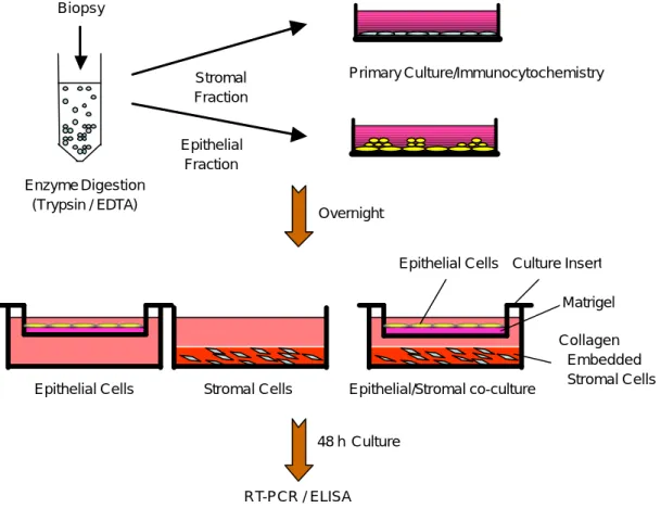
Design: In the co-culture, endometrial epithelial cells cultured in the matrigel-coated cell culture insert are seeded on top of the endometrial stromal cells cultured within a collagen gel. The co-culture was maintained for 48 hours under the following hormonal conditions: progesterone dominant condition (100 nM P4 and 1 nM E2) or estrogen-dominant condition (100 nM E2 and 1 nM P4). 10 ng/ ml HGF and/or 10 ng/ml TGF-ß1 are added.
Methods: RT-PCR is utilized to detect mRNAs quantitatively. Enzyme-linked immunosorbent assay (ELISA) and immunohistochemical staining are utilized to detect proteins in the tissue.
Results: Prolactin mRNA is expressed in the co-cultured stromal cells under the progesterone dominant condition. TGF-ß1 and its receptors are expressed in both the co-cultured epithelial and stromal cells irrespective of the steroid present, which is in contrast with no or negligible expression of TGF-ß1 or its receptor in cells separately cultured. Both estrogen and progesterone significantly elevate the concentration of hepatocyte growth factor (HGF) in the conditioned medium of the co-culture with the value of 4,325 pg/ml in E2-dominant and 2,000 pg/ml in P4-dominant condition compare to 150 pg/
ml in no hormone. In separately cultured stromal cells, administration of HGF induces the expression of TGF receptor 1 in both hormonal conditions, but induction of TGF receptor 2 is only manifest in the P4-dominant condition. Administration of TGF-ß and HGF directly induce the decidualization marker
연 락 저 자: 박동욱, 우) 442-749 경기도 수원시 팔달구 원천동 산5번지 아주대학교의과대학 산부인과학교실 Tel: (031) 219-5300, Fax: (031) 219-5245, e-mail: [email protected]주관책임자: 김미란, 우) 442-749 경기도 수원시 팔달구 원천동 산5번지, 아주대학교의과대학 산부인과학교실 Tel: (031) 219-5300, Fax: (031) 219-5245, e-mail: [email protected]
본 논문은 korea health industry development grant (by Churl K Min)에 의해 이루어짐 (01-PJ11-PG9-01NT00-0036 to C.K.M).
prolactin mRNA in separately cultured stromal cells.
Conclusion: It is likely that steroid hormones induces prolactin mRNA indirectly by promoting the cell to cell communication between the stromal and the epithelial cells. TGF-ß and HGF are two possible paracrine mediators in the human endometrial decidualization.
Key Words: Decidualization, TGF-ß, HGF, Co-culture, Paracrine
인간자궁내막은 생리주기를 통해 증식, 분화 및 탈락을 거치게 된다. 후기 분비기로 분화된 자궁내 막 기질세포 (endometrial stromal cell; ESC)는 형태 와 기능적으로 독특한 특징을 지니게 된다. 이러한 특징적인 현상을 탈락막화 (decidualization)라고 하 며 탈락막화 된 세포는 포배 (blastocyst)의 착상 및 임신에 필요한 기능층 (functional layer)으로 작용한 다. 현재까지 성호르몬,1 cAMP,2 gonadotropin,3 PGE2
등4이 자궁내막 기질세포의 탈락막화를 유도하는 것 으로 보고 되었다.
Transformin growth factor-ß (TGF-ß)는 25 kD의 homodimeric 단백질이며 세포의 성장5 및 분화6에 다양한 역할을 하는 것으로 알려져 있다. 이외에도 스테로이드 생합성,7 혈관형성8 및 종양의 침윤과 전이에도 관여하는 것으로 보고되어 왔다.9 특히, 인 간자궁내막조직에서는 TGF-ß mRNA의 양이 중기 및 후기 분비기에 상승하며 이렇게 생성된 TGF-ß는 생리 후 내막조직의 재생성과정에서 혈관형성에 관 여할 것으로 보고되었으며,10 progesterone과 함께 자 궁내막에서 matrix metalloproteinase의 발현을 억제 하는 것으로 알려져 있다.11 또한 자궁기질세포에서 분비된 TGF-ß는 상피세포의 MMP 발현을 조절하 는 paracrine mediator로 작용한다고 보고되기도 하 였다.12
TGF-ß의 활성화과정은 비활성화 상태 (latent pro- form)로 세포에서 분비된 후 plasmin에 의한 prote- olytic cleavage를 거쳐 활성화 된다.13 이 과정에는 TGF-ß의 세포내 신호전달에 참여하는 다양한 분자 들이 이용되는데 TGF-ß type 2 수용체와 결합하여 type 1 수용체를 활성화시킨다. 이후 활성화 된 type 1 수용체는 세포질단백질인 Smad-2와 -3를 인산화 시켜 Smad-4와 결합하게 한다. 이렇게 형성된 복합 체는 핵내부로 이동하여 다양한 유전자의 발현을 조절하게 된다.14 Ando 등15은 TGF-ß의 각 subtype (TGF-ß1, ß2, ß3) 및 수용체 type-1, type-2 mRNA가
인간의 first trimester decidua 및 villous에서 발현되 는 것으로 보아 TGF-ß가 탈락막화 및 태반형성과정 (placentation)에 역할을 하리라 가정하였다.
HGF (Hepatocyte growth factor)는 최초로 일차 배양된 토끼 간세포에서 강력한 세포분열 촉진인자 로서 발견되었다.16 HGF는 인간자궁에서는 자궁내 막 기질세포의 분열 및 내강형성 (lumen formation) 을 조절하며,17 영양배엽 (trophoblast)의 침윤에 관 여하는 것으로 알려져 있다.18 또한, 인간 혈청내에 서 HGF는 중기 황체기에서 후기 황체기에 증가하 여 생리기에 최고 농도를 보이며 임신중에는 그 농 도를 유지하는 것으로 알려져 있다.19
현재까지 여러 연구자들이 배양된 기질세포에 성 호르몬을 첨가하여 탈락막화를 유도할 수 있음을 보고하였다. Irwin 등은20 일차 배양된 기질세포에 progesterone과 estrogen을 첨가하여 decidualization을 유도할 수 있다고 하였다. 그러나 이러한 실험 결과 는 일차 배양한 기질세포에 progesterone을 첨가한 후 장시간 (10일 이상) 배양하여 유도된 결과이므로 체내에서의 탈락막화 (in vivo decidualization) 기전을 완벽히 재현했다고 보기 어렵다. 뿐만 아니라, 2차원 적으로 단층 배양된 자궁내막세포들은 증식은 이루 어지나 기능적인 분화 특성을 유지하지 못하는 단점 을 가지고 있다.21 Bentin-Ley 등22은 이러한 2차원 배양의 단점을 보완하기 위하여 collagen gel을 이용 하여 자궁내막세포를 3차원적으로 배양하는 방법을 개발하였으며, 이어서 3차원적으로 배양된 자궁내막 세포가 형태학적으로 체내의 자궁내막조직과 유사한 구조를 하고 있음을 보고하였다.
본 연구진은 TGF-ß 및 수용체 mRNA의 발현을 RT-PCR을 이용하여 관찰하였고, 3차원적으로 공배 양한 상피세포와 기질세포에 성호르몬의 투여가 배 양액내 HGF의 농도를 증가시키는지를 ELISA를 이 용하여 관찰하였다. 이러한 실험을 통하여 인간자궁 내막의 탈락막화과정이 상피세포와 기질세포의 상
호작용에 의한 것인지를 확인해 보고, 세포간 상호 작용이 있다면 TGF-ß 및 HGF에 의하여 중재되는 지를 알아보고자 하였다.
연구 대상 및 방법
1. 자궁내막조직의 획득
아주대병원 산부인과를 내원한 30세에서 45세 사 이의 중기 및 후기 분비기에 자궁내막질환 이외의 양성질환 (자궁 근종 및 자궁 선근종)으로 전자궁적 출술을 시행한 후 자궁 후저부로부터 자궁내막조직 을 획득하였다.
2. 자궁내막 상피세포와 기질세포의 배양 채취된 자궁내막조직은 PBS가 들어 있는 conical
tube (Falcon, USA)에 담아 실험실로 운반했으며 다 시 PBS로 여러 번 세척하여 남아 있는 혈액을 제 거하였다. 배양 접시에 Dulbeco's Modified Eagle's Media (DMEM; Gibco, USA) 2~3 ml을 부어준 후 조 직을 넣고 멸균된 가위를 이용하여 1~2 mm의 크기 로 잘랐다. 잘게 잘린 조직은 다시 cornical tube에 용액과 함께 부어준 후 원심 분리하여 상층액을 따 라내고 trypsin-EDTA (Gibco, USA) 5 ml을 첨가하여 37℃에서 회전 배양하였다. 1시간 경과 후 10% fetal bovine serum (FBS; Gibco, USA)이 포함된 DMEM 배양액을 5 ml 첨가하여 효소작용을 정지시켰다. 기 질세포들이 포함된 용액 상층부 5 ml과 상피세포들 이 포함된 하층부 5 ml을 각각 나누어 60 mm 배양 접시 (Falcon, USA)에 분주한 후 24시간 배양하였다.
배양된 상피세포를 단일세포로 분리하기 위하여 새
Epithelial Cells Biopsy
Enzyme Digestion (Trypsin / EDTA)
Stromal Fraction
Epithelial Fraction
Overnight
Primary Culture/Immunocytochemistry
Stromal Cells
Collagen Embedded Stromal Cells Epithelial Cells Culture Insert
Matrigel
Epithelial/Stromal co-culture
48 hCulture
RT-PCR / ELISA
Figure 1. A schematic drawing of the cultrue system. Endometrial biopsy was minced and separated into respective
epithelial and stromal fractions. Each fraction was cultured overnight. In the co-culture, endometrial epithelial cells cultured in the matrigel-coated cell culture insert are seeded on top of the endometrial stromal cells cultured within a collagen gel. The cultures was maintained for 48 hours under each hormone conditions.로운 배양액으로 세척한 후 덩어리를 이루고 있는 상피세포를 배지로 강하게 세척하여 배양 접시로부 터 탈락시켰다. 분리한 상피세포 덩어리를 1:4로 희 석한 matrigel (Biocoat, USA)이 coating된 cell culture insert (Sigma, USA)에 접종한 후 6 well 배양 접시 (Nunc A/S, Denmark)에 위치시켜 10% FBS가 들어 있는 배지에서 24시간 배양하였다. 이와 동시에 배 양된 기질세포는 배양 접시에 배양액을 제거한 후 trypsin-EDTA 2 ml을 첨가하여 10분 경과 후 새로운 배양액 5 ml를 첨가하여 400 rpm에서 5분간 원심 분리하였다. 단일세포로 분리된 기질세포를 5×10⁶ cells/ml의 농도로 collagen (Biocoat, USA) 용액과 섞 어 6 well 배양 접시 (Nunc A/S, Roskilde, Denmark) 에 500 µl를 부은 후 37℃에서 1시간 배양하여 col- lagen이 중합되도록 하였다. 3차원적으로 중합된 기 질세포를 10% FBS (Gibco, USA)가 첨가된 배지에서 24시간 배양하였다. 이렇게 각각 배양된 상피세포와 기질세포를 실험 조건에 따라 48시간 동안 공배양 또는 분리 배양하였다 (Figure 1). 실험에 사용한 성 호르몬의 농도는 progesterone 우세 환경일 경우 100 nM progesterone (Sigma, USA), 1 nM estrogen (Sigma, USA)을 첨가하였으며, esterogen 우세 환경일 경우 100 nM estrogen, 1 nM progesterone의 농도로 정하 였다. 10 nM/ml의 TGF-ß1 (Sigma, USA)과 10 nM/
ml의 HGF (Sigma, USA)를 실험에 사용하였다.
3. 면역화학적 염색
Cytokeratin, vimentin 및 Met 에 대한 면역조직화 학적 염색은 먼저 배양된 세포에서 배지를 제거한 후 PBS로 3회 세척하였다. 세척한 세포를 4% PBS 로 희석된 parafrormaledhyde와 차가운 methanol로 고정하였다. 여기에 4% H2O2 용액을 5분간 전 처리 하여 조직에 남아 있는 peroxydase를 제거하였다. 세 포를 다시 PBS로 세척한 후 1/400으로 희석한 cyto- keratin에 대한 일차 항체 (Anti-human mouse mono- clonal; Santa Cruz Biotechnology Inc., USA), vimentin 에 대한 일차 항체 (Anti-human goat polyclonal; Santa Cruz Biotechnology, USA) 및 Met 에 대한 일차 항체 (Anti-human rabbit polyclonal; Santa Cruz Biotechno- logy Inc., USA)로 상온에서 1시간 동안 항습 cham- ber에서 반응시킨 후 LSAB-kit (DAKO A/S, Den- mark)에 포함된 이차 항체를 사용하여 15분간 처리 하였다. 표본은 PBS로 세척한 후 diaminobenzidine (DAB; DAKO A/S, Denmark)을 이용하여 발색시킨 후 관찰하였다.
4. RT-PCR
TRIsol reagent (Invitrogen life technology, Nether- land)를 이용하여 배양한 세포로부터 모든 RNA를 추출하였다. 추출한 RNA는 260 nm와 280 nm 파장
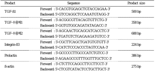
Table 1. PCR primers
Product Sequence Product size
Forward : 5'-CACGTGGAGCTGTACCAGAA-3' TGF-ß1
Backward: 5'-GTCCAGGCTCCAAATGTAGG-3' 500 bp Forward : 5'-ACGGCGTTACAGTGTTCTG-3'
TGF-수용체1
Backward: 5'-GGTGTGGCAGATATAGACC-3'
358 bp Forward : 5'-AGCAACTGCAGCATCACCTC-3'
TGF-수용체2
Backward: 5'-TGATGTCTGAGAAGATGTCC-3' 688 bp Forward : 5'-CGCTTCAGCTGATGTGTGTT-3'
Integrin-ß3
Backward: 5'-CATCTCCCACCCTAGTCCAA-3' 224 bp Forward : 5'-GCCCCCTTGCCCATCTGTCC-3'
Prolactin
Backward: 5'-AGAAGCCGTTTGGTTTGCTCC-3' 386 bp Forward : 5'-CTCTTCCAGCCTTCCTTCCT-3'
ß-actin
Backward: 5'-CTCGTCATACTCCTGCTTGCT-3' 275 bp
에서 흡광도를 측정하여 이들의 비가 1.9 이상일 경 우에만 사용하였다. cDNA의 합성은 M -MLV reverse transcriptase kit (Bioneer corporation, Korea)를 사용하 여 수행하였다. 실험에 사용한 모든 primer는 primer designing software Primer3 (available at steve@genome.
wi.mit.edu)를 이용하여 제작하였다.
실험에 사용한 primer와 PCR 산물의 크기는 다음 과 같다 (Table 1). 모든 primer는 주문 제작 및 정제 를 거쳐 사용하였다. 모든 PCR 과정은 Accupower PCR premix kit (Bioneer, Korea)을 이용하였으며, PCR에 사용한 증폭과정은 다음과 같다. 94℃에서 3분간 1 cycle, 94℃에서 30초간 35 cycle, 72℃에서 1분간 1 cycle, 그리고 72℃에서 5분간 1 cycle이다.
각 PCR은 동일한 조건의 각기 다른 3개의 시료에서 측정하였으며 3번 모두 동일한 결과가 나오는 것만 을 결과로 인정하였다.
5. 세포 배양액 내에서 HGF 농도의 측정 배양액내 HGF의 농도는 기질세포와 상피세포를 각 호르몬 조건 (No hormone, progesterone 우세 환 경, estrogen 우세 환경)에서 48시간 배양한 후 배양 액을 채취하여 실험일까지 -75℃에서 보관 후 사용 하였다. 보관된 배양액을 상온에서 해동한 후 Quan- tikine human HGF immunoassay kit (R&D systems, Inc., USA)를 사용하여 수행하였으며, SpectraMAX
190 (Molecular device, USA)을 이용하여 450 nm에 서 흡광도를 측정하였다. 각 측정은 동일한 조건의 각기 다른 3개의 시료에서 측정하였다.
결 과
1. 배양된 자궁내막세포에서 cytokeratin과 vi- mentin의 발현
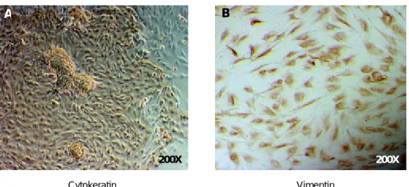
상피세포층과 기질세포층으로 나누어 배양한 자궁 내막세포를 상피세포 표지자인 cytokeratin과 기질세 포 표지자인 vimentin으로 각각 면역화학적 염색을 수행하였다. 일차 배양된 상피세포층의 경우 장방형 의 모양을 보였으며 동시에 다세포 과립이 관찰되었 다. 또한 95% 이상의 세포가 cytokeratin 양성 소견 을 보였다 (Figure 2A). 반면에 일차 배양된 기질세 포층의 경우 섬유세포 모양의 세포들이 관찰되었으 며, 95% 이상의 세포가 vimentin 양성 소견을 보여 (Figure 2B) 자궁내막 상피세포와 기질세포가 성공적 으로 분리 배양되었음을 알 수 있다.
2. 공배양 또는 분리 배양된 자궁내막 상피세포 와 기질세포에서 성호르몬 유무에 따른 TGF-ß1, 수용체 1, 2, integrin ß3, prolactin mRNA의 발현 양상
RT-PCR을 이용한 mRNA 분석 결과 상피세포와
200X
A
200X B
Cytokeratin Vimentin
Figure 2. Immunocytochemical staining of primary cultures of human endometrial epithelial (A) and stromal (B)
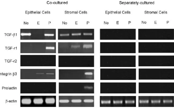
cells. Epithelial cells and stromal cells were separated as described. Epithelial cells were cytokeratin-positive while stromal cells were vimentin-positive.기질세포가 공배양된 경우 TGF-ß1은 상피세포에서 는 호르몬이 없을 때와 progesterone 우세 조건일 때 발현되었으며 기질세포에서는 모든 조건에서 발현 되었다. 이에 대한 수용체 type 1은 상피세포에서는 progesterone 우세 조건일 때에 발현되었으며, 기질 세포의 경우에는 호르몬이 없는 조건 및 estrogen
우세 조건에서 매우 약한 발현이 관찰되었으나 pro- gesterone 우세 조건일 때 더욱 강한 발현을 보였다.
수용체 type 2의 경우에는 상피세포와 기질세포 모 두에서 발현을 관찰할 수 없었다. Integrin ß3 소단위 는 상피세포에서 estrogen 우세 환경과 progesterone 우세 환경 모두에서 발현이 관찰되었으며, 기질세포
TGF-ß1
ß-actin
Epithelial Cells N E P
Stromal Cells N E P
Figure 4. HGF administration induces TGF-ß1 mRNA expression in separately cultured epithelial and stromal cells.
HGF (10 nM/ml) was administrated in separately cultured epithelial and stromal cells. In epithelial cells, TGF-ß1 mRNA was detected in all hormone conditions. In stromal cells, TGF-ß1 mRNAs were detected only in the progesterone domi- nant condition.
Figure 3. Expression of TGF-ß1, its receptor 1, 2, integrin ß3 subunit and prolactin mRNAs in co-cultured or sepa-
rately cultured epithelial and stromal cells in three different hormonal conditions. TGF-ß1, its receptor 1 (TGF-r1), integrin ß3 mRNAs were detected in the co-cultred epithelial and stromal cells. Prolactin mRNA was shown in the co-cultured stromal cells under the progesterone dominant condition. In contrast, there was no significant expression of these mRNAs in separately cultured cells.에서는 progesterone 우세 환경일 경우에만 발현이 관찰되었다. Prolactin은 기질세포에서만 progesterone 우세 환경일 때 발현이 관찰되었다 (Figure 3).
3. 분리 배양된 상피세포와 기질세포에 HGF 첨가 후 TGF-ß mRNA의 발현 양상
RT-PCR을 이용한 mRNA 분석 결과 분리 배양 한 상피세포에서는 HGF를 첨가하면 TGF-ß1의 경 우 estrogen 우세 환경을 제외한 모든 실험 조건에서 강하게 발현하였다. 이와는 대조적으로 분리 배양한 기질세포에서는 TGF-ß1, mRNA 모두 progesterone 우세 환경에서만 관찰되었다 (Figure 4).
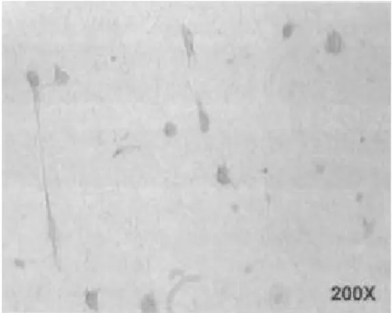
4. 분리 배양된 기질세포에서의 Met의 발현 단독 배양된 기질세포에서 HGF의 수용체인 Met
의 발현을 관찰한 결과 Met 단백질이 세포질에서 발현하고 있음을 관찰할 수 있었다 (Figure 5). Met의 발현 양상은 사용한 호르몬 조건과 상관없이 동일한 발현 양상을 보였다 (data not shown).
5. 공배양한 상피세포와 기질세포 배양액 내에서 호르몬 첨가 유무에 따른 HGF 단백질의 분비 양상
ELISA 방법으로 배양액내의 HGF 단백질의 농 도를 측정한 결과 호르몬이 없는 조건일 때 평균 150±24 pg/ml (n=3), estrogen 우세 환경일 때 평균 4,325±436 pg/ml (n=3), progesterone 우세 환경일 때 평균 2,000±540 pg/ml (n=3)의 농도로 측정되었다 (Table 2).
6. 분리 배양된 기질세포에 HGF 및 TGF -ß 첨가 후 TGF-ß 수용체 mRNA의 발현 양상
분리 배양한 기질세포에 HGF 및 TGF-ß1을 각각 첨가하면 모든 호르몬 환경에서 TGF 수용체 type 1 mRNA가 발현하였다. TGF 수용체 type 2 mRNA의 경우에는 HGF 첨가 시에는 progesterone 우세 환경 에서만 발현되었으며, TGF-ß1의 첨가 시에는 성호 르몬을 첨가한 모든 조건에서 발현하였으나 estrogen 우세 환경일 경우가 progesterone 우세 환경에 비해 강하게 발현되었다 (Figure 6). 그러나, 분리 배양한 상피세포의 경우에는 HGF 및 TGF-ß1의 첨가에 의 한 수용체 mRNA의 발현이 모든 호르몬 환경에서 관찰되지 않았다 (data not shown).
7. 분리 배양된 기질세포에 HGF 및 TGF - beta 첨가 후 prolactin mRNA의 발현 양상
분리 배양한 기질세포에 HGF 및 TGF-ß1을 각각
Table 2. The concentration of HGF in conditioned medium was determined by enzyme-linked immunosorbent assay
(ELISA). Cells grown under either the estrogen dominant or the progesterone dominant condition (n=3; 4325±436 pg/ml in the estrogen dominant; 2000±540 pg/ml in the progesterone dominant) revealed higher concentrations of HGF than no hormone-treated groups (n=3; 150±24 pg/ml) (Mean±S.D.)Hormone conditions HGF concentration (pg/ml)
No hormone 150±24
Estrogen dominant 4,325±436
Progesterone dominant 2,000±540
Note. Results are expressed as mean concentration of three different conditioned medium (n=3; Mean±S.D.) Figure 5. Immunocytochemical staining of 3-dimen-
sionally cultured human endometrial stromal cells. Stro- mal cells were separated and 3-dimensionally cultured in collagen gel as described. Met positive stromal cells were shown brown color.
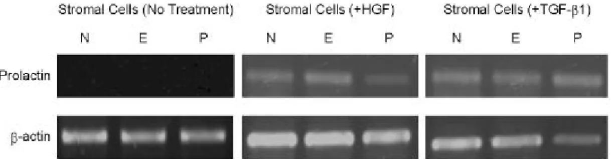
첨가하면 모든 호르몬 환경에서 prolactin mRNA가 발현되었다. 대조적으로 어느 호르몬도 첨가하지 않 은 환경에서는 prolactin mRNA의 발현을 관찰할 수 없었다 (Figure 7).
고찰 및 결론
인간자궁내막의 decidualization은 생리주기의 마지 막 주에 일어나며, 임신이 일어나지 않더라도 성호 르몬에 의해 유도된다.1 현재까지 자궁내막세포의 생리주기에 따른 변화 양상은 단순한 호르몬의 영향 이외에도 상피세포와 기질세포의 상호작용이 중요한 역할을 함이 보고되어 왔다. Wegner 등23은 백서 자 궁내막 상피세포와 기질세포를 분리 배양하여 이들
의 conditioned medium을 이용한 실험에서 상피세포 에서 분비되는 수용성 단백질이 기질세포의 역할 조 절에 관여함을 보고했다. 이러한 연구 결과는 자궁 내막 구성인자간의 상호작용이 내막의 분화 및 기능 조절에 중요한 역할을 수행함을 시사하며 in vitro에 서 in vivo의 조절 기전을 재현할 수 있음을 제시한 다. 이러한 결과를 바탕으로, 본 연구는 인간자궁내 막세포를 3차원적으로 공배양하여 기질세포의 탈락 막화 기전에 상피세포와 기질세포간의 상호작용에 대하여 조사하였다.
공배양 조건에서 TGF-ß1 및 수용체 type 1 mRNA 의 발현을 관찰할 수 있었다. 이와는 반대로 상피세 포와 기질세포를 각각 분리 배양했을 때 대부분의 mRNA는 관찰할 수 없었으며, 기질세포에서 TGF-ß TGF-r1
TGF-r2
ß-actin
Stromal Cells (+HGF) N E P
Stromal Cells (+TGF-ß1) N E P
Figure 6. HGF and TGF-ß1 administration induces TGF-r1 and -r2 mRNA expression in separately cultured stromal
cells. Administration of HGF at 10 nM/ml induces TGF-r1 expression in all hormone conditions, while TGF-r2 was expressed in the progesterone dominant condition. TGF-ß1 administration (10 nM/ml) induces TGF-r1 and -r2 expres- sion in all hormone conditions except the expression of TGF-r2 is detected in no hormone condition.Figure 7. HGF and TGF-ß1 administration induces prolactin mRNA expression in separately cultured stromal cells.
Prolactin mRNA was induced by either HGF or TGF-ß1 administration in all hormone conditions.
및 TGF-ß 수용체 mRNA가 매우 약하게 발현됨을 관찰할 수 있었다 (data not shown). 이 결과로 미루 어 TGF-ß1 및 수용체의 발현에 상피세포와 기질세 포의 상호작용이 중요한 역할을 함을 알 수 있다.
Integrin ß3 소단위의 경우 중기 및 후기 implantation window의 표지자로 알려져 있다.24 본 실험에서도 integrin ß3 소단위 mRNA가 공배양 조건에서 pro- gesterone 우세 환경에서만 발현되었다. 따라서, 본 연구자들이 사용한 공배양 조건하에서 배양한 자궁 내막세포들이 생체내 분비기 자궁내막과 유사하게 분화함을 알 수 있다. Prolactin은 탈락막화된 세포의 표지자로 알려져 있다.25 Prolactin mRNA의 발현은 공배양한 기질세포에서 progesterone 우세 환경에서 관찰되었으며, 이 결과로 미루어 자궁내막 기질세포 의 탈락막화에 상피세포와 기질세포의 공배양 환경 이 중요하게 작용함을 알 수 있다.
HGF는 현재까지 생체 내에서 다양한 역할이 알 려져 있다. 그중 인간자궁에서는 자궁내막 기질세포 의 분열 및 내강형성을 조절하며,17 영양배엽 침윤 에 관여하는 것으로 알려져 있다.18 뿐만 아니라, 인 간 혈청내 농도가 중기에서 후기 황체기에 증가 하 에 생리기에 최고 농도를 보이며 임신기에는 그 농 도를 유지하는 것으로 알려져 있다.19 따라서, 본 연 구자들은 자궁내막 기질세포의 탈락막화에도 HGF
가 paracrine factor로 간여할 것으로 가정하였다. 가 설을 확인하기 위하여 본 연구자들은 기질세포에서 HGF의 수용체인 Met 의 발현을 관찰하였으며 공배 양한 조건에서 호르몬 환경에 따른 배양액내 HGF 의 농도를 측정하였다. 단독 배양된 기질세포에서 HGF 수용체인 Met의 발현을 관찰한 결과 Met 단백 질이 세포질에서 발현함을 관찰할 수 있었다. ELISA 를 이용하여 측정한 결과 호르몬이 없는 조건일 때 150±24 pg/ml (n=3), estrogen 우세 환경일 때 4,325±
436 pg/ml (n=3), progesterone 우세 환경일 때 2,000±
540 pg/ml (n=3)의 농도로 측정되었다. 따라서, 공배 양된 자궁내막세포에 성호르몬의 배양된 세포로 부 터 HGF의 분비를 촉진시킴을 알 수 있었다. 또한, HGF의 첨가가 분리 배양된 상피세포와 기질세포에 서 TGF-ß mRNA의 발현을 유도하는지를 확인하였 다. 각각 분리 배양한 상피세포와 기질세포에 10 nM/
ml 농도의 HGF를 첨가한 결과 상피세포에서 TGF- ß1은 estrogen 우세 환경을 제외한 모든 환경에서 mRNA의 발현을 관찰할 수 있었으며, TGF-ß2의 경 우에는 모든 호르몬 환경에서 mRNA의 발현을 관 찰할 수 있었다. 반면에 기질세포에서는 progesterone 우세 환경에서만 이 두 가지 cytokine의 발현이 관찰 되었다. 뿐만 아니라 이들의 수용체의 발현에 있어 서는 HGF의 첨가가 상피세포내 TGF 수용체 mRNA
Figure 8. A proposed mechanism for the stomal decidualization. Progesterone induces HGF expression in stromal
cells, then HGF plays a paracrine role in epithelial cells by inducing TGF-ß expression and an autocrine role in stromal cells by inducing the expression of TGF-ß receptor 1 and 2. Consequently, HGF and TGF-ß induce the stromal decidu- alization under the progesterone dominant condition in vitro.의 발현은 유도하지 않았다. 그러나, 기질세포에서는 HGF 첨가에 의해 TGF 수용체 type 1 mRNA는 모 든 호르몬 조건에서 발현되었으며 TGF 수용체 type 2의 경우에는 progesterone 우세 환경일 경우에만 발 현하였다. 공배양한 상피세포와 기질세포에서는 TGF 수용체 type 2의 발현을 관찰할 수 없었는데 (Figure 3) 이것은 아마도 type 2 수용체의 발현양이 type 1 수용체에 비하여 매우 적었기 때문인 것으로 사료된 다. TGF-ß의 세포내 신호전달의 과정은 TGF 수용체 type 2와 TGF-ß가 결합하면 결과적으로 TGF 수용 체 type 1을 인산화시켜 활성화시킴으로서 시작하게 된다.9 따라서, 본 연구에서 HGF에 의한 TGF 수용 체 type 2의 발현은 progesterone 우세 환경에서 TGF- ß에 의한 세포내 신호전달이 작동함을 의미한다.
본 연구에서 HGF 또는 TGF-ß1의 첨가가 각기 배양된 기질세포에서 호르몬 환경에 상관없이 pro- lactin mRNA의 발현을 유도하는 것을 관찰할 수 있 었다. 이 결과로 미루어 TGF-ß 및 HGF가 기질세포 의 decidualization을 직접적으로 유도할 수 있는 것 으로 생각된다.
Makrigiannakis 등26은 progesterone에 의해 유도된 decidualization에 있어서 progesterone에 의해 발현된 CRH (corticotrophin-releasing hormone)가 이 과정을 중재함을 보고하였다. 따라서, 단독 배양된 기질세포 의 탈락막화는 progesterone에 의한 직접적인 탈락막 화과정이라고 단정하기는 어렵다.
본 연구자들은 인간자궁내막 상피세포와 기질세포 를 3차원적으로 공배양하여 48시간 이내에 proge- sterone 우세 환경에서 decidualization maker인 pro- lactin mRNA의 발현을 관찰하였다. 그리고, 이러한 과정은 성호른몬에 의해 유도된 HGF와 HGF에 의 해 유도된 TGF-ß 및 TGF 수용체 type 1, 2에 의해 중재되는 것으로 사료된다 (Figure 8).
결론적으로, progesterone에 의한 기질세포의 deci- dualization에 있어서 progesterone은 간접적으로 작 용하며 이러한 과정에는 HGF와 TGF에 의해 중재 되는 기질세포와 상피세포간의 상호작용이 중요한 역할을 하는 것으로 사료된다. 또한 이를 좀더 확실 히 증명하기 위한 단백질 수준의 연구가 더 진행돼 야 할 것으로 사료된다.
참 고 문 헌
1. Noyes RW, Hertig AT, Rock J. Dating of endome- trial biopsy. Fertil Steril 1950; 1: 3-25.
2. Tang B, Guller S, Gurpid E. Cyclic adenosine 3' 5'-monophosphate induces prolactin expression in stromal cells isolated from human proliferative endometrium. Endocrinology 1993; 133: 2197-203.
3. Tang B, Grupide E. Direct effect of gonodotropins on decidualization of human endometrial stromal cells. J Steroid Biochem Mol Biol 1993; 47: 115- 21.
4. Frank GR, Brar AK, Cedars MI, Handwerger S.
Prostaglandin E2 enhances human endometrial cell differentiation. Endocrinology 1994; 134: 258-63.
5. Moses HL, Coffey RJ Jr, Leof EB, Lyons RM, Keski-Oja J. Transforming growth factor beta regu- lation of cell proliferation. J Cell Physiol Suppl 1987; 5: 1-7.
6. Moses HL, Serra R. Regulation of differentiation by TGF-beta. Curr Opin Genet Dev 1996; 6: 581-6.
7. Herrmann M, Scholmerich J, Straub RH. Influence of cytokines and growth factors on distinct steroi- dogenic enzymes in vitro: a short tabular data col- lection. Ann N Y Acad Sci 2002; 966: 166-86.
8. Renner U, Lohrer P, Schaaf L, Feirer M, Schmitt K, Onofri C, et al. Transforming growth factor-beta stimulates vascular endothelial growth factor pro- duction by folliculostellate pituitary cells. Endocrin- ology 2002; 143: 3759-65.
9. Akhurst RJ, Derynck R. TGF-beta signaling in cancer-a double-edged sword. Trends Cell Biol 2001; 11: s44-51.
10. Casslén B, Sandberg T, Gustavsson B, Willén R, Nilbert M. Transforming growth factor beta1 in the human endometrium. Cyclic variation, increased expression by estradiol and progesterone, and regu- lation of plasminogen activators and plasminogen activator inhibitor-1. Biol Reprod 1998; 58: 1343- 50.
11. Bruner KL, Eisenberg E, Gorstein F, Osteen KG.
Progesterone and transforming growth factor-beta coordinately regulate suppression of endometrial matrix metalloproteinases in a model of experimen- tal endomeriosis. Steroid 1999; 64: 648-53.
12. Bruner KL, Roders WH, Gold LI, Korc M, Harg- rove JT, Matrisian LM, et al. Transforming growth factor beta mediates the progesterone suppression on an epithelial metalloproteinase by adjacent stroma in the human endometrium. Proc. Natl Acad Sci USA 1995; 92: 7362-6.
13. Odekone S, Matt D, Strom S, Rifkin DB. Require- ment for receptor-bound urokinase in plasmin- dependent cellular conversion of latent TGF-beta to TGF-beta. J Cell Physiol 1994; 158: 398-407.
14. Rotraud W. The transforming growth factor-beta signaling pathway in tumorigenesis. Current Opinion in Oncology 2001; 13: 70-7.
15. Ando N, Hirahara F, Fukushima J, Kawamoto S, Okuda K, Funabashi T, et al. Differential gene expression of TGF-beta isoforms and TGF-beta receptors during the first timester of pregnancy at the human maternal-fetal interface. Am J Reprod Immunol 1998; 40: 48-56.
16. Nakamura T, Nishizawa T, Hagiya M, Seki T, Shi- monishi M, Sugimura A, et al. Molecular cloning and expression of human hepatocyte growth factor.
Nature 1989; 42: 440-3.
17. Sugawara J, Fukaya T, Murakami T, Yoshida H, Yajima A. Hepatocyte growth factor stimulated proliferation, migration, and lumen formation of human endometrial epithelial cells in vitro. Biol Reprod 1997; 57: 936-42.
18. Kauma SW, Bae-Jump V, Walsh SW. Hepatocyte growth factor stimulates trophoblast invasion: A potential mechanism for abnormal placentation in
preeclampsia. J Clin Endocrin Metab 1999; 84:
4092-6.
19. Negami AI, Sasaki H, Kawakami Y, Kamitani N, Kotsuji F, Tominaga T, Nakamura T. Serum human hepatocyte growth factor in human menstrual cycle and pregnancy: a novel serum marker on regene- ration and reconstruction of human endometrium.
Horm Res 1995; 44 suppl 2: 42-6.
20. Irwin JC, Kirk D, King RJB, Quigley MM, Gwatkin RBL. Hormonal regulation of human endometrial stromal cells in culture: an in vivo model for deci- dualization. Fertil Steril 1989; 52: 761-8.
21. Hearn JP. The embryo-maternal dialogue during early pregnancy in primates. J Reprod Fertil 1986; 76:
809-19.
22. Bentin-Ley U, Pedersen B, Lindenberg S. Isolation and culture of human endometrial cells in a three- dimensional culture system. J Reprod Fertil 1994;
101: 327-32.
23. Wegner CC, Carson DD. Mouse uterine stromal cells secrete a 30 kilodalton protein in response to cocu- lture with uterine epithelial cells. Endocrinology 1992; 131: 2565-72.
24. Lessey BA, Castelbaum AJ, Wolf L, Greene W, Paulson M, Meyer WR, et al. Use of integrins to date the endometrium. Fertil Steril 2000; 73: 779- 87.
25. Telgmann R, Gellersen B. Marker genes of decidu- lization: activation of the decidual prolactin gene.
Hum Reprod Update 1998; 4: 472-9.
26. Makrigiannakis A, Margioris AN, Chatzaki E, Zou- makis E, Chrousos GP, Gravanis A. The decidu- alizing effect of progesterone may involve direct transcriptional activation of corticotrophin-releasing hormone from human endometrial stromal cells.
Mol Hum Reprod 1999; 5: 789-96.