가톨릭대학교 의과대학 이비인후과학교실
김지은, 박경호, 김민주, 김 민
Department of Otolaryngology-Head & Neck Surgery, College of Medicine, The Catholic University of Korea, Seoul, Korea
Ji-Eun Kim, MD, Kyoung-Ho Park, MD, PhD, Min-Ju Kim, MD, Min Kim, MD
외측 측두골절제술을 시행한 외이도 편평세포암 2례
Lateral temporal bone resection for squamous cell carcinoma of the external auditory canal : Report of 2 cases
J Korean Skull Base Society 8권 1호 : 23~28, 2013
Malignant tumors of the external auditory canal (EAC) are uncommon. It is a rare malignant neoplasm with an aggressive nature and an overall poor prognosis. The primary goal of the treatment of EAC tumors is to remove the primary tumor without recurrence and with maximal preservation of function. At this point, lateral temporal bone resection can achieve preservation of hearing and facial nerve function when the tumor is confined to the EAC.
We report two cases of squamous cell carcinoma of EAC treated by lateral temporal bone resection. In the first case, the patient underwent surgery, and subsequently he underwent radiotherapy. In the second case, the patient underwent only surgical treatment without radiation therapy. The 2 patients showed no evidence of recurrence during the follow-up period. In this report, we compare treatment of malignant tumors limited to the EAC by lateral temporal bone resection only and surgery with adjuvant radiotherapy
논문 접수일 : 2013년 4월 25일 심사 완료일 : 2013년 5월 20일
주소 : Department of Otolaryngology-Head
& Neck Surgery, College of Medicine, The Catholic University of Korea, Seoul, Korea
222 Banpo-daero, Seocho-gu, Seoul 137-701, Korea Tel : +82-2-2258-6213 Fax : +82-2-595-1354 E-mail : khpent@catholic.ac.kr
Kyoung-Ho Park, MD, PhD
교신저자
External auditory canal cancer, Squamous cell carcinoma, Lateral temporal bone resection, Radiotherapy
Key Words
종설1 증례1 증례2 증례3 증례4
▒ Introduction
The malignant tumor of the external auditory canal is a rare disease that occurs as one case per million population per year.1) Squamous cell carcinoma has been reported to be the most common histopathologic and other primary histologic types of neoplasms arising in the external auditory canal and temporal bone, including adenoid cystic carcinoma, basal cell carcinoma, and melanoma.2)
This is an aggressive disease with the prognosis dependent on the stage of disease and primary treatment.
For the treatment of squamous cell carcinoma of the external auditory canal, surgery is most preferable. The type of surgery (lateral temporal bone resection, subtotal, total temporal bone resection) depends on the cancer stage.3) Radiotherapy is known to be an effective adjuvant treatment.
However, squamous cell cancer cases that occur in the external auditory canal are rare; therefore, there is controversy regarding the effects of radiation therapy.
Further, the need for postoperative radiation therapy in early stage patients is still controversial.
We want to discuss the effects of radiotherapy after lateral temporal bone resection in squamous cell carcinoma of external auditory canal, comparing 2 different cases.
Appearance of otoscopy A. Case I : The left
external auditory canal was occupied by the tumor bulging from the posterior wall. The tumor was reddish and irregular.
B. Case II : reddish and firm mass occupying in the right external auditory canal. We have seen commitant otorrhea.
A B
Fig. 1
Axial (A) and coronal (B) CT of the temporal bone (case I)
demonstrating soft tissue density filling the left EAC, MEC and mastoid air cells without definite bony destruction.
Axial (C) and coronal (D) CT of the right temporal bone (case II) demonstrating an EAC mass with limited soft tissue involvement.
Fig. 2 A
B
C D
▒ Case report 1
- radiotherapy after lateral temporal bone resection
A 65-year-old male was presented to a local clinic with a 6 month history of left ear otalgia and hearing impairment.
EAC was seen in otoscopic examination. He was referred to our hospital because of malignancy in a biopsy report.
The patient described otorrhea and earfullness. He did not have cranial nerve palsy his past history; he is on medication due to diabetes mellitus and craniotomy by traffic accident.
Physical examination of the left ear showed that the external auditory canal was eclipsed 1.0×1.0cm sized mass (figure 1A). Pure tone audiometry showed 90dB air conduction loss and 55dB bone conduction loss in the left ear, but the right ear was normal. It was scale out in the left ear speech audiometry but the right ear was normal. Palpation of
the neck demonstrated no lymphadenopathy.
A computer tomographic scan of the left ear of temporal bone showed opacification of the left EAC and middle ear cavity, as well as the mastoid air cell. However bony erosion was not shown in the mastoid process(Figure 2A). Given the limited soft tissue involvement on CT, the lesion was classified as T2N0M0 by the University of Pittsburgh staging system.
Magnetic resonance imaging (MRI) scan revealed T1 low T2 high signal intensity, filling in the middle ear cavity, mastoid antrum and mastoid air cell. Middle ear cavity and some portion of the left mastoid antrum revealed enhancing soft tissue lesion after enhancement(Figure 3A). Re-biopsy was done at our hospital. It revealed squamous cell carcinoma and PET-CT reported negative distant metastasis. Radiologic CT imaging of the left ear showed the tumor was not destructed
Axial (A) and coronal (B) view of the magnetic resonance imaging showing heterogeneous signal abnormality in the left middle ear cavity, mastoid antrum, and mastoid air cell.
Axial view (C, D) of the magnetic resonance imaging showing about 1.6x6cm size T1 iso and T2 with slightly high signal intensity mass filling the right EAC subtle enhancement after contrast infusion.
A
C D
B Fig. 3
EAC bone and no invasion of the middle ear cavity mucosa.
Therefore, we scheduled lateral temporal bone resection.
In surgery, there was no facial nerve invasion. Left near total parotidectomy and anterior based temporalis muscle, as well as sternocleidomastoid muscle rotational flap reconstruction was done(Figure 4A). Selective neck dissection (level II-III) was also done.
All surgical resection margins were free of tumor and there was no evidence of lympatic, vein and perineural invasion.
There was no evidence of metastasis in 13 level II and III lymph nodes.
After surgery, facial palsy was developed as H-B grade II level. Radiotherapy started 5 weeks after surgery. Total 6600cGy radiated at the operation site. After radiotherapy, the operation wound was clear.
After 10 days, radiotherapy complication was revealed, such as facial edema and erythema around the left ear, and the left post occipital area hair loss was revealed. Leukopenia appeared . Therefore, he took LTFS.
After 3 months, facial edema and erythema were decreased and leukopenia also improved to grade I. However, tinnitus newly appeared.
He lost about 6kg of weight compared to 3 months ago.
However, follow-up MRI, neck CT, temporal bone CT, PET- CT revealed no signs of recurrence and distant metastasis 6
months and 1 year later.
▒ Case report 2
- only lateral temporal bone resection
A 62-year-old male complained of intermittent otorrhea in the right ear for 5 month duration. During this period, the patient had hearing impairment, not otalgia, tinnitus, or vertigo. According to the information given, he was diagnosed with squamous cell carcinoma in the right external auditory canal before 1 year ago at the local clinic. However, he did not receive treatment.
In the past history, the male patient is on medication due to diabetes mellitus and hypertension since last year. When reviewing his previous chart, he had chemoradiation therapy at 1990 because of squamous cell carcinoma in the right neck (unknown origin).
Our first physical examination revealed granulation and otorrhea in the right external ear canal. The tympanic membrane could not be seen directly with an otoscopy(Figure 1B). Other clinical examinations, including the head and neck region, and laboratory findings were within normal limits.
Pure tone audiogram (PTA) showed obvious right side air- bone gap. Auditory brainstem response was scaled out in the right ear, but the left ear was normal.
Lateral temporal bone resection with near total parotidectomy, selective neck dissection. (A) case I, (B) case II
A B
Fig. 4
A CT scan of her right external auditory canal showed opacification, not bony destructive changes of the mastoid cortex(Figure 2B). Given the limited soft tissue involvement on a CT, the lesion was classified as T2N0M0 by the University of Pittsburgh staging system for external auditory meatus carcinoma.
A magnetic resonance imaging scan with contrast revealed approximately 1.6×6cm mass occupying external auditory canal with T1 iso T2 slightly high signal intensity(Figure 3B).
PET-CT reported negative distant metastasis.
Radiologic CT imaging of the right ear showed that the tumor was not destructed EAC bone and no invasion to the middle ear cavity mucosa. Based on these results, the patient underwent a left lateral temporal bone resection, parotidectomy, and selective neck dissection.
In surgery, there was no facial nerve invasion. Left near total parotidectomy and anterior based temporalis muscle, as well as the sternocleidomastoid muscle rotational flap reconstruction was done(Figure 4B). Selective neck dissection (level I-III) was also done.
All surgical resection margins were free of tumor and there was no evidence of carcinoma invading the underlying bone.
There was no evidence of metastasis in level I~III lymph nodes.
Twenty months after surgery, the patient is in good health with no evidence of complication or recurrence. Follow-up MRI, neck CT, temporal bone CT, PET-CT revealed no sign of recurrence and distant metastasis to 6 months and 1 year later.
▒ Discussion
SCC of EAC is an uncommon but ominous malignancy.
Most common symptoms of EAC cancer are otorrhea (47.2%), otalgia (39.6%), and hearing impairment (28.3%). Therefore, it is confused with chronic otitis media in the early stage.4) Historically, study of this disease process has been limited by small groups of patients leading to disparity in staging, workup, treatment, and outcomes.1)
Pittsburgh tumor staging system (Table 1.) is used most commonly in the EAC cancer staging system. M. B. Gillespie and colleagues expressed that preoperative CT scan is chart review of 15 patients treated for squamous cell carcinoma and reported that the University of Pittsburgh staging system correlated with that of patient outcome and was more sensitive than the preoperative radiological staging.5)
An article was reported that a prognosis depends on the stage. Chi et al. showed 5-year survival rates in 72 patients to be T1 -100%, T2 -66.7%, T3 -21.1%, and T4- 14.3%.6) Morris and colleagues evaluated 72 patients with malignancy of the temporal bone (31 of which had squamous cell carcinoma), and found that 5-year DFS (disease free survival) was 87.5% for T1-2 tumors and 63.4% for T3-4 tumors.7)
The extension of resection indicated the radical surgical treatment; for example, en bloc resections of the EAC in T1 tumors, partial temporal bone resections for T2 tumors, subtotal temporal bone resections, or total temporal bone resections for advanced tumors. Both of our cases were T2N0 stage II by the Pittsburgh staging system(Table 1). Therefore we decided on lateral temporal bone resection.
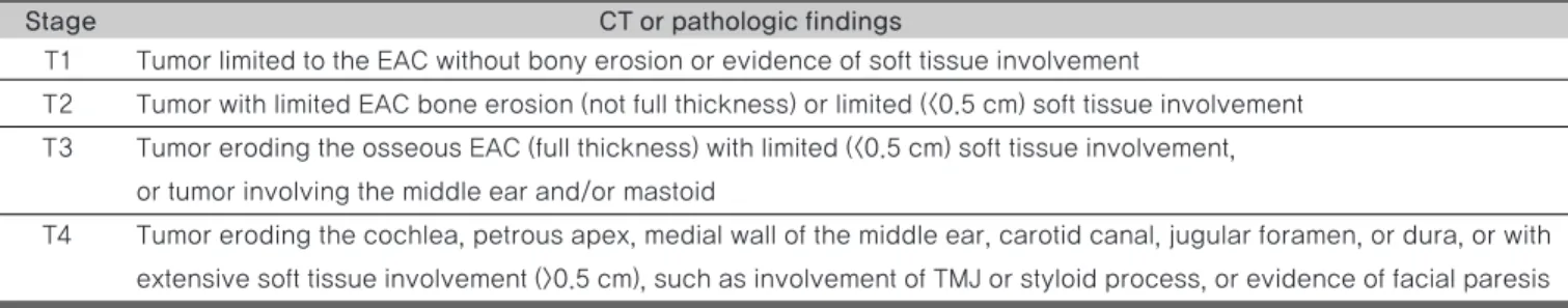
T1 Tumor limited to the EAC without bony erosion or evidence of soft tissue involvement
T2 Tumor with limited EAC bone erosion (not full thickness) or limited (<0.5 cm) soft tissue involvement T3 Tumor eroding the osseous EAC (full thickness) with limited (<0.5 cm) soft tissue involvement,
or tumor involving the middle ear and/or mastoid
T4 Tumor eroding the cochlea, petrous apex, medial wall of the middle ear, carotid canal, jugular foramen, or dura, or with extensive soft tissue involvement (>0.5 cm), such as involvement of TMJ or styloid process, or evidence of facial paresis
Stage CT or pathologic findings
Table 1. Pittsburgh tumor staging system (modified)
(EAC : external auditory canal, TMJ : temporomastoid joint)
A lateral temporal bone resection (LTBR) involves resection of the bony EAC, the tympanic membrane, the malleus, and the incus, with, medial limit defined at the level of the incudostapedial joint. The bony labyrinth and facial nerve are preserved. A lateral temporal bone resection is considered as an adequate surgical approach for tumors of the postauricular sulcus, concha, or EAC, or when there is bony erosion of the posterior EAC or mastoid cortex without involvement of the middle ear or mastoid air cells.8)
In some cases, tumor is limited in the external auditory canal. However, in other cases., it metastasizes to the temporomandibular joint, parotid gland, infratemporal fossa and neck lymph node through the foramen of Huschke, Santorini fissure of cartilage portion of EAC.9) As such,if resection margin is positive in a frozen biopsy, radical resection involved with parotid gland, infratemporal fossa, and neck dissection are also considered. In our case, near total parotidectomy and prophylactic selective neck dissection are also involved. After that surgery, margins are negative.
Ogawa et al. suggested surgery and postoperative radiotherapy as the standard treatment in T2 stage. Further, as the standard treatment for T3 and advanced stage, combination of surgery and radiotherapy is suggested.
Treatment with only radiotherapy decreases the treatment outcome. However it is useful as an adjuvant therapy in surgical margin positive after surgery.
However in some articles, radiotherapy is not effective in a surgical margin positive case.10)In addition tumors involving only EAC, radiotherapy combined with surgery are not any more effective than only surgery.
In our case, T2 stages of two patients did not undergo recurrence for 2 years. However, surgery with radiotherapy patient had some complication with weight loss, hair loss, tinnitus, and facial edema. Therefore, we question the need for radiation therapy in T2 stage patients.
Squamous cell carcinoma in the external auditory canal is a rare tumor and the standard treatment is controversial.
Therefore it is difficult to decide on a treatment plan. The most important prognostic factor is the stage of the tumor.
In our case, surgical free margin is an important prognosis factor. So, if we can get an appropriative negative margin after surgery, radiotherapy is not effective. On the contrary, radiotherapy prevents patient's quality of life.
However squamous cell carcinoma in the external auditory canal is a rare disease. Therefore we need further studies and discussions to get a firm treatment plan.
References
1. Kuhel WI, Hume CR, Selesnick SH. Cancer of the external auditory canal and temporal bone. Otolaryngol Clin North Am 1996 Oct;29:827- 52
2. Shih L, Crabtree JA. Carcinoma of the external auditory canal: An update. Laryngoscope 1990;100:1215-8
3. Moody SA, Hirsch BE, Myers EN. Squamous cell carcinoma of the external auditory canal: An evaluation of a staging system. Am J Otol 2000;21:582-8
4. Park KT, Song JJ, Jang JH, Oh SH, et al. The Analysis of Prognostic Factor and Treatment Outcome of Malignancies of the External Auditory Canal. Korean J otohinolayngol-Head Neck Surg 2010;53:275-83
5. Gillespie MB, Francis HW, Chee N, Eisele DW, Squamous cell carcinoma of the temporal bone: a radiographicpathologic correlation.
Arch Otolaryngol Head and Neck Surg, 2001l;127:803-7
6. Chi FL, Gu FM, Dai CF, et al. Survival outcomes in surgical treatment of 72 cases of squamous cell carcinoma of the temporal bone. Otol Neurotol 2011;32:665-9
7. Morris LG, Mehra S, Shah JP, et al. Predictors of survival and recurrence after temporal bone resection for cancer. Head Neck 2012;34:1231-9
8. Moore MG, Deschler DG, et al. Management outcomes following lateral temporal bone resection for ear and temporal bone malignancies. Otolaryngol Head and Neck Surg 2007;137:893-8 9. Gacek RR, Goodman M. Management of malignancy of the temporal
bone. Laryngoscope 1977;87:1622-34
10. Goodwin WJ, Jesse RH. Malignant neoplasms of the external auditory canal and temporal bone. Arch Otolaryngol 1980;106:675-9