ISSN 1975-7425(Print) / ISSN: 2288-016X(Online) Korean J Urogenit Tract Infect Inflamm 2013;8(2):121-124
121
Case Report
신유두괴사 환자에서 급성신우신염과 진통제 남용에 의해 발생된 신배파열
허정식, 김성대, 박경기, 김영주
제주대학교 의학전문대학원 비뇨기과학교실
Renal Papillary Necrosis with Calyceal Rupture: Caused by Acute Pyelonephritis and Analgesic Abuse
Jung-Sik Huh, Sung-Dea Kim, Kyung Kgi Park, Young-Joo Kim Department of Urology, Jeju National University School of Medicine, Jeju, Korea
Spontaneous renal rupture is a rare condition. Renal rupture most often occurs as a result of traumatic injury, a rare entity of obstructive uropathy with stones, and spontaneous causes such as malignancy. We report on a rare case of renal rupture caused by a ureter stone measuring 5 mm in size with acute pyelonephritis (APN) in a patient with renal papillary necrosis (RPN). The patient, who suffers from attacks of gouty arthritis, frequently used analgesic for pain relief. The patient was treated with temporary percutaneous drainage and antibiotics. This case demonstrates that RPN with APN can induce renal rupture even when ureter stones are small. Thus, consideration of all medical problems is important when deciding on treatment of patients with ureter stones.
Keywords: Rupture; Calculi; Kidney papillary necrosis; Pyelonephritis
Copyright 2013, Korean Association of Urogenital Tract Infection and Inflammation. All rights reserved.
This is an open access article distributed under the terms of the Creative Commons Attribution Non-Commercial License (http://creativecommons.org/licenses/by-nc/3.0) which permits unrestricted non-commercial use, distribution, and reproduction in any medium, provided the original work is properly cited.
Received: 21 May, 2013 Revised: 30 August, 2013 Accepted: 31 August, 2013
Correspondence to: Young-Joo Kim
Department of Urology, Jeju National University School of Medicine, 102, Jejudaehak-ro, Jeju 690-756, Korea
Tel: +82-64-717-1476, Fax: +82-64-717-1131 E-mail: [email protected]
No potential conflict of interest relevant to this article was reported.
Multiple predisposing conditions have been associated with the development of renal papillary necrosis (RPN), particularly diabetes, analgesic drug abuse such as, nonste- roidal drugs and obstruction.1,2
RPN is not common in the urologic department. This case, the patient who suffers from attack of gouty arthritis frequent used analgesic for pain relief. We report a spontaneous renal rupture with RPN and acute pyelone- phritis (APN) due to a small ureter stone.
CASE REPORT
A 60-year-old man was admitted to the emergency department with acute left abdominal pain and high fever.
His medical history included hypertension and gout for 13 years ago. From several decades ago, he suffered from painful acute attack of gouty arthritis. Thus, he had almost always taken analgesic for pain relief. The patient had nausea but no vomiting. There were no other urinary symptoms. His vital signs were as follows: blood pressure, 100/60 mmHg and body temperature, 40.2oC. Clinical
122 Jung-Sik Huh, et al. Renal Papillary Necrosis with Calyceal Rupture
Korean J Urogenit Tract Infect Inflamm Vol. 8, No. 2, October 2013
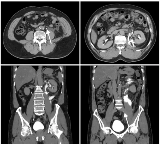
Fig. 1. An abdominal computed tomo- graphic scan showed about 5 mm sized upper ureter stone (arrows) with hydro- nephroureterosis and perinephric fluid collection.
examination revealed diffuse pain in the left abdomen with tenderness.
Urinalysis showed pH 5.0 and many white cells per field under high-power magnification. Complete blood cell count results were as follows: 26,600/mm3 (band-45%). Serum urea level, 26.3 mg/dl; a creatinine level, 2.1 mg/dl; and a C-reactive protein level of 11.45 mg/dl. An abdominal computed tomographic (CT) scan was performed, which showed about 5 mm sized upper ureter stone with hydronephroureterosis and perinephric fluid collection (Fig.
1). The fornices of the calyx become irregular, widened and swollen. It showed radiologic findings of early stage of RPN.
We diagnosed this case as a urosepsis with calyceal rupture in RPN. A 8.5 F pigtail percutaneous nephrostomy catheter was inserted into the renal pelvis, and then antegrade pyelography (AGP) performed through the nephrostomy catheter showed extravasation of radio- contrast from the irregular shaped upper calyx.
In a few days, fever was controlled. He used to be treated for gout with benzbromarone and frequent used to abuse
of analgesic for acute attack of gouty arthritis. Benzbro- marone leads to excretion of uric acid urine higher, then gout patients are at greater risk of forming uric acid stones for using this medicine. We planned to change over the gout medicine for reducing serum uric acid level.
After 7 days, His vital signs were normalized. Urine culture and blood culture showed
E. coli
. We treated this patient with sensitive antibiotics such as ciprofloxacin orally for more than 7 days.After a week, urine culture and blood culture showed no growth of pathogen and urinalysis showed normal range.
AGP performed through the nephrostomy cathteter showed no extravasation of radiocontrast (Fig. 2). A repeat plain abdominal X-ray confirmed the stone was still in the ureter.
We attempted to extract the stone, using the extra corporeal shock wave (ESWL). The stone was passed out after ESWL.
After 1 weak, the nephrostomy catheter was removed. A follow-up intravenous pyelography was performed 2 months later. It showed no urinary leakage and no obstructing lesions.
Jung-Sik Huh, et al. Renal Papillary Necrosis with Calyceal Rupture 123
Korean J Urogenit Tract Infect Inflamm Vol. 8, No. 2, October 2013 Fig. 2. Antegrade pyelography showed no extravasation of
radiocontrast.
DISCUSSION
RPN is a spectrum disease. Multiple predisposing condi- tions have been associated with the developing of RPN, particular diabetes mellitus, analgesic abuse and obstruc- tion. We report uncommon case of RPN accompanied by APN and analgesic abuse. An accurate diagnosis of analgesic abuse-induced RPN is difficult because of few specific symptoms and signs. Moreover, Radiologic diagnosis of RPN is difficult, but the CT and ultrasound help being able to increasing diagnostic sensitivity.1 We performed CT because patient admitted to a emergency room. RPN in diabetic patients had been associated with the urinary tract obstruction and infection such as APN and so on. But, most common cause was analgesic drug abuse such as, the nonsteroidal, anti-inflammatory drugs.1,2 Nontraumatic spontaneous renal rupture is rare in nature.3 Moreover, rupture combined with RPN and APN was extremely rare like this case.
Gout may increase the risk for calcium oxalate stone formation, the most common type of urinary stones.4,5 Also, gout patients are at greater risk of forming uric acid stones.
Patient with chronic tophaceous gout suffers from accumulation of urate crystals in joints. Thus analgesic abuse was common situation. Urinary stone and gout are common disorders. RPN in patients who suffered from painful acute attack of gouty arthritis, had been associated with the analgesic abuse. RPN accompanied by acute ureteral
obstruction is a urologic emergency. At the moment lately, it was treated successfully by the insertion of a double-J ureteric stent or other minimally invasive techniques such as percutaneous drainage under fluoroscopy, open surgical intervention is rarely indicated.6 We performed percuta- neous nephrostomy, after 7 days, healing was successfully.
The mechanism of RPN due to analgesics is not completely understood. Analgesic abuse-induced RPN mechanism which is a direct toxic effect on cells in the medulla, is controversial. NSAIDs inhibit renal cyclooxygenase and leading to renal vasoconstriction and renal papillary necrosis sequentially.7,8 Thus, it may be prudent for physicians to warn an individual with gout of the increased risk for urinary stone formation and to warn abused analgesics of the increased risk for RPN. In this case, the patient was managed with benzbromarone and used to abuse analgesic for pain.
In general, the candidate for a uricosuric agent such as benzbromarone is the gout patient who is younger than 60 years old, has a creatinine clearance greater than 80 ml/min, a 24-hour urinary uric acid excretion of less than 800 mg on a general diet, and no history of renal calculi unlike this case.9 Thus, we changed over the gout medicine for reducing serum uric acid level.
Rupture combined with RPN and APN of gout patient was extremely rare. But urinary stones, gout and APN are common disorders. Analgesic abuse was considered of acute attack of gouty arthritis.
Thus, it is importance to be taken all the medical problem into consideration when decided to treat with ureter stone.
REFERENCES
1. Bach PH, Nguyen TK. Renal papillary necrosis--40 years on.
Toxicol Pathol 1998;26:73-91.
2. Carlton WW, Engelhardt JA. Experimental renal papillary necrosis in the Syrian hamster. Food Chem Toxicol 1989;27:
331-40.
3. Kaplan LM, Farrer JH, Lupu AN. Spontaneous rupture of ureter.
Urology 1987;29:313-6.
4. Johnson CM, Wilson DM, O'Fallon WM, Malek RS, Kurland LT.
Renal stone epidemiology: a 25-year study in Rochester, Minnesota. Kidney Int 1979;16:624-31.
5. Coe FL, Parks JH, Asplin JR. The pathogenesis and treatment of kidney stones. N Engl J Med 1992;327:1141-52.
6. Stravodimos K, Adamakis I, Koutalellis G, Koritsiadis G, Grigoriou I, Screpetis K, et al. Spontaneous perforation of the ureter: clinical presentation and endourologic management. J
124 Jung-Sik Huh, et al. Renal Papillary Necrosis with Calyceal Rupture
Korean J Urogenit Tract Infect Inflamm Vol. 8, No. 2, October 2013 Endourol 2008;22:479-84.
7. Moghal NE, Hegde S, Eastham KM. Ibuprofen and acute renal failure in a toddler. Arch Dis Child 2004;89:276-7.
8. Silverman LR, Khan KN. "Have you seen this?" Nonsteroidal anti-inflammatory drug-induced renal papillary necrosis in a
dog. Toxicol Pathol 1999;27:244-5.
9. Harris M, Bryant LR, Danaher P, Alloway J. Effect of low dose daily aspirin on serum urate levels and urinary excretion in patients receiving probenecid for gouty arthritis. J Rheumatol 2000;27:2873-6.