161
© Copyright The Korean Academy of Asthma, Allergy and Clinical Immunology • The Korean Academy of Pediatric Allergy and Respiratory Disease http://e-aair.org INTRODUCTION
Idiopathic hypereosinophilic syndrome (HES) is character- ized by persistent blood and tissue eosinophilia. HES involves multiple organ systems, predominantly the skin, heart, lung, gastrointestinal tract, and nervous system.1 Often, the present- ing manifestation is thrombosis. Sporadic cases of intra-abdom- inal, cerebral, and cutaneous thrombosis have been reported.2 Disseminated intravascular coagulation (DIC) associated with HES is rare, and few cases have been reported.3-6 Here we re- port a unique case of HES presenting with pulmonary, renal, splenic, and intracerebral thrombosis associated with DIC.
CASE REPORT
A 23-year-old woman was referred to the emergency depart- ment of our hospital with fever and hemoptysis. She had bilat- eral costovertebral angle tenderness. A simple chest X-ray dem- onstrated patchy consolidation on the right middle lobe and peribronchial opacity on the lower bilateral lung fields (Fig. 1A).
A chest computed tomography (CT) scan revealed a heavy bur- den of pulmonary embolism in the bilateral pulmonary arteries and their branches, with bland infarction in both lungs (Fig. 1B-
A Case of Hypereosinophilic Syndrome Presenting With Multiorgan Infarctions Associated With Disseminated Intravascular Coagulation
Sun-Mi Park, Ji-Won Park, Sung-Min Kim, Eun-Hee Koo, Jin-Young Lee, Chul-Su Lee, Dong-Chull Choi, Byung-Jae Lee*
Division of Allergy, Department of Medicine, Samsung Medical Center, Sungkyunkwan University School of Medicine, Seoul, Korea
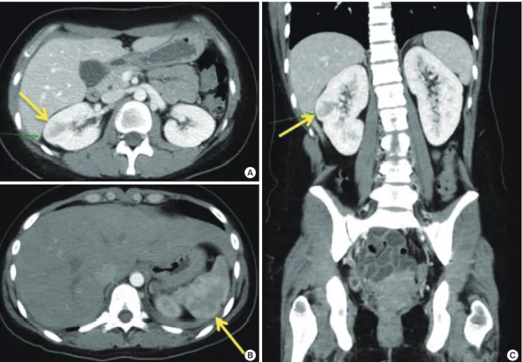
D). In an abdominal-pelvic CT scan to evaluate the patient’s flank pain, a focal wedge-shaped low-density lesion in the right kidney midpole and spleen was found (Fig. 2), suggesting in- farction. A duplex ultrasonogram of the lower extremities for the edematous right lower leg revealed a totally occluding thrombus of the right femoropopliteal vein and a partially oc- cluding thrombus of the right posterior tibial vein.
White blood cell count was elevated (11,430/μL) with 21% eo- sinophils (2,550/μL), hemoglobin 11 g/dL, and thrombocyto- penia of 39,000/μL. The prothrombin time (PT) was prolonged to 15 seconds and activated partial thromboplastin time was 42 seconds. Fibrinogen was in the normal range (156 mg/dL), but D-dimer was increased to 46.14 μg/mL. Total IgE was 2,296 IU/
mL and ECP was 78.6 ng/mL. Anti-neutrophil cytoplasmic and cardiolipin antibodies were negative. A test for parasites in the
Case Report
Allergy Asthma Immunol Res. 2012 May;4(3):161-164.
http://dx.doi.org/10.4168/aair.2012.4.3.161 pISSN 2092-7355 • eISSN 2092-7363
Thromboembolism is one of the most critical complications of hypereosinophilic syndrome (HES). We report here a case of multi-organ infarctions related to HES. A 23-year-old woman was referred to our hospital with hemoptysis. Not only pulmonary, but also renal and splenic infarctions were detected on computed tomography images. Blood tests showed profound peripheral eosinophilia. She was diagnosed with HES with disseminated intravascular coagulation (DIC). We initiated infusion of corticosteroids, which effectively suppressed peripheral eosinophilia. However, consumptive coagulopathy did not improve and intracerebral hemorrhage related to thrombosis then developed. Addition of interferon-alpha resulted in the cor- rection of the DIC associated with HES.
Key Words: Hypereosinophilic syndrome; eosinophilia; thromboembolism; disseminated intravascular coagulation; consumptive coagulopathy; in- terferon-alpha
This is an Open Access article distributed under the terms of the Creative Commons Attribution Non-Commercial License (http://creativecommons.org/licenses/by-nc/3.0/) which permits unrestricted non-commercial use, distribution, and reproduction in any medium, provided the original work is properly cited.
Correspondence to: Byung-Jae Lee, MD, PhD, Division of Allergy, Department of Medicine, Samsung Medical Center, Sungkyunkwan University School of Medicine, 50 Irwon-dong, Gangnam-gu, Seoul 135-710, Korea.
Tel: +82-2-3410-3427; Fax: +82-2-3410-3849; E-mail: [email protected] Received: September 28, 2011; Revised: November 11, 2011;
Accepted: November 22, 2011
•There are no financial or other issues that might lead to conflict of interest.
Park et al.
Allergy Asthma Immunol Res. 2012 May;4(3):161-164. http://dx.doi.org/10.4168/aair.2012.4.3.161 Volume 4, Number 3, May 2012
162 http://e-aair.org
stool and serum was negative. A bone marrow biopsy showed normal cellularity with increased eosinophils. Fip1-like1 and platelet-derived growth factor receptor alpha gene (FIP1L1- PDGFRA) fusion was not detected. An echocardiogram showed no abnormal findings.
She was diagnosed as idiopathic HES manifested as multiple thromboembolisms involving the lung, kidney, spleen, and lower extremities. She was treated with corticosteroids (methyl- prednisolone 1 mg/kg) and heparin on the second day of ad- mission. Eosinophil count normalized within 3 days. On the fifth day of treatment, she complained of a persistent and wors- ening headache. A brain CT scan revealed left frontal and tem- poral lobe intracerebral hemorrhage. D-dimer remained ele- vated at 18.93 μg/mL, and the platelet count was low at 30,000/
μL. Due to suspicion of hemorrhage associated with intracere- bral thrombosis, we continued corticosteroid and anticoagula- tion therapy. On the ninth day of treatment, a follow-up brain CT scan showed no interval change of hemorrhage. Although
the peripheral eosinophil count was within the normal range, thrombocytopenia (48,000/μL) and D-dimer elevation (60 μg/
mL) remained abnormal. We started subcutaneous injection of interferon-alpha (INF-α) (3 MU/day), after which the thrombo- cytopenia and elevation of D-dimer gradually normalized (Fig.
3). The patient’s headache and abdominal pain were then com- pletely absent. INF-α injections were stopped after 5 days and corticosteroids were gradually tapered off without any recur- rence of symptoms or abnormal laboratory findings.
DISCUSSION
Since 1975, three criteria have been used to define HES: blood eosinophilia >1,500/μL for longer than 6 months; lack of evi- dence of parasitic, allergic, or other known causes of eosinophil- ia; and presumptive signs of organ involvement.7 Our case ful- filled the diagnostic criteria of HES with the exception of dura- tion. However, the relative importance of HES duration is con- Fig. 1. Chest x-ray showed patchy consolidation on the right middle lobe
and increased peribronchial opacity on lower bilateral lung fields (A). Chest CT showed large thrombus (arrows) in the right interlobar artery (B) and combined pulmonary infarction (arrowhead) (C). Another thrombus (arrows) was seen in the left interlobar artery (D).
A
B
C
D
Hypereosinophilic Syndrome
Allergy Asthma Immunol Res. 2012 May;4(3):161-164. http://dx.doi.org/10.4168/aair.2012.4.3.161 AAIR
163 http://e-aair.org troversial. Simon et al.8 emphasized the importance of effective
therapies to halt progression of organ damage that can occur with HES, rather than waiting if the criterion of duration has not been met.
The patient initially presented with pulmonary, renal, and splenic infarctions associated with eosinophilia and consump- tive coagulopathy. Consumptive coagulopathy, also called DIC, is characterized by abnormal activation and consumption of clotting factors. Thromboembolism is one of the most serious complications of HES. About 25% of HES patients develop thromboembolism, and 5%-10% die as a result.2 The mecha- nisms underlying the thrombotic diathesis in HES are not fully understood, but the four main granule proteins released by eo- sinophils-major basic protein (MBP), eosinophil derived neu- rotoxin (EDN), eosinophil cationic protein (ECP), and eosino- phil peroxidase (EPO)-are thought to cause hypercoagulation.
ECP has been reported to promote coagulation through a fac- tor XII-dependent mechanism.9 MBP and EPO are known to activate platelets.10 MBP can inhibit the anticoagulant activities of the endothelial membrane by binding to thrombomodu- lin.11,12 Moreover, hypothiocyanous acid (HOSCN), the predom- inant oxidant product of EPO, has been shown to stimulate tis- sue factor expression, thus promoting thrombosis.13
Corticosteroids are the first-line therapy for FIP1L1-PDGFRA- negative HES, and are very effective for reducing peripheral eo- Fig. 2. Abdominal-pelvic CT scan showed focal wedge shaped low density lesion (arrows) in the right kidney midpole (A, C) and spleen (B) suggesting infarctions.
A
C B
Fig. 3. Disseminated intravascular coagulation profiles according to the patient progress. Platelet count, D-dimer, and fibrinogen level were gradually recovered after addition of interferon-alpha. *Gray bar, the duration of corticosteroid treat- ment; yellow bar, the duration of interferon-alpha treatment; INF-α, interferon- alpha; HD, hospital day.
Corticosteroids 300
250 200 150 100 50 0
60 50 40 30 20 10 0
(Platelet, 103/μL) (Fibrinogen, mg/dL) (D-dimer, μg/mL)
HD 1 HD 6 HD 11 HD 16 HD 21 HD 26 HD 31 INF-α
D-dimer Fibrinogen Platelet
Park et al.
Allergy Asthma Immunol Res. 2012 May;4(3):161-164. http://dx.doi.org/10.4168/aair.2012.4.3.161 Volume 4, Number 3, May 2012
164 http://e-aair.org
sinophils.14 If corticosteroid-resistance is observed, second-line therapies such as INF-α, hydroxyurea, and anti-IL-5 monoclo- nal antibodies are warranted. In this case, peripheral eosinophil counts were normalized immediately by corticosteroid admin- istration. However, the consumptive coagulopathy was not cor- rected, and intracranial hemorrhage developed, which must have been related to thrombosis. Peripheral eosinophil count does not always parallel tissue damage. Kobayashi et al.15 re- ported a case of idiopathic HES manifesting as acute abdomi- nal pain caused by thrombosis of the mesenteric arteries. Al- though the peripheral eosinophilia was profoundly reduced by corticosteroid administration, the patient needed emergency laparotomy for the intestinal perforation. There is another re- port of progressive ischemia in the extremities with HES requir- ing amputation of the legs, despite the use of corticosteroids.16
In our case, administration of INF-α resulted in a restoration of platelet count and a decreased D-dimer level. INF-α is effec- tive for treatment of corticosteroid-resistant HES.17-20
In summary, the case we present here shows that activated eosinophils can provoke thromboembolism in various organs in HES. If consumptive coagulopathy is not controlled by ad- ministration of corticosteroids alone, addition of an immuno- modulatory therapy, such as INF-α, should be considered.
ACKNOWLEDGMENTS
This study was supported by a Samsung Medical Center Clini- cal Research Development Program grant, #CRS1092011.
REFERENCES
1. Klion A. Hypereosinophilic syndrome: current approach to diag- nosis and treatment. Annu Rev Med 2009;60:293-306.
2. Ogbogu PU, Rosing DR, Horne MK 3rd. Cardiovascular manifesta- tions of hypereosinophilic syndromes. Immunol Allergy Clin North Am 2007;27:457-75.
3. Yeung TF, Lau SW, Wong K. An unusual case of hypereosinophilic syndrome and disseminated intravascular coagulation. Chin Med J (Engl) 2005;118:1582-4.
4. Miyagi J, Ichimiya M, Ozaki K, Goto T, Fujino O, Nagata J, Hiasa Y.
Hypereosinophilic syndrome complicated by disseminated intra- vascular coagulation (DIC), deep venous thrombosis and pulmo- nary embolism. Nihon Naika Gakkai Zasshi 2004;93:364-6.
5. Nagashima M, Nishizawa M, Yamauchi T, Mori S, Honma Y. A case of the idiopathic hypereosinophilic syndrome presenting with mononeuritis multiplex, multiple thrombosis, and disseminated intravascular coagulation. Rinsho Shinkeigaku 1986;26:698-703.
6. Fukuta A, Hara T, Tsurumi H, Moriwaki H. Hypereosinophilic syn- drome with DIC treated successfully with a combination of high- dose methylprednisolone and cyclosporin A. Rinsho Ketsueki 2001;
42:1145-7.
7. Chusid MJ, Dale DC, West BC, Wolff SM. The hypereosinophilic syndrome: analysis of fourteen cases with review of the literature.
Medicine (Baltimore) 1975;54:1-27.
8. Simon HU, Rothenberg ME, Bochner BS, Weller PF, Wardlaw AJ, Wechsler ME, Rosenwasser LJ, Roufosse F, Gleich GJ, Klion AD. Re- fining the definition of hypereosinophilic syndrome. J Allergy Clin Immunol 2010;126:45-9.
9. Venge P, Dahl R, Hällgren R. Enhancement of factor XII dependent reactions by eosinophil cationic protein. Thromb Res 1979;14:641-9.
10. Rohrbach MS, Wheatley CL, Slifman NR, Gleich GJ. Activation of platelets by eosinophil granule proteins. J Exp Med 1990;172:1271-4.
11. Slungaard A, Vercellotti GM, Tran T, Gleich GJ, Key NS. Eosinophil cationic granule proteins impair thrombomodulin function. A po- tential mechanism for thromboembolism in hypereosinophilic heart disease. J Clin Invest 1993;91:1721-30.
12. Mukai HY, Ninomiya H, Ohtani K, Nagasawa T, Abe T. Major basic protein binding to thrombomodulin potentially contributes to the thrombosis in patients with eosinophilia. Br J Haematol 1995;90:
892-9.
13. Wang JG, Mahmud SA, Thompson JA, Geng JG, Key NS, Slungaard A. The principal eosinophil peroxidase product, HOSCN, is a uniquely potent phagocyte oxidant inducer of endothelial cell tis- sue factor activity: a potential mechanism for thrombosis in eosin- ophilic inflammatory states. Blood 2006;107:558-65.
14. Park YM, Bochner BS. Eosinophil survival and apoptosis in health and disease. Allergy Asthma Immunol Res 2010;2:87-101.
15. Kobayashi M, Komatsu N, Kuwayama Y, Bandobashi K, Kubota T, Uemura Y, Taguchi H. Idiopathic hypereosinophilic syndrome pre- senting acute abdomen. Intern Med 2007;46:675-8.
16. Ferguson GT, Starkebaum G. Thromboangiitis obliterans associat- ed with idiopathic hypereosinophilia. Arch Intern Med 1985;145:
1726-8.
17. Ceretelli S, Capochiani E, Petrini M. Interferon-alpha in the idio- pathic hypereosinophilic syndrome: consideration of five cases.
Ann Hematol 1998;77:161-4.
18. Yamada O, Kitahara K, Imamura K, Ozasa H, Okada M, Mizoguchi H. Clinical and cytogenetic remission induced by interferon-alpha in a patient with chronic eosinophilic leukemia associated with a unique t(3;9;5) translocation. Am J Hematol 1998;58:137-41.
19. Fruehauf S, Fiehn C, Haas R, Doehner H, Hunstein W. Sustained remission of idiopathic hypereosinophilic syndrome following al- pha-interferon therapy. Acta Haematol 1993;89:91-3.
20. Terrier B, Piette AM, Kerob D, Cordoliani F, Tancrède E, Hamidou L, Lebbé C, Blétry O, Kahn JE. Superficial venous thrombophlebitis as the initial manifestation of hypereosinophilic syndrome: study of the first 3 cases. Arch Dermatol 2006;142:1606-10.