Received February 19, 2016, Revised April 22, 2016, Accepted for publication May 23, 2016
*The content has not been published or submitted elsewhere except as a brief abstract in the proceedings of a scientific meeting or symposium (Korean Dermatological Association 67th Fall Meeting).
Corresponding author: Hei Sung Kim, Department of Dermatology, The Catholic University of Korea, Incheon St. Mary’s Hospital, 56 Dongsu-ro, Bupyeong-gu, Incheon 21431, Korea. Tel: 82-32-280-5105, Fax: 82-32- 506-9514, E-mail: hazelkimhoho@gmail.com
This is an Open Access article distributed under the terms of the Creative Commons Attribution Non-Commercial License (http://creativecommons.
org/licenses/by-nc/4.0) which permits unrestricted non-commercial use, distribution, and reproduction in any medium, provided the original work is properly cited.
Copyright © The Korean Dermatological Association and The Korean Society for Investigative Dermatology
Ann Dermatol Vol. 29, No. 1, 2017 https://doi.org/10.5021/ad.2017.29.1.39
ORIGINAL ARTICLE
Intense Pulsed Light Alone and in Combination with Erbium Yttrium-Aluminum-Garnet Laser on
Small-to-Medium Sized Congenital Melanocytic Nevi:
Single Center Experience Based on Retrospective Chart Review
Mi So Lee, Hee Jin Jun, Sang Hyun Cho1, Jeong Deuk Lee1, Hei Sung Kim1
Department of Dermatology, Seoul St. Mary’s Hospital, Seoul, 1Department of Dermatology, Incheon St. Mary’s Hospital, Incheon, Korea
Background: Treatment of congenital melanocytic nevi (CMN) with intense pulsed light (IPL) has recently produced promising results. Objective: To evaluate the clinical and his- tological outcomes of small-to-medium sized CMN treated with IPL alone and in combination with erbium: yttrium-alu- minum-garnet (Er: YAG) laser. Methods: We performed a ret- rospective chart review of 26 small-to-medium sized CMN treated as described above. The reduction in visible pigmen- tation, signs of recurrence and any adverse skin changes were evaluated by two independent clinicians. Results:
Seventeen patients completed treatment and were followed- up. Nine were not able to complete treatment due to work, change in residence, and treatment related stress. Ten pa- tients received IPL alone (mean: 10.5 sessions) and 7 under- went treatment with IPL (mean: 7.7 sessions) and Er: YAG/IPL combination therapy (mean: 4.7 sessions). The initial treat- ment outcome was cleared in 5 patients and excellent in 12.
Fourteen patients (82.4%) showed CMN recurrence one year after treatment completion. The histological results from a patient with an excellent clinical outcome showed remnant nevus cells nests in the deep dermis. Conclusion: IPL treat- ment alone and in combination with Er: YAG laser are not de- finitive treatments for CMN and should not be considered as first-line treatment. (Ann Dermatol 29(1) 39∼47, 2017) -Keywords-
Erbium YAG laser, Intense pulsed light therapy, Nei, Outcome
INTRODUCTION
Congenital melanocytic nevi (CMN) are benign nevomela- nocytic proliferations present at birth or which arise within the first few weeks of life1. CMN are classified according to their diameter as small (<1.5 cm), medium (1.5∼19.9 cm) or large (≥20 cm)2, where size is a predictor of malig- nant transformation3. The estimated prevalence of CMN is 0.5% to 31.7%3, and is a common presentation for der- matologists. Treatment of CMN varies depending on the patients’ age, psychological effects, lesion size and location.
Over the past decade, many CMN treatments have been described4. Excision has been the first-line treatment, espe- cially for lesions with a high risk of malignant trans- formation5. While bulk removal of the nevus cells is possi- ble, the associated risks include sepsis, scarring and re- strictions in joint mobility6. The incidence rate of melano- ma in small and medium CMN is <1%7-10. Therefore, in
these cases, the main aim of treatment is to reliably re- move the pigmented skin with minimal scarring. With the evolution of laser and light technology, a number of lasers and intense pulsed light (IPL) have been applied to small and medium sized CMN4,11-16.
IPL therapy using non-coherent broad-spectrum light has been effectively used for hair removal and for treating su- perficial pigmented lesions and vascular lesions17-22. Although effective, removal of the deep-seated nevus cells in CMN is expected to be difficult using IPL alone. Recent case reports have described successful treatment of small sized and medium sized CMN combining erbium: yt- trium-aluminum-garnet (Er: YAG) lasers with long pulsed laser or IPL12,15. The exposure of deep-seated nevomelano- cytes to IPL or long pulse laser forms the basis of combina- tion treatment, along with the additional removal of super- ficial nevus cells by Er: YAG laser ablation.
We herein present the clinical and histological outcomes of small-to-medium sized CMN treated by IPL alone and in combination with Er: YAG laser.
MATERIALS AND METHODS
The study was approved by the institutional review board of the Catholic Medical Center, Seoul, Korea (IRB no.
XC15RISI0049KO). We retrospectively screened all pa- tients who received IPL alone or in combination with Er:
YAG laser under the diagnosis of small-to-medium sized CMN. Patients with incompatible clinical assessment or biopsy results were excluded. Data regarding patient age at referral, sex, location and size of the nevi, details of treatment, and complications were collected.
Laser treatment
Patients were counseled regarding the risks and benefits of IPL and informed consent for treatment was obtained from the patient or parents (in cases where the patient was un- der 20 years of age). CMN lesions were photographed pre-treatment. All procedures were performed under local anesthesia and by a single dermatologist. Patients under- went treatment with a non-coherent IPL source (Lumenis OneTM; Lumenis Inc., San Jose, CA, USA) at 2∼3 week in- terval with a spot size of 15 mm×35 mm, 590 nm filter (590∼1,200 nm), fluence of 12∼13 J/cm2, double pulse trains of 4 ms with a pulse delay of 20 ms and 755 nm fil- ter (755∼1,200 nm), fluence energy 17∼18 J/cm2, triple pulse trains of 4 ms with pulse delays of 80 ms. Two or three passes were applied to the CMN. Initially, all pa- tients were treated only with IPL, but in cases where the pigment reduction was less than 15% following treatment, IPL was combined with Er: YAG laser (JouleTM; Sciton Inc.,
Palo Alto, CA, USA). Ablation was performed at settings of 2.5∼5 J/cm2 and 8 Hz, with a 2 mm spot size, followed by simultaneous IPL (755 nm filter [755∼1,200 nm], flu- ence energy 17 J/cm2, triple pulse trains of 4 ms with pulse delays of 80 ms) once monthly. IPL fluence and treatment passes were adjusted if judged appropriate by the operator. Further treatments were not carried out if sites of previous treatment had not healed.
Follow-up and clinical evaluation
Patients received IPL treatment at 2∼4 week interval until the patient and clinician were satisfied with the results or treatment was deemed complete (defined as “completion of treatment”). Patients were further reviewed one year or more after the last treatment session to check recurrence.
At each treatment visit, the CMN were evaluated for the healing status, pigmentation, and complications. Clinical photographs were taken at all visits under identical cam- era settings (EOS 5D mark II; Canon, Tokyo, Japan) and light conditions. The treatment results were independently evaluated by comparing the pre and post-procedural (taken at the time of treatment completion) photographs by two physicians who were blinded to the study. The de- gree of improvement and lightening was evaluated using the 5-point scale established by Kilmer et al.23: 1=poor (no change, with lightening of ≤15%); 2=fair (slight im- provement, with lightening of 16%∼50%); 3=good (im- provement, with lightening of 51%∼75%); 4=excellent (lightening of 76%∼95%); and 5=clear (near-complete disappearance of the lesion, with lightening of ≥95%).
Recurrence was defined as the re-appearance of residual pigmentation after completion of treatment in the same area of skin.
Histological evaluation
Punch biopsies (3 mm) were obtained mostly at initial visit to confirm the diagnosis of CMN and to examine the his- tological melanin content and depth of the nevus cells.
Additional biopsies were obtained during or after treat- ment completion to assess the effect of IPL on CMN and to evaluate the degree of elimination of the nevomela- nocytes. Specimens were fixed in 10% formaldehyde sol- ution, embedded in paraffin, sectioned, and stained with hematoxylin-eosin and s-100.
Statistical analysis
Data were analyzed using software (SPSS ver. 22.0 for Windows; IBM Co., Armonk, NY, USA). An independent 2-sample t-test was used to evaluate difference in treat- ment number and treatment outcomes between the treat- ment-completed small and medium sized nevi. A p-value
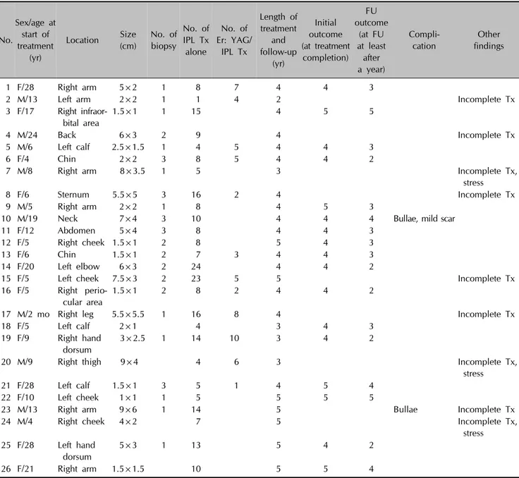
Table 1. Overview of CMN patients who received IPL treatment (IPL alone or in combination with Er: YAG laser)
No.
Sex/age at start of treatment
(yr)
Location Size (cm)
No. of biopsy
No. of IPL Tx alone
No. of Er: YAG/
IPL Tx
Length of treatment
and follow-up
(yr)
Initial outcome (at treatment
completion) FU outcome
(at FU at least
after a year)
Compli- cation
Other findings
1 F/28 Right arm 5×2 1 8 7 4 4 3
2 M/13 Left arm 2×2 1 1 4 2 Incomplete Tx
3 F/17 Right infraor- bital area
1.5×1 1 15 4 5 5
4 M/24 Back 6×3 2 9 4 Incomplete Tx
5 M/6 Left calf 2.5×1.5 1 4 5 4 4 3
6 F/4 Chin 2×2 3 8 5 4 4 2
7 M/8 Right arm 8×3.5 1 5 3 Incomplete Tx,
stress
8 F/6 Sternum 5.5×5 3 16 2 4 Incomplete Tx
9 M/5 Right arm 2×2 1 8 4 5 3
10 M/19 Neck 7×4 3 10 4 4 4 Bullae, mild scar
11 F/12 Abdomen 5×4 3 8 4 4 3
12 F/5 Right cheek 1.5×1 2 8 5 4 3
13 F/6 Chin 1.5×1 2 7 3 4 4 3
14 F/20 Left elbow 6×3 2 24 4 4 2
15 F/5 Left cheek 7.5×3 2 23 5 5 Incomplete Tx
16 F/5 Right perio- cular area
1.5×1 2 8 2 4 4 2
17 M/2 mo Right leg 5.5×5.5 1 16 8 4 Incomplete Tx
18 F/5 Left calf 2×1 4 3 4 3
19 F/9 Right hand dorsum
3×2.5 1 14 10 3 4 2
20 M/9 Right thigh 9×4 4 6 3 Incomplete Tx,
stress
21 F/28 Left calf 1.5×1 3 5 1 4 5 4
22 F/10 Left cheek 1×1 1 5 5 5 5
23 M/13 Right arm 9×6 1 14 5 Bullae Incomplete Tx
24 M/4 Right cheek 4×2 7 5 Incomplete Tx,
stress 25 F/28 Left hand
dorsum
5×3 1 13 5 4 2
26 F/21 Right arm 1.5×1.5 10 5 5 4
Treatment outcome 5-point scale: 1=poor (no change, with lightening of 15% or less); 2=fair (slight improvement, with lightening of 16%∼50%); 3=good (improvement, with lightening of 51%∼75%); 4=excellent (lightening of 76%∼95%); and 5=clear (near-complete disappearance of the lesion, with lightening of 95% or more).
CMN: congenital melanocytic nevi, IPL: intense pulsed light, Er: YAG: erbium: yttrium-aluminum-garnet, Tx: treatment, FU: follow-up, F: female, M: male.
of less than 0.05 was considered statistically significant.
RESULTS
Twenty six patients with 26 small-to-medium sized CMN who preferred non-surgical treatment were included. The mean age of the subjects at the time of their first IPL treat- ment was 11.9±8.4 years. Nine (34.6%) of the CMN were located on the upper extremities, 8 (30.8%) on the
face, 5 (19.2%) on the lower extremities, 3 (11.5%) on the trunk and one (3.8%) on the neck (Table 1).
Seventeen patients (65.4%) completed treatment and were followed-up. Nine (34.6%) were not able to complete treatment due to school, work, change in residence, and treatment related stress. Among the treatment completed patients, 10 (58.8%) received IPL alone with a mean of 10.5±5.8 sessions. Seven patients (41.2%) underwent treatment with IPL (mean: 7.7±3.2 sessions) and Er: YAG/IPL
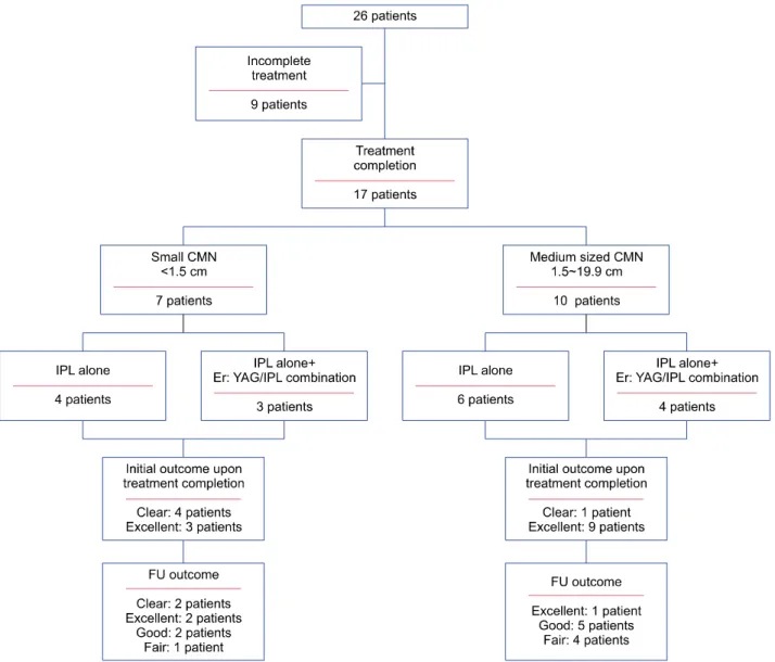
Fig. 1. Flow chart of patients with congenital melanocytic nevi (CMN) treated with intense pulsed light (IPL) alone and in combination with erbium: yttrium-aluminum-garnet (Er: YAG) laser. FU: follow-up.
combination therapy (mean: 4.7±3.1 sessions) (Fig. 1, Table 2).
Of the 17 treatment complete nevi, 7 (41.2%) were small and 10 (58.8%) were medium in size. Four patients (57.1%) with small CMN received IPL alone with a mean of 9.5±4.2 sessions. Three (42.9%) small CMN were treated with IPL (mean: 6.7±1.5 sessions) and Er: YAG/IPL combination therapy (mean: 2±1 sessions). Six patients (60.0%) with medium sized CMN received IPL alone with a mean of 11.2±6.9 sessions. The remaining four (40%) medium sized CMN were treated with IPL (mean:
8.5±4.1 sessions) and Er: YAG/IPL combination therapy (mean: 6.8±2.4 sessions) (Fig. 1, Table 2).
The treatment outcome measured at treatment completion was clear in 5 patients and excellent in 12 (Fig. 2). In the small CMN group, 4 were scored clear and 3 excellent
(mean: 4.6±0.5), whereas in the medium-sized CMN group, one was scored clear and 9 excellent (mean: 4.1±0.3).
Follow-up evaluation, which was generally made at least one year after treatment completion, showed recurrence in 14 cases (82.4%) (Fig. 3). The overall follow-up scores were 2 clear, 3 excellent, 7 good and 5 fair. The small CMN scored as follows: 2 clear, 2 excellent, 2 good and one fair (mean: 3.7±1.1). The medium-sized CMN scored as follows: 1 excellent, 5 good and 4 fair (mean: 2.7±0.7) (Fig. 1, Table 3).
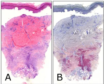
Histologically, the skin biopsy specimens showed features of a compound nevus with nevomelanocytic cell infiltra- tion in the deep dermis and around the hair follicles.
Epidermal detachment and profound collagen degener- ation in the superficial and mid-dermis were the findings of a biopsy specimen taken immediately after IPL (Fig. 4).
Fig. 2. (A) Photo before treatment. (B) Treatment outcome measured at treatment completion is clear in patient 3.
Fig. 3. (A) Photo before treatment. (B) The outcome at treatment completion is clear in patient 26, (C) but she later on shows recurrence at follow-up evaluation, which is made 14 months after treatment completion.
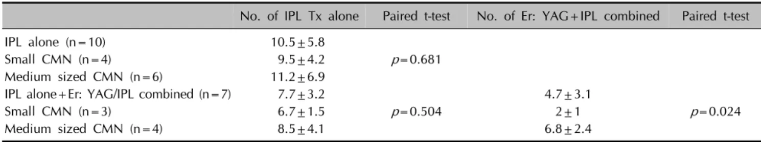
Table 2. Number of IPL treatment alone and the number of Er: YAG/IPL combined between the small and medium sized CMN No. of IPL Tx alone Paired t-test No. of Er: YAG+IPL combined Paired t-test
IPL alone (n=10) 10.5±5.8
Small CMN (n=4) 9.5±4.2 p=0.681
Medium sized CMN (n=6) 11.2±6.9
IPL alone+Er: YAG/IPL combined (n=7) 7.7±3.2 4.7±3.1
Small CMN (n=3) 6.7±1.5 p=0.504 2±1 p=0.024
Medium sized CMN (n=4) 8.5±4.1 6.8±2.4
Values are presented as mean±standard deviation.
IPL: intense pulsed light, Er: YAG: erbium: yttrium-aluminum-garnet, CMN: congenital melanocytic nevi, Tx: treatment.
In terms of complication, 2 patients (7.7%) experienced bullae from IPL therapy (Fig. 5) and one patient (3.8%) ex- perienced focal scarring later on. Treatment related crust- ing, erythema and post-inflammatory hyperpigmentation and changes in skin texture were experienced by all, but faded with time and was not so significant by the time of their follow-up visit post treatment completion.
One patient who received 23 sessions of IPL and 5 ses- sions of Er: YAG/IPL combination therapy achieved im- provement of CMN but was not able to complete treat- ment due to change in residence. In the follow-up photos sent from her mother, significant re-pigmentation was
noticed. We recommended staged excision which was successfully performed by a derm-surgeon (Fig. 6).
DISCUSSION
IPL emits non-coherent, broad-spectrum light, usually in the 500∼1,200 nm range. Apart from wavelength, a wide range of treatment parameters including pulse duration, pulse sequences and pulse delay time are flexible with IPL, allowing it to eliminate hair, treat vascular lesions and remove pigment18-22,24. Treatment of pigmented melano- cytic lesions by means of IPL has been the object of recent
Table 3. Initial treatment outcome and the follow-up outcome between the small CMN and the medium sized CMN
Small CMN (n=7)
Medium sized CMN (n=10)
Paired t-test Initial outcome at
treatment completion
4.6±0.5 4.1±0.3 p=0.037
FU outcome 3.7±1.1 2.7±0.7 p=0.033
Values are presented as mean±standard deviation. Treatment outcome 5-point scale: 1=poor (no change, with lightening of 15% or less); 2=fair (slight improvement, with lightening of 16%∼50%); 3=good (improvement, with lightening of 51%∼
75%); 4=excellent (lightening of 76%∼95%); and 5=clear (near-complete disappearance of the lesion, with lightening of 95% or more).
CMN: congenital melanocytic nevi, FU: follow-up.
Fig. 4. Epidermal detachment and profound collagen degeneration in the superficial and mid dermis are the findings of a biopsy specimen from patient 1 taken immediately after intense pulsed light treatment (×50; A: H&E, B: periodic acid-Schiff [PAS]).
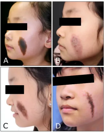
Fig. 5. (A) Photo before treatment. (B) Patient 10 experiences bullae following intense pulsed light therapy. (C) congenital melanocytic nevi is cleared at the time of treatment completion with mild scarring and erythema. (D) Follow-up 29 months after treatment completion.
interest and its success has been described12,13,17,18,21,22,24. The target chromophore of IPL in the treatment of melano- cytic lesions is the melanosome. Selective thermal damage has been observed in a wide range of wavelengths, from 351 nm to 1,064 nm25. Short wavelengths (500∼600 nm) reach right beneath the epidermis and target superficial
melanocytic lesions while sparing the dermis. Longer wavelengths penetrate deeper into the dermis and are suit- able for dermal melanocytic lesions17,18. Since most CMN
Fig. 6. (A) Photo before treatment. (B) Patient 15, who receives 23 sessions of intense pulsed light (IPL) and 5 sessions of erbium:
yttrium-aluminum-garnet/IPL combination therapy achieves improvement of congenital melanocytic nevi but is not able to complete treatment due to change of residence. (C) Follow-up (FU) photo sent from her mother, 5 months after the last treatment shows significant re-pigmentation. (D) Photo FU after the first staged excision.
are compound nevi, we presumed that the broad wave- length of IPL would be beneficial in CMN treatment. We have applied a 590 nm filter (590∼1,200 nm) and a 755 nm filter (755∼1,200 nm) to our CMN patients, targeting both the superficial and deep melanosomes. However, to our disappointment, a skin biopsy taken immediately after IPL treatment revealed the effective depth of IPL to be the mid-dermis, sparing the deep nevus cells.
Pulse width is also an important factor to consider. The IPL pulse duration is on the order of ms whereas the ther- mal relaxation time of melanosomes is 70∼250 ns. The long pulse duration of IPL allows less spatially selective, but more gentle, heating of the target chromophores (melanosomes), making it more effective than Q-switched mode lasers for the treatment of melanocytic nevi as they target individual cells and clusters of nevus cells.
However, with the high density of melanosomes in CMN, heat accumulation can be excessive at times, resulting in bullae formation.
Among our patients, 2 (7.7%) experienced bullae for-
mation; both patients had dark colored (heavily pig- mented) CMN. We advise reducing the fluence energy in treating such patients. As for the results, a Er: YAG/IPL combination was used less often for small CMN, but the number of IPL sessions required for completion of treat- ment was similar between the small and medium sized CMN. The findings were slightly different from what we had expected, and suggest that the depth rather than the size of the CMN is the primary determinant of treatment number. The small CMN were found to have a better out- come (measured at the time of treatment completion) and follow-up outcome (generally made at least one year after treatment completion) with a comparatively lower re- currence rate (71.4%) to medium sized CMN (90%). Re- currence was observed in the majority of treatment com- pleted patients (82.4%) suggesting the presence of a rem- nant nevi. In fact, a skin biopsy from one of our treatment completed CMN patient showed remnant nevus cells in the deep dermis confirming the hypothesis (Fig. 7).
Three patients (ages 4, 8, and 9 years) were not able to complete treatment due to treatment related stress. For an- esthesia, we have routinely applied EMLA cream with li- docaine 2.5% and prilocaine 2.5% under occlusion for 40∼50 minutes. In some cases, lidocaine injection was added. Many Asian parents disapprove of the use of gen- eral anesthesia and its avoidance was initially thought as an advantage of IPL treatment over surgery. However, our results suggest that local anesthesia may not be sufficient for IPL on CMN, especially in young children.
Unlike most lasers, IPL has a large spot size (15 mm×35 mm in case of Lumenis OneTM) and is convenient in treat- ing bigger lesions22. From our experience, it took less than 5 minutes to treat small to medium sized CMN with IPL.
Initially, the combination of broad wavelength, the ms pulse duration, and the large spot size of IPL was felt very much suitable for CMN treatment. IPL alone or in combi- nation with Er: YAG laser effectively decreased the clinical pigmentation and destroyed the superficial nevus cells without significant scarring. Unfortunately, due to the per- sistence of the deep nevus cells, there was high recurrence.
Compared to CMN, acquired melanocytic nevi are more superficially located, and may be successfully removed with IPL alone or in combination with Er: YAG18,24.
There are a number of limitations to our study. The small sample size and restriction to Asians limit the general- izability of our results. In addition, our study was also not able to present the effect of IPL alone on CMN in a num- ber of cases due to the limited efficacy of IPL on its own.
Also, there has been no objective standardized assessment tool of clinical images for pigment reduction and also lacks a control group.
Fig. 7. (A) The clinical photo (patient 11) and (B, C) histology before the start of treatment (×50; B: H&E, C: periodic acid-Schiff [PAS]). (D) A clinical photo after treatment completion (the focal scarring seen here is due to repeated punch biopsy). (E, F) The follow-up skin biopsy shows remnant nevus cells in the deep dermis (×50; E: H&E, F: PAS).
Despite the limitations, we feel that our study is mean- ingful in that it is the first large-scale study to evaluate the efficacy and safety of IPL application on CMN. The find- ings of this study suggest that IPL alone and in combina- tion with Er: YAG laser are not definitive treatments and should not be considered as the first-line treatment.
ACKNOWLEDGMENT
This study was supported by a grant of the Korean Healthcare technology R & D project, Ministry of Health
& Welfare, Republic of Korea (Grant No. HN15C0105).
REFERENCES
1. Krengel S. Nevogenesis--new thoughts regarding a classical problem. Am J Dermatopathol 2005;27:456-465.
2. Kopf AW, Bart RS, Hennessey P. Congenital nevocytic nevi and malignant melanomas. J Am Acad Dermatol 1979;
1:123-130.
3. Alikhan A, Ibrahimi OA, Eisen DB. Congenital melanocytic nevi: where are we now? Part I. Clinical presentation, epidemiology, pathogenesis, histology, malignant transfor- mation, and neurocutaneous melanosis. J Am Acad Dermatol 2012;67:495.e1-e17.
4. Ibrahimi OA, Alikhan A, Eisen DB. Congenital melanocytic nevi: where are we now? Part II. Treatment options and approach to treatment. J Am Acad Dermatol 2012;67:515.
e1-e13.
5. Marghoob AA, Agero AL, Benvenuto-Andrade C, Dusza SW. Large congenital melanocytic nevi, risk of cutaneous melanoma, and prophylactic surgery. J Am Acad Dermatol 2006;54:868-870.
6. Kinsler VA, Chong WK, Aylett SE, Atherton DJ. Com- plications of congenital melanocytic naevi in children:
analysis of 16 years' experience and clinical practice. Br J Dermatol 2008;159:907-914.
7. Dawson HA, Atherton DJ, Mayou B. A prospective study of congenital melanocytic naevi: progress report and evaluation after 6 years. Br J Dermatol 1996;134:617-623.
8. Berg P, Lindelöf B. Congenital melanocytic naevi and cutaneous melanoma. Melanoma Res 2003;13:441-445.
9. Tannous ZS, Mihm MC Jr, Sober AJ, Duncan LM. Congenital melanocytic nevi: clinical and histopathologic features, risk of melanoma, and clinical management. J Am Acad Dermatol 2005;52:197-203.
10. Zaal LH, Mooi WJ, Klip H, van der Horst CM. Risk of malignant transformation of congenital melanocytic nevi: a retrospective nationwide study from The Netherlands. Plast Reconstr Surg 2005;116:1902-1909.
11. Al-Hadithy N, Al-Nakib K, Quaba A. Outcomes of 52 patients with congenital melanocytic naevi treated with UltraPulse Carbon Dioxide and Frequency Doubled Q-Swit- ched Nd-Yag laser. J Plast Reconstr Aesthet Surg 2012;
65:1019-1028.
12. Lee JM, Kim IH, Rhyu IJ, Ryu HJ. Combined intense pulsed light and Er:YAG laser treatment of congenital melanocytic nevus. J Cosmet Laser Ther 2015;17:162-164.
13. Moreno-Arias GA, Ferrando J. Noncoherent-intense-pulsed light for the treatment of relapsing hairy intradermal mela- nocytic nevus after shave excision. Lasers Surg Med 2001;29:142-144.
14. Minakawa S, Takeda H, Korekawa A, Kaneko T, Urushidate S, Sawamura D. Q-switched ruby laser therapy and long- term follow-up evaluation of small to medium-sized con- genital melanocytic naevi. Clin Exp Dermatol 2012;37:
438-440.
15. Lee SE, Choi JY, Hong KT, Lee KR. Treatment of acquired and small congenital melanocytic nevi with combined Er:
YAG laser and long-pulsed alexandrite laser in Asian skin.
Dermatol Surg 2015;41:473-480.
16. Arora H, Falto-Aizpurua L, Chacon A, Griffith RD, Nouri K.
Lasers for nevi: a review. Lasers Med Sci 2015;30:1991- 2001.
17. Moreno Arias GA, Ferrando J. Intense pulsed light for melanocytic lesions. Dermatol Surg 2001;27:397-400.
18. Martín JM, Monteagudo C, Bella R, Reig I, Jordá E.
Complete regression of a melanocytic nevus under intense pulsed light therapy for axillary hair removal in a cosmetic center. Dermatology 2012;224:193-197.
19. Raulin C, Hellwig S, Schönermark MP. Treatment of a nonresponding port-wine stain with a new pulsed light source (PhotoDerm VL). Lasers Surg Med 1997;21:203-208.
20. Weiss RA, Weiss MA, Marwaha S, Harrington AC. Hair removal with a non-coherent filtered flashlamp intense pulsed light source. Lasers Surg Med 1999;24:128-132.
21. Kawada A, Shiraishi H, Asai M, Kameyama H, Sangen Y, Aragane Y, et al. Clinical improvement of solar lentigines and ephelides with an intense pulsed light source. Dermatol Surg 2002;28:504-508.
22. Goldberg DJ. Current trends in intense pulsed light. J Clin Aesthet Dermatol 2012;5:45-53.
23. Kilmer SL, Lee MS, Grevelink JM, Flotte TJ, Anderson RR.
The Q-switched Nd:YAG laser effectively treats tattoos. A controlled, dose-response study. Arch Dermatol 1993;129:
971-978.
24. Bjerring P, Christiansen K. Intense pulsed light source for treatment of small melanocytic nevi and solar lentigines. J Cutan Laser Ther 2000;2:177-181.
25. Anderson RR, Parrish JA. Selective photothermolysis:
precise microsurgery by selective absorption of pulsed radiation. Science 1983;220:524-527.