Recurrent Plastic Bronchitis in a Child with 2009 Influenza A (H1N1) and Influenza B Virus Infection
Plastic bronchitis is an uncommon disorder characterized by the formation of bronchial casts. It is associated with congenital heart disease or pulmonary disease. In children with underlying conditions such as allergy or asthma, influenza can cause severe plastic bronchitis resulting in respiratory failure. A review of the literature showed nine cases of plastic bronchitis with H1N1 including this case. We report a case of a child with recurrent plastic bronchitis with eosinophilic cast associated with influenza B infection, who had recovered from plastic bronchitis associated with an influenza A (H1N1) virus infection 5 months previously. To the best of our knowledge, this is the first case of recurrent plastic bronchitis related to influenza viral infection. If patients with influenza virus infection manifest acute respiratory distress with total lung atelectasis, clinicians should consider plastic bronchitis and early bronchoscopy should be intervened. In addition, management for underlying disease may prevent from recurrence of plastic bronchitis.
Key Words: H1N1 Subtype Influenza A Virus; Influenza B virus; Bronchitis; Bronchial Hyperreactivity
Sun Kim1, Hwa Jin Cho2, Dong Kyun Han2, Yoo Duk Choi3, Eun Seok Yang4, Young Kuk Cho2, and Jae Sook Ma2
Departments of 1Family Medicine, 2Pediatrics, and
3Pathology, Chonnam National University Medical School, Chonnam National University Hospital, Gwangju; 4Department of Pediatrics, Chosun University College of Medicine, Gwangju, Korea Received: 9 February 2012
Accepted: 28 May 2012 Address for Correspondence:
Young Kuk Cho, MD
Department of Pediatrics, Chonnam National University Hospital, 42 Jebong-ro, Dong-gu, Gwangju 501-757 Korea
Tel: +82.62-220-6646, Fax: +82.62-222-6103 E-mail: [email protected]
http://dx.doi.org/10.3346/jkms.2012.27.9.1114 • J Korean Med Sci 2012; 27: 1114-1119
INTRODUCTION
Plastic bronchitis is a rare disease characterized by recurrent formation of bronchial casts (1). Plastic bronchitis can be asso- ciated with inflammatory diseases of the lungs such as asthma and pulmonary infection (1). Influenza is one of major etiologic agents of acute viral lower respiratory tract infections in hospi- talized children (2). Although the latest H1N1 (here after referred to as H1N1) epidemic was declared over by the World Health Organization on August 10, 2010, the WHO has cautioned that H1N1 may circulate as a seasonal influenza for years (3). Dur- ing the H1N1 pandemic, a higher incidence and mortality due to H1N1 infection was evident among children compared with seasonal influenza, and pneumonia was the most common com- plication of H1N1 infection (4, 5). In children with pneumonia accompanied by influenza viral infection, severe plastic bron- chitis can occur. We report a child with recurrent plastic bron- chitis associated with H1N1 and influenza B virus.
CASE DESCRIPTION
First attack with new influenza (H1N1) virus infection A 7-yr-old boy was admitted to a local hospital because of a 1-day history of cough, fever and aggravating dyspnea on November
15, 2009. Real-time polymerase chain reaction (RT-PCR) for H1N1 was positive, and the patient was treated with oral oselta- mivir. On physical examination, breath sounds were decreased in the left lung. A chest radiograph revealed complete atelectasis in the left lung and over-inflation of the right lung (Fig. 1A). A chest computed tomography (CT) scan showed left main bron- chial obstruction with low attenuated materials and atelectasis of the left lung (Fig. 2A, C). Laboratory studies revealed as he- moglobin of 13.2 g/dL, white blood cell count of 10,600/μL (poly- morphonuclear cells, 95.8%; lymphocytes, 1.5%; and eosino- phils, 2.7%), and platelet count of 318,000/μL. The C-reactive protein level was 2.3 mg/dL, erythrocyte sedimentation rate was 6 mm/hr, and antistreptolysin O titer was 105 IU/mL. Anti-my- coplasma antibody was negative. Total IgE exceeded 3,000 IU/
mL. Electrolytes, and liver and kidney function tests were in the normal range. The arterial blood gas analysis was as follows: pH 7.42, pCO2 34.5 mmHg, pO2 67.5 mmHg, and HCO3 22.3 mmHg.
Gram-stain, acid fast stain, potassium hydroxide mounts, Myco- bacterium tuberculosis culture, culture for other sputum bacte- ria, and fungus culture of sputum were negative. After supply- ing O2 via an oxygen mask, dyspnea was relieved and aeration of the left lung on the chest radiograph was also improved. The patient received mucolytics, chest physiotherapy, and antibiot- ics. Although amoxicillin-clavulanate was chosen due to the pa-
tient’s history of allergy to ceftriaxone, an urticarial rash devel- oped after amoxicillin-clavulanate administration. The rash sub- sided with antihistamine use. On day 4 following admission, the patient underwent a bronchoscopy because of aggravating dyspnea. The bronchoscopy showed total obstruction of the left main bronchus by a rubbery cast. The chest radiograph and CT scan after extraction of the cast showed the left lung recovered with good aeration (Fig. 2B, D). The patient was discharged 12 days after admission.
Second attack with influenza B virus infection
The same patient was referred to the emergency department of our hospital because of acute respiratory distress 5 months later after the first attack. The patient was hospitalized at a local hos- pital 2 days before because of a 4-day history of cough and mild fever. The patient was managed with antibiotics and mucolytics under the diagnosis of pneumonia. However, shortness of breath was aggravated suddenly and a chest radiograph showed total atelectasis of the left lung (Fig. 1B). On arrival at our emergency room, the patient presented with tachypnea, deceased breath sounds in the left lung field, and chest retraction. SpO2 was 85%
on 100% face mask oxygen, and the arterial blood gas analysis was as follows: pH 7.33, pCO2 37.9 mmHg; pO2 68.8 mmHg, and HCO3, 19.5 mmHg. The body temperature was 36.7°C, pulse rate was 164/min, respiratory rate was 48/min, and blood pressure was 100/60 mmHg. The patient was intubated and mechanical ventilation was applied in the emergency room due to respira- tory failure. Laboratory studies revealed hemoglobin of 14.4 g/dL, white blood cell count of 23,200/μL (polymorphonuclear cells,
95.4%; lymphocytes, 2.0%; and eosinophils, 0.4%), and platelet count of 357,000/μL. The C-reactive protein level was 5.2 mg/dL, erythrocyte sedimentation rate was 11 mm/hr, lactate dehydro- genase was 941 U/L, and antistreptolysin O titer was 85 IU/mL.
Urine pneumococcal antigen and the anti-mycoplasma anti- body were negative. The rapid nasal swab influenza antigen test was negative for influenza A but positive for influenza B virus.
RT-PCR for H1N1 and influenza B virus were negative and pos- itive, respectively. Shell vial culture for influenza A, parainflu- enza, adenovirus, and respiratory syncytial virus were all nega- tive, but only for influenza B was positive. To exclude allergic bronchial fungal disease, we also did tests for fungal infection.
Each fungus culture with specimens obtained from endotrache- al aspiration and bronchial lavage was negative. Aspergillus an- tigen and antibody immunoglobulin G were negative. Electro- lytes, liver and renal function tests were within normal range. A chest CT scan demonstrated low attenuated materials filling the left main and lobar bronchi, and total consolidation or atelecta- sis in the left lung. An electrocardiogram and echocardiography were normal. On the admission day, an emergent bronchosco- py was performed and thick rubbery material was extracted out from the left main bronchus. The patient received a 5-day course of oral oseltamivir and intravenous methylprednisolone for 5 days. Antibiotics (cefotaxime, netilmycin, clindamycin,and rox- ithromycin) and oral leukotriene modulator were administered.
Massive chest physiotherapy with inhaled corticosteroids, bron- chodilator, and mucolytics was carried out. In spite of the bron- choscopic removal and medical treatment, the patient experi- enced respiratory distress again on day 2 of hospitalization. Flex-
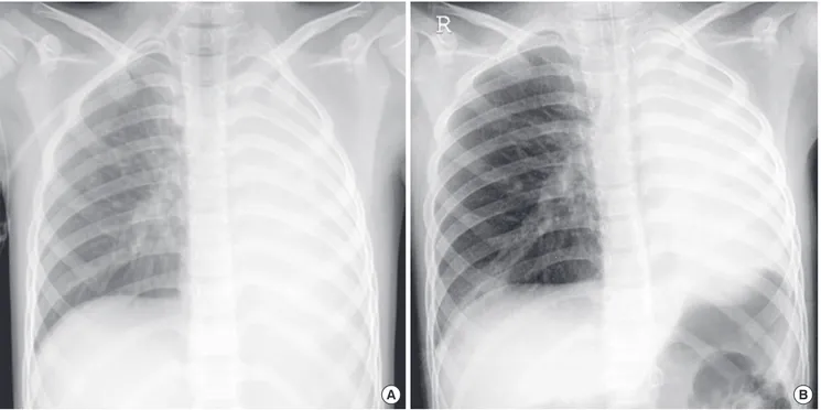
A B
Fig. 1. Chest radiographs at the first attack with H1N1 infection in November, 2009 (A) and second attack with influenza B infection in April, 2010 (B) shows total atelectasis in the left lung and hyperaeration in the right lung.
A B
C D
Fig. 2. Chest computed tomography (CT) at the first attack (A, C) reveals left main bronchial obstruction with low attenuated materials and atelectasis of the left lung. Chest CT after bronchoscopic removal of bronchial casts (B, D) shows recovered left lung with good aeration.
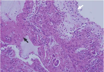
Fig. 3. Casts extracted from the left main bronchus at the second attack showed
preserved anatomy of the bronchial tree. Fig. 4. The specimen composed of denudated bronchial epithelium, fibrin clot (black arrow), mucin material (white arrow), and inflammatory exudates. The inflammatory cells were eosinophils and neutrophils (H&E stain, × 200).
ible bronchoscopy at the bedside in the intensive care unit re- vealed a gelatinous yellow plug in the left bronchus. Thus, a sec- ond bronchoscopic removal of bronchial casts was performed on day 3 of hospitalization (Fig. 3). After removal of the cast, dys- pnea resolved and chest radiography revealed a marked im- provement. On histologic examination, the firm and rubbery cast was composed of fibrinous clot with eosinophil-dominant inflammatory exudates (Fig. 4). There were no Charcot-Leyden crystals, which are frequently seen in sputum from patients with bronchial asthma. The patient was extubated on day 4 of hospi- talization and discharged after 2 weeks without any complica- tions. The patient also underwent allergic evaluations. Total IgE was 3,560 IU/mL. Some specific IgE reactions using UniCAP® (Pharmacia, Uppsala, Sweden) were positive: Dermatophagoi- des pteronyssi of 3.01 k/U, D. farinae of 15.9 kU/L, and Alternar- ia tenuis of 29.6 k/U. Unicap of cat fur, ragweed, mugwort, and Aspergillus fumigatus were negative. Eosinophil cationic pro- tein was 12.1 ng/mL. After tachypnea was improved, spirome- try was performed. The forced expiratory volume in one second
(FEV1), forced vital capacity (FVC), and FEV1/FVC ratio, were 1.19 L, 1.20 L, and 0.99 respectively. The forced expiratory flow 25%-75% was 1.38 L/s. The serial spirometry was within normal limit on follow-up visits after discharge. A methacholine provo- cation test was performed 3 months later after discharge in our out-patient clinic. The provocative concentration that resulted in a 20% fall in FEV1 was 8.0 mg/mL, which means bronchial hyperresponsiveness. Therefore, we recommended that the pa- tient use inhaled corticosteroids for 3 months and get vaccinat- ed for H1N1 and seasonal influenzas during influenza season.
The patient has been in a good condition without respiratory infection, asthma attack, and recurrence of plastic bronchitis for 12 months since discharge.
DISCUSSION
Most patients with H1N1 were self-limited and recovered with- out complications. However, it can complicate severe lower re- spiratory illness including plastic bronchitis in young age groups
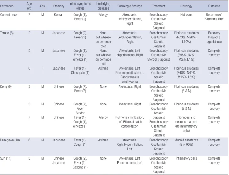
Table 1. Clinical characteristics of the children of plastic bronchitis associated with new influenza A (H1N1) virus infection Reference Age
(yr) Sex Ethnicity Initial symptoms (days)
Underlying
diseases Radiologic findings Treatment Histology Outcome
Current report 7 M Korean Cough (1),
Fever (1)
Allergy Atelectasis, Left Hyperinflation,
Right
Bronchoscopy Oseltamivir
Steroid β agonist
Not done Recurrence*
5 months later
Terano (8) 2
5
6 M
M
F
Japanese
Japanese
Japanese
Cough (2), Fever (1)
Cough (1), Fever (1), Wheeze (1)
Fever (1), Chest pain (1)
None, but wheeze on common
cold None, but wheeze on common
cold Asthma
Atelectasis, Left Hyperinflation,
Right Atelectasis, Left Hyperinflation, Right
Atelectasis, Left Pneumomediastinum,
Subcutaneous emphysema
Bronchoscopy Oseltamivir
Steroid β agonist Bronchoscopy
Oseltamivir Steroid β agonist
Bronchoscopy Oseltamivir
Steroid β agonist
Fibrinous exudates (N70%, M20%,
L10%) Fibrinous exudates
(E95%, N2%, M2%, L1%) Fibrinous exudates
(E40%, N40%, M15%, L5%)
Recovery Inhaled β agonist use Complete recovery
Complete recovery
Deng (9) 3
3
7 M
M
M
Chinese
Chinese
Chinese
Cough (7), Fever (7)
Cough (7), Fever Stridor Fever (1), Cough (1), Wheeze (1)
None
None
Allergy
Atelectasis, Right
Atelectasis, Right
Pulmonary infiltration, Left Bilateral patch
consolidation
Bronchoscopy Oseltamivir
Steroid β agonist Bronchoscopy
Oseltamivir Steroid β agonist Bronchoscopy
Oseltamivir Steroid β agonist
Fibrinous exudates (E & N)
Fibrinous exudates (E & N) Fibrinous and necrotic material (no inflammatory
cells)
Complete recovery
Complete recovery Complete
recovery
Hasegawa (10) 6 M Japanese Fever (1),
Cough (1)
Asthma Atelectasis, Right Hyperinflation,
Left
Bronchoscopy Oseltamivir
Steroid β agonist
Mucoid substance (E > 90%)
Complete recovery
Sun (11) 5 M Chinese
Japanese Cough (2), Fever (1), Gasping (1)
None Atelectasis, Left
Pneumothorax, Left Bronchoscopy Oseltamivir
Steroid β agonist
Inflamatory cells Complete recovery
*Recurrence associated with influenza B virus infection. E, eosinophils; N, neutrophils; M, monocytes; L, lymphocytes; Sex, M male, F female.
(4, 5). Plastic bronchitis might occur more frequently in children than in adults associated with H1N1 infection (6, 7).
We reviewed nine cases, including the present case, of plastic bronchitis associated with H1N1 in the English literature (Table 1) (8-11). The median age was 5 yr (range 2-7 yr) with a predom- inance of boys (89%). Interestingly, all nine cases were Asian, Japanese, Chinese, or Korean. Cough and fever were most com- mon initial symptoms. The left lung was more frequently involved (6/9 cases, 67%) than the right lung (3/9 cases, 33%). The most common radiographic finding was atelectasis of the affected lung (8/9 cases, 89%). They were children with allergy (2/9 cases, 22%), asthma (2/9 cases, 22%), and no underlying disease, but with a wheeze on common cold (2/9 cases, 22%). The remainder were previously healthy (3/9 cases, 34%). All the patients underwent bronchoscopic removal of casts. Histology of the casts showed mostly inflammatory cells (7/8 cases, 88%) and mucinous sub- stance with no inflammatory cells (1/8 case, 12%). Good prog- nosis was shown as complete recovery (7/9 cases, 78%) and re- covery following inhaled β agonist use (1/9 case, 11%). Only this present case had recurrent plastic bronchitis with influenza B virus infection 5 months later after recovery from plastic bron- chitis with H1N1.
Although plastic bronchitis can occur in previously healthy children, pediatric patients with allergy or asthma are at high risk of plastic bronchitis (12). Our patient had no history of ato- py such as atopic dermatitis, allergic rhinitis, and asthma. How- ever, the methacholine provocation test revealed bronchial hy- perresponsiveness. Considering the patient’s known allergy to some antibiotics, high total IgE and elevated specific IgE to house dust mites, the patient seemed to have atopy. Although it is not clear whether influenza virus has a causal link for the develop- ment of asthma, pandemic H1N1 can induce severe asthma at- tack in atopic children who have no history of asthma. In a re- cent study, H1N1 caused asthma attack more than seasonal in- fluenza did (13). Another study explained that repeated viral infections cause prolongation of airway hyperresponsiveness in atopic subjects (14). Our patient experienced two attacks of se- vere plastic bronchitis with H1N1 and influenza B infection. The patient was infected with influenza B 5 months later after re- covery from H1N1. H1N1 might induce prolongation of bron- chial hyperresponsiveness, in which a sticky inflammatory plug obstructs airways more easily and rapidly before the casts are expectorated spontaneously. Children with bronchial hyperre- sponsiveness may be prone to marked increase in airway resis- tance by inflammatory exudate. When large bronchial casts blocks the main bronchus, it acts as a stop valve and results in total lung atelectasis (15).
In plastic bronchitis with an underlying atopic condition, asth- matic and infectious conditions can prompt the use of anti-in- flammatory regimens, including inhaled and oral steroids (1).
In this case, we prescribed inhaled steroids to prevent recurrence
of bronchial casts. According to Hasegawa et al. (13), 20 (90.9%) among 22 patients who experienced severe asthma attack dur- ing the H1N1 pandemic did not receive long-term treatment.
The treatment of the underlying pulmonary disease such as asth- ma may decrease or prevent cast formation (13).
Plastic bronchitis in children can complicate severe hypoxic damage and lead to death. Bronchoscopic intervention should be performed as early as possible because obstruction of the major airways by bronchial casts may proceed very rapidly. In some cases, repeated bronchoscopy may be required to find bronchial casts (8). Anti-viral agents may less effective for the treatment of plastic bronchitis (8). Besides bronchoscopic re- moval of bronchial casts, therapeutic options include chest phys- iotherapy, oseltamivir, antibiotics, mucolytics, and steroids. Also, macrolides have emerged as an immunomodulator, and treat- ment with low-dose azithromycin in idiopathic plastic bronchi- tis has been successful (1).
Immunization is the most effective strategy of preventing complicated influenza infection (17). Kwon et al. (18) reported that none of children with severe neurologic complication from H1N1 in his study had been immunized for H1N1 and seasonal influenzas.
In conclusion, if patients with influenza virus infection mani- fest acute respiratory distress with total lung atelectasis, clini- cians should consider plastic bronchitis and early bronchosco- py should be carried out. To prevent recurrence of plastic bron- chitis in patients with atopy or bronchial hyperresponsiveness, inhaled corticosteroids can be used besides influenza vaccina- tion.
REFERENCES
1. Eberlein MH, Drummond MB, Haponik EF. Plastic bronchitis: a man- agement challenge. Am J Med Sci 2008; 335: 163-9.
2. Neuzil KM, Mellen BG, Wright PF, Mitchel EF Jr, Griffin MR. The effect of influenza on hospitalizations, outpatient visits, and courses of antibi- otics in children. N Engl J Med 2000; 342: 225-31.
3. World Health Organization. H1N1 in post-pandemic period. Available at: http://www.who.int/mediacentre/news/statements/2010/h1n1_
vpc_20100810/en/index.html. [Accessed on 10 August 2010].
4. Hasegawa M, Okada T, Sakata H, Nakayama E, Fuchigami T, Inamo Y, Mugishima H, Tajima T, Iwata S, Morozumi M, et al. Pandemic (H1N1) 2009-associated pneumonia in children, Japan. Emerg Infect Dis 2011;
17: 279-82.
5. Kim HS, Kim JH, Shin SY, Kang YA, Lee HG, Kim JS, Lee JK, Cho B. Fa- tal cases of 2009 pandemic influenza A (H1N1) in Korea. J Korean Med Sci 2011; 26: 22-7.
6. Shin SY, Kim JH, Kim HS, Kang YA, Lee HG, Kim JS, Lee JK, Kim WK.
Clinical characteristics of Korean pediatric patients critically ill with in- fluenza A (H1N1) virus. Pediatr Pulmonol 2010; 45: 1014-20.
7. Lee E, Seo JH, Kim HY, Na S, Kim SH, Kwon JW, Kim BJ, Hong SJ. Clini- cal characteristics and outcomes among pediatric patients hospitalized
with pandemic influenza A/H1N1 2009 infection. Korean J Pediatr 2011;
54: 329-34.
8. Terano C, Miura M, Fukuzawa R, Saito Y, Arai H, Sasaki M, Ariyasu D, Hasegawa Y. Three children with plastic bronchitis associated with 2009 H1N1 influenza virus infection. Pediatr Infect Dis J 2011; 30: 80-2.
9. Deng J, Zheng Y, Li C, Ma Z, Wang H, Rubin BK. Plastic bronchitis in three children associated with 2009 influenza A (H1N1) virus infection.
Chest 2010; 138: 1486-8.
10. Hasegawa M, Inamo Y, Fuchigami T, Hashimoto K, Morozumi M, Ubu- kata K, Watanabe H, Takahashi T. Bronchial casts and pandemic (H1N1) 2009 virus infection. Emerg Infect Dis 2010; 16: 344-6.
11. Sun DJ, Yang YS, Wang BC. Plastic bronchitis associated with severe in- fluenza A (H1N1) in children: a case report and review of the literature.
Zhonghua Jie He He Hu Xi Za Zhi 2010; 33: 837-9.
12. Brogan TV, Finn LS, Pyskaty DJ Jr, Redding GJ, Ricker D, Inglis A, Gibson RL. Plastic bronchitis in children: a case series and review of the medical literature. Pediatr Pulmonol 2002; 34: 482-7.
13. Hasegawa S, Hirano R, Hashimoto K, Haneda Y, Shirabe K, Ichiyama T.
Characteristics of atopic children with pandemic H1N1 influenza viral infection: pandemic H1N1 influenza reveals ‘occult’ asthma of childhood.
Pediatr Allergy Immunol 2011; 22: e119-23.
14. Xepapadaki P, Papadopoulos NG, Bossios A, Manoussakis E, Manou- sakas T, Saxoni-Papageorgiou P. Duration of postviral airway hyperre- sponsiveness in children with asthma: effect of atopy. J Allergy Clin Im- munol 2005; 116: 299-304.
15. Bowen A, Oudjhane K, Odagiri K, Liston SL, Cumming WA, Oh KS.
Plastic bronchitis: large, branching, mucoid bronchial casts in children.
AJR Am J Roentgenol 1985; 144: 371-5.
16. Ko JH, Kim JH, Kang JH, Kim JH, Eun BW, Kim KY, Hong JY, Oh SH.
Characteristics of hospitalized children with 2009 pandemic influenza A(H1N1): a multicenter study in Korea. J Korean Med Sci 2012; 27:
408-15.
17. Fiore AE, Uyeki TM, Broder K, Finelli L, Euler GL, Singleton JA, Iskan- der JK, Wortley PM, Shay DK, Bresee JS, Cox NJ; Centers for Disease Control and Prevention (CDC). Prevention and control of influenza with vaccines: recommendations of the Advisory Committee on Immu- nization Practices (ACIP), 2010. MMWR Recomm Rep 2010; 59: 1-62.
18. Kwon S, Kim S, Cho MH, Seo H. Neurologic complications and outcomes of pandemic (H1N1) 2009 in Korean children. J Korean Med Sci 2012;
27: 402-7.