INTRODUCTION
Posterior cervical foraminotomy is an effective surgical tech- nique for the treatment of radicular pain arising from com- pression in the foramen secondary to bony stenosis or soft disc herniation (1-3). In 1943, Semmes and Murphey first des- cribed cervical radicular root syndromes (4), and Murphey followed up with a description of 648 cases in 1973 (5).
Various surgical techniques can be used for the treatment of cervical radiculopathy. Although anterior cervical proce- dures have gained prominence, posterior cervical foramino- tomy has proven benefits. It has been found to relieve symp- toms in 82% to 97% of patients who have radiculopathy caused by foraminal stenosis or posterolateral herniated discs (1, 3, 6-9). Moreover, a posterior procedure avoids the com- plications associated with anterior approaches to the cervical spine, particularly vascular injury, esophageal injury, dyspnea, recurrent laryngeal nerve injury, dysphagia, and the acceler- ated degeneration of adjacent motion segments after fusion, known as adjacent segment disease (10-12). However, post- operative neck pain and spasm are disadvantages of a poste- rior procedure. A wider incision and an extensive periosteal muscle dissection for adequate visualization can induce neck discomfort, which can result in a slower recovery. Minimal- ly invasive spine surgery is necessary to meet the demands of modern society for faster recoveries, which allow patients to resume their normal activities as soon as possible.
Currently, among the various minimally invasive spinal surgery techniques, the tubular retractor system is a well devel- oped system that enables posterior cervical foraminotomy and discectomy through a tubular retractor under microscopic visualization. The present study was performed to compare the clinical parameters and surgical outcomes between open foraminotomy/discectomy (OF/OFD) and tubular retractor assisted foraminotomy/discectomy (TAF/TAFD) in the treat- ment of cervical radiculopathy.
MATERIALS AND METHODS Patients and evaluation
A total of 41 patients with radiating pain in the upper arm caused by foraminal stenosis or cervical posterolateral disc herniation were enrolled in the study from January 2003 to June 2005. They were randomly assigned to have surgical treatment by either OF/OFD or TAF/TAFD. Inclusion cri- teria were the presence of a foraminal stenosis or a posterolat- eral disc herniation on magnetic resonance imaging (MRI) and persistent radiating pain in the upper arm after 6 weeks of conservative treatment. Patients were excluded if they had neoplasm, traumatic injury, myelopathy, or recurrent cervi- cal disc disease.
After the inclusion criteria were met and informed consent
649
Kyoung-Tae Kim and Young-Baeg Kim
Department of Neurosurgery, College of Medicine, Chung-Ang University, Seoul, Korea
Address for correspondence Young-Baeg Kim, M.D.
Department of Neurosurgery, Chung-Ang Unveristy Hospital, 224-1 Heukseok-dong, Dongjak-gu, Seoul 156-755, Korea
Tel : +82.2-6299-1598, Fax : +82.2-821-8409 E-mail : [email protected]
DOI: 10.3346/jkms.2009.24.4.649
Comparison Between Open Procedure and Tubular Retractor Assisted Procedure for Cervical Radiculopathy: Results of a Randomized
Controlled Study
Posterior cervical foraminotomy is an effective surgical technique for the treatment of radicular pain caused by foraminal stenosis or posterolateral herniated discs. The present study was performed to compare the clinical parameters and surgical out- comes of open foraminotomy/discectomy (OF/OFD) and tubular retractor assisted foraminotomy/discectomy (TAF/TAFD) in the treatment of cervical radiculopathy.
A total of 41 patients were divided into two groups: 19 patients in Group 1 under- went OF/OFD and 22 patients in Group 2 underwent TAF/TAFD. Among the vari- ous clinical parameters, skin incision size, length of hospital stay, analgesic using time, and postoperative neck pain (for the first 4 weeks after the operation) were favorable in Group 2. Surgical outcomes were not different between the two groups.
In conclusion, TAF/TAFD should increase patient’s compliance and is as clinically effective as much as the OF/OFD.
Key Words : Radiculopathy; Tubular Retractor Assisted Foraminotomy; Open Foraminotomy
Received : 21 July 2008 Accepted : 3 February 2009
was obtained, patients were randomly allocated into one of two groups based on the surgical technique: Group 1 under- went OF/OFD (19 patients); and Group 2 underwent TAF/
TAFD (22 patients). All of these operations were performed by the same spinal neurosurgeon.
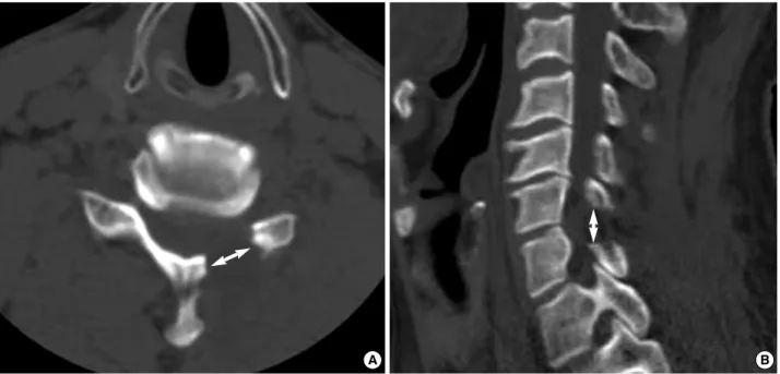
The preoperative and postoperative evaluation consisted of a radiologic finding, a neurological examination, and a pain scoring for the upper arm and neck using a Visual analogue scale (VAS). Postoperative evaluations were done on days 1 and 5, week 4, and months 3, 6, 12, and 24 after the opera- tion. The clinical outcomes were evaluated using modified Odom et al. criteria (13) (Table 1). All patients underwent a preoperative MRI as well as preoperative and postoperative computed tomography (CT) scans. The postoperative CT scan was done on 5 days after the operation. We calculated the vertical and transverse diameters of the foraminotomy using the postoperative CT scan (Fig. 1).
Surgical technique
OF/OFD
All patients were operated on in a prone position. A radi- olucent three pin-head fixed instrument was used. A verti- cal 3 cm midline incision was made after determining the correct level on a lateral radiograph. The lateral lamina/medi- al facet joints were exposed. Under a surgical microscope, a partial hemilaminectomy and foraminotomy with partial face- tectomy of the target level was performed using high-speed drills. The extent of the facet resection was based on the extent of the foraminal pathology on that half of the facet joint. In cases of pure disc herniation, the proximal root was adequate- ly visualized in order to remove the compressing disc mate- rial. However, in cases of foraminal stenosis, bony decompres- sion and skeletonization of the proximal root were carefully performed using a 2-mm bullet burr, cervical curette and punch. Patients wore a soft collar for 2-4 weeks after the oper- ation, and were given adequate medication.
TAF/TAFD
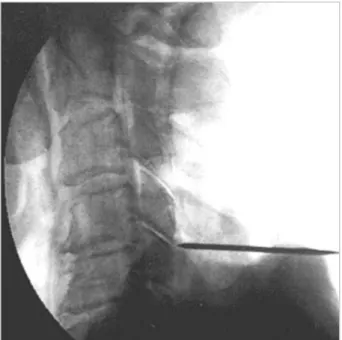
The technique used for TAF or TAFD was similar to that used for OF or OFD. Although the procedures use a tubular retractor (METRx system, Medtronic Sofamor Danek, Mem- phis, TN, U.S.A.), the principles are the same regardless of the specific type of retractor system that was used. A skin incision was initially made approximately 5 mm off the mid- line ipsilateral to and at the target level. For two-level pro- cedures, the incision should be placed midway between the target levels. After an initial skin incision, the K-wire is slow- ly advanced through the musculature. This is done under continuous fluoroscopic guidance in order to avoid malposi-
Grading Definition
Excellent All preoperative symptoms and abnormal findings improved.
Good Minimal persistence of preoperative symptoms (neck tenderness only, otherwise no symptoms). Abnormal findings improved.
Fair Definite relief of some preoperative symptoms. Other symptoms slightly improved (residual root irritation with transient pain).
Poor Symptoms and signs unchanged or worse.
Table 1. Modified Odom’s criteria
Fig. 1. Postoperative CT shows the foramintomy defect. (A) Transverse diameter (B) Vertical diameter.
A B
tion of the K-wire. The K-wire is docked on bone at the infer- omedial edge of the rostral lateral mass at the target level (Fig.
2). At this point, the skin incision was extended above and below the K-wire entry point for a total length of approxi- mately 2 cm and the wire is removed. The cervical fascia is incised equal to the length of the skin incision using monopo- lar cautery. The tubular muscle dilators are placed serially.
After dilation is complete, a final working channel (16-mm or 18-mm tubular retractor) is placed over the dilators and fixed over the laminofacet junction with a table-mounted flex- ible retractor arm, and the dilators are removed. The addition- al skin incision can be needed in order to avoid skin necrosis, if the final working channel is tightly packed the skin inci- sion. Under the surgical microscope, a partial hemilaminec- tomy and foraminotomy with partial facetectomy of the tar- get level was performed using high-speed drills. Although the techniques of foraminotomy and discectomy were the same as those for OF/OFD, the tubular retractor assisted pro- cedure needs a more detailed review of preoperative radiologi- cal findings in order to achieve adequate neural decompres- sion.
Postoperative pain control
All patients received the same postoperative pain manage- ment medication, which included talniflumate (1,110 mg/
day) and afloqualone (60 mg/day) as the postoperative anal- gesic and muscle relaxant, respectively. These medications were discontinued if both VAS scores for radicular and neck pain were less than 3. Patient-controlled analgesia was used for the first 2 days after the operation.
Statistical analysis
The SPSS 12.0 statistical software package (SPSS, Inc., Chica- go, IL, U.S.A.) was used for statistical analysis. Data were analyzed using the chi-square, Fisher exact, Student’s t, and Mann-Whitney tests, as appropriate. A P value of <0.05 was considered to be statistically significant.
RESULTS Clinical parameters
A total of 41 patients were divided into two groups: 19 patients in Group 1 underwent OF/OFD and 22 patients in Group 2 underwent TAF/TAFD (Table 2). The mean follow- up period was 34.2 months in Group 1 (range, 24-66 months) and 33.1 months in Group 2 (range, 24-64 months). The clinical parameters (age, sex, duration of symptoms, surgical time, skin incision size, length of hospital stay, analgesic using
Fig. 2. The K-wire is advanced slowly through the musculature under fluoroscopic guidance and docked on bone at the infero- medial edge of the rostral lateral mass at the target level.
Variable Group 1
(19 patients)
Group 2 (22 patients)
P value Age (range) 54.1±15. 2 yr 54.4±14.7 yr NS
(31-72) (28-72)
Sex (male:female) 12:7 14:8 NS
Duration of symptoms 6.4±5.1 mon 6.5±4.9 mon NS
(range) (1-70) (1-69)
Surgical time (range) 76.5±19. 2 min 78.5±18. 2 min NS (55-154) (52-131)
Skin incision size (range) 3.6±0.4 cm 3.2±0.2 cm <0.05 (2.4-4.4) (2.0-4.1)
Length of hospital stay 6.7±2.1 ds 4.1±1.7 ds <0.05
(range) (5-14) (3-15)
Analgesic using time 3.6 wk (1-11) 2.6 wk (1-9) <0.05 (range)
Total levels 28 31
No. patients of 1 level (%) 12 (63.2%) 13 (59.1%) No. patients of 2 levels (%) 7 (36.8%) 9 (40.9%)
Affected lesion NS
No. C4-5 (%) 3 (10.7) 4 (12.9)
No. C5-6 (%) 12 (42.9) 13 (41.9)
No. C6-7 (%) 12 (42.9) 12 (38.7)
No. C7-T1 (%) 1 (0.35) 2 (6.5)
Diagnosis NS
HNP±Foraminal stenosis 7 (36.8%) 8 (36.4%) Foraminal stenosis only 12 (63.2%) 14 (63.6%)
Foraminotomy defect NS
Transverse diameter 11.2±0.2 mm 11.5±0.1 mm
(range) (9-13) (9-13)
Vertical diameter (range) 9.6±0.2 mm 9.5±0.2
(8-11) (8-11)
Table 2. Comparison between clinical parameters of Group 1 (OF/OFD) and 2 (TAF/TAFD)
Group 1, Open foraminotomy/discectomy (OF/OFD); Group 2, Tubular retractor assisted foraminotomy/discectomy (TAF/TAFD).
NS, not significant.
time, number and location of affected lesion, diagnosis and foraminotomy defect diameter) are shown in Table 2. The skin incision size, length of hospital stay and analgesic using time were significantly shorter in Group 2 compared to Group 1 (P<0.05).
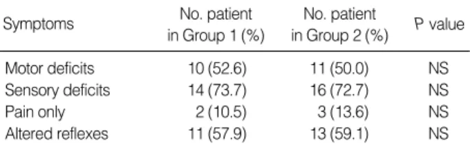
Five patients (12.2%) experiended only pain as a present- ing symptom. All other patients had an additional motor and/or sensory deficit prior to the operation. The preopera- tive neurological status of the patients is shown in Table 3 and there were no differences in neurological status between the two groups.
Changes in degree of pain after the operation
The pain evaluations were done using the VAS. We used the VAS score to check for radicular pain and neck pain on days 1 and 5, week 4, and months 3, 6, 12, and 24 after the operation (Table 4). The degree of radicular pain did not differ between the two groups. However, the degree of neck pain was different. VAS scores for neck pain from day 1 to week 4 after the operation were more severe in Group 1 than in Group 2.
Surgical outcome
The total success rate was 85.4% using modified Odom’s criteria. Each surgical outcome is shown in Table 5. There were no differences in surgical outcomes between the two groups, and there were no reported complications due to the surgeries.
DISCUSSION
Various mechanical causes, such as disc herniation, bony spur and foraminal stenosis, can induce radiculopathy. There are various surgical techniques to treat radiculopathy (1, 3, 5, 7, 14, 15). The effectiveness of posterior foraminotomy/dis- cectomy for treating foraminal stenosis and posterolateral disc herniation is well established in the literature (1, 16-19).
The advantages of posterior foraminotomy/discectomy in- clude the avoidance of complications associated with anteri- or approaches to the cervical spine and no need for cervical fusion and instrumentation. However, posterior procedures also have some problems, such as a limitation in surgical indi- cation (e.g., central disc herniation) and postoperative neck discomfort. A wider incision and an extensive periosteal mus- cle dissection for adequate visualization can induce neck pain, which can result in a slower recovery. Modern society demands faster recovery times, which necessitates minimally invasive spine surgery. Foley and Smith (20) first described the tubular retractor assisted endoscopic discectomy for lumbar disc her- niation in 1997. They believed that this procedure is a less invasive and more effective technique for treating lumbar steno- sis and disc herniation. This concept has since been adapted to treatments of the cervical spine.
In this study, we compared the various clinical parameters of TAF/TAFD and OF/OFD (Table 2). The skin incision size, length of hospital stay, and analgesic using time were significantly shorter in Group 2 than in Group 1 (P<0.05).
In addition, the postoperative neck pain reported during the 4 weeks after the operation was significantly lower in Group
Group 1, Open foraminotomy/discectomy; Group 2, Tubular retractor assisted foraminotomy/discectomy.
NS, not significant.
Symptoms No. patient
in Group 1 (%)
No. patient
in Group 2 (%) P value
Motor deficits 10 (52.6) 11 (50.0) NS
Sensory deficits 14 (73.7) 16 (72.7) NS
Pain only 2 (10.5) 3 (13.6) NS
Altered reflexes 11 (57.9) 13 (59.1) NS
Table 3. Preoperative neurological status
Group 1, Open foraminotomy/discectomy; Group 2, Tubular retractor assisted foraminotomy/discectomy.
NS, not significant.
Group 1 (19 patients)
Group 2
(22 patients) P value
Excellent 11 (57.9%) 13 (59.1%)
Good 5 (26.3%) 6 (27.3%)
Fair 3 (15.8%) 3 (13.6%)
Poor 0 (0.0%) 0 (0.0%)
Success (excellent+good) 16 (84.2%) 19 (86.4%) NS Table 5. Long-term outcome after operation (Modified Odom’s Criteria)
Group 1, Open foraminotomy/discectomy; Group 2, Tubular retractor assisted foraminotomy/discectomy.
VAS, Visual Analogue Scale; NS, not significant.
Variable Group 1 Group 2 P value
VAS of radicular pain
Preoperative (range) 7.4 (6-10) 7.3 (6-10) NS 1 day after operation (range) 2.9 (1-4) 3.0 (1-4) NS 5 day after operation (range) 3.4 (1-5) 3.2 (1-4) NS 4 wk after operation (range) 2.2 (0-4) 2.3 (1-4) NS 3 mon after operation (range) 1.9 (0-3) 1.8 (0-3) NS 6 mon after operation (range) 1.8 (0-3) 1.7 (0-3) NS 12 mon after operation (range) 1.7 (0-3) 1.8 (0-3) NS 24 mon after operation (range) 1.6 (0-3) 1.7 (0-3) NS VAS of neck pain
Preoperative (range) 2.9 (1-4) 3.0 (1-5) NS 1 day after operation (range) 5.9 (4-8) 4.7 (3-7) <0.05 5 day after operation (range) 5.8 (4-8) 4.5 (3-7) <0.05 4 wk after operation (range) 4.4 (2-6) 3.5 (2-6) <0.05 3 mon after operation (range) 2.1 (1-4) 2.0 (0-4) NS 6 mon after operation (range) 1.4 (0-4) 1.5 (0-3) NS 12 mon after operation (range) 1.5 (0-4) 1.4 (0-3) NS 24 mon after operation (range) 1.4 (0-3) 1.4 (0-3) NS Table 4. Changes of pain degree after operation (VAS)
2. These results show the advantages of TAF/TAFD. TAF/
TAFD is a minimally invasive procedure using a tubular retractor system, which allows for a smaller skin incision and far less muscle injury (21). It also reduces the amount of post- operative discomfort and shortens the length of hospital stays and the postoperative analgesic using time.
Our surgical outcomes for the two groups were not statis- tically different. Fessler et al. described the use of the tubu- lar retractor assisted endoscopic system for posterior cervical laminoforaminotomy in human cadaveric spines in 2000 (10).
They demonstrated that the average vertical and transverse diameters of the foraminotomy defect were greater in the TAF group compared to the OF group. In our study, the average vertical and transverse diameters of the foraminotomy defect were the same in the two groups. These results suggest that the TAF is at least as clinically effective as the OF in reliev- ing the neural compression and thereby reducing radicular symptoms. Our total success rate was 85.4% using modified Odom’s criteria. This outcome is similar to the success rates (82-97%) reported in other studies (1, 3, 6-9).
The complication rate in other studies was 0-9%. Duro- tomy and CSF leak were the most commonly reported com- plications (5, 6, 8, 9, 13, 18). Although we did not experi- ence complications after the posterior procedure, our study had a small number of patients and more complications may arise in a large study group. However, it is clear that the cer- vical posterior procedures (TAF/TAFD and OF/OFD) can be safely performed in patients with foraminal stenosis and posterlateral disc herniation.
The superiority of TAF/TAFD over OF/OFD has been demonstrated in some studies (10, 19), but until now, there has not been a randomized clinical study comparing TAF/
TAFD and OF/OFD. To our knowledge, this is the first pro- spective randomized clinical study comparing the clinical outcomes of these techniques.
Some parameters (skin incision size, length of hospital stay, analgesic using time, and postoperative neck pain during the first four postoperative weeks) were favorable in Group 2 and these factors should affect the willingness of a patient to under- go this operation. In conclusion, TAF/TAFD should increase patient’s compliance and is as clinically effective as OF/OFD.
REFRENCES
1. Henderson CM, Hennessy RG, Shuey HM Jr, Shackelford EG. Pos- terior-lateral foraminotomy as an exclusive operative technique for cervical radiculopathy: a review of 846 consecutively operated cases.
Neurosurgery 1983; 13: 504-12.
2. Caglar YS, Bozkurt M, Kahilogullari G, Tuna H, Bakir A, Torun F, Ugur HC. Keyhole approach for posterior cervical discectomy: expe- rience on 84 patients. Minim Invasive Neurosurg 2007; 50: 7-11.
3. Ducker TB, Zeidman SM. The posterior operative approach for cer-
vical radiculopathy. Neurosurg Clin N Am 1993; 4: 61-74.
4. Semmes RE, Murphey F. Syndrome of unilateral rupture of the sixth, cervical intervertebral disk, with compression of the seventh cervi- cal nerve root. Report of four cases with symptoms simulating coro- nary disease. JAMA 1943; 121: 1209-14.
5. Murphey F, Simmons JC, Brunson B. Surgical treatment of lateral- ly ruptured cervical disc. Review of 648 cases, 1939 to 1972. J Neu- rosurg 1973; 38: 679-83.
6. Aldrich F. Posterolateral microdisec tomy for cervical monoradicu- lopathy caused by posterolateral soft cervical disc sequestration. J Ne- urosurg 1990; 72: 370-7.
7. Korinth MC, Kruger A, Oertel MF, Gilsbach JM. Posterior foramino- tomy or anterior discectomy with polymethyl methacrylate interbody stabilization for cervical soft disc disease: results in 292 patients with monoradiculopathy. Spine 2006; 31: 1207-14.
8. Jodicke A, Daentzer D, Kastner S, Asamoto S, Boker DK. Risk fac- tors for outcome and complications of dorsal foraminotomy in cer- vical disc herniation. Surg Neurol 2003; 60: 124-9.
9. Onimus M, Destrumelle N, Gangloff S. Surgical treatment of cervi- cal disk displacement. Anterior or posterior approach? Rev Chir Orthop Reparatrice Appar Mot 1995; 81: 296-301.
10. Fessler RG, Khoo LT. Minimally invasive cervical microendoscop- ic foraminotomy: an initial clinical experience. Neurosurgery 2002;
51: S37-45.
11. Hilibrand AS, Robbins M. Adjacent segment degeneration and adja- cent segment disease: the consequences of spinal fusion? Spine J 2004; 4: 190S-4S.
12. Ishihara H, Kanamori M, Kawaguchi Y, Nakamura H, Kimura T.
Adjacent segment disease after anterior cervical interbody fusion.
Spine J 2004; 4: 624-8.
13. Odom GL, Finney W, Woodhall B. Cervical disk lesions. JAMA 1958; 166: 23-8.
14. Roh SW, Kim DH, Cardoso AC, Fessler RG. Endoscopic foramino- tomy using MED system in cadaveric specimens. Spine 2000; 25:
260-4.
15. O’Toole JE, Sheikh H, Eichholz KM, Fessler RG, Perez-Cruet MJ.
Endoscopic posterior cervical foraminotomy and discectomy. Neu- rosurg Clin N Am 2006; 17: 411-22.
16. Zeidman SM, Ducker TB. Posterior cervical laminoforaminotomy for radiculopathy: review of 172 cases. Neurosurgery 1993; 33: 356-62.
17. Krupp W, Schattke H, Muke R. Clinical results of the foraminoto- my as described by Frykholm for the treatment of lateral cervical disc herniation. Acta Neurochir (Wien) 1990; 107: 22-9.
18. Parker WD. Cervical laminoforaminotomy. J Neurosurg 2002; 96:
254.
19. Gala VC, O’Toole JE, Voyadzis JM, Fessler RG. Posterior minimally invasive approaches for the cervical spine. Orthop Clin N Am 2007;
38: 339-49.
20. Foley KT, Smith MM. Microendoscopic discectomy. Tech Neuro- surg 1997; 3: 301-7.
21. Shin DA, Kim KN, Shin HC, Yoon DH. The efficacy of microendo- scopic discectomy in reducing iatrogenic muscle injury. J Neurosurg Spine 2008; 8: 39-43.