Bickerstaff 뇌줄기 뇌염 양상으로 발현된 신경정신성 전신 홍반성 루푸스 1례
전남대학교 의과대학 소아과학교실
최하영・송태양・천경렬・정금희・박수민・우영종・김영옥
Submitted: 21 July, 2015 Revised: 20 September, 2015 Accepted: 24 September, 2015
Correspondence to Young Ok Kim, MD, PhD
Department of Pediatrics, Chonnam National University Medical School, 42, Jebongro, Dong-gu, Gwangju, 61469, Korea
Tel: +82-62-220-6646, Fax: +82-62-220-6646 E-mail: ik052@jnu.ac.kr
Neuropsychiatric Systemic lupus Erythematosus Presenting as Bickerstaff’s Brainstem Encephalitis: a Case Report
Neuropsychiatric events are commonly reported in patients with systemic lupus erythematosus and they are an important contributor to both morbidity and mortality. Neuropsychiatric systemic lupus erythematosus (NPSLE) mainly involves the central nervous system, and rarely invades the peripheral nervous system.
A 12-year-old girl presented with sudden-onset motor weakness in her lower extremities and face, as well as drowsiness. Brain magnetic resonance imaging and angiography produced no abnormal findings, and a cerebrospinal fluid examination revealed only pleocytosis. Reticular erythema in the extremities, oral ulcers, and a dry mouth were noted at her first visit, but a malar rash appeared on the third day with positivity for serum antinuclear and anti-dsDNA antibodies. Therefore a high- dose steroid was administered based on a diagnosis of NPSLE. An ophthalmologic examination, salivary gland scan, and biopsy of minor salivary gland resulted in the additional diagnosis of Sjögren’s syndrome. The reported patient represents a rare pediatric case of NPSLE accompanied with Sjögren’s syndrome, presenting as Bickerstaff’s brainstem encephalitis and being successfully treated with early steroid pulse therapy.
Key Words: Lupus erythematosus, Systemic, Sjögren’s syndrome, Brainstem,
encephalitis, Child
Ha Yeong Choe, MD, Tae Yang Song, MD, Kyeong Ryeol Cheon, MD, Ku Mi Jeong, MD, Soo Min Park, MD, Young Jong Woo, MD, Young Ok Kim, MD
Department of Pediatrics, Chonnam National University Medical School, Gwangju, Republic of Korea
Copyright © 2015 by The Korean Child Neurology Society
http://www.cns.or.kr
Introduction
Systemic lupus erythematosus (SLE) is an autoimmune disease involving multiple organs with various clinical presentations1). The prevalence of SLE in the United States has been estimated at approximately 50 per 100,000 people2). According to a nomenclature system for neuropsychiatric SLE (NPSLE) the con
cept of NPSLE includes not only involvement of the organs but also complica
tions of treatment24). Neuropsychiatric events have been reported in up to 70%
of patients with SLE, and these are a major cause of morbidity and mortality57). A few cases of NPSLE syndromes involving the peripheral nervous system (PNS) have been reported and Bickerstaff’s brainstem encephalitis (BBE) is quite rarely
mentioned in patients with NPSLE810).
GuillainBarré syndrome (GBS) is an acute ascending inflam
matory demyelinating polyneuropathy9). Antibodies to ganglio
sides invading the node of Ranvier are thought to contribute to acute peripheral neuropathy9). Variants of GBS include BBE and Fisher syndrome11). BBE is characterized by alteration of cons
ciousness and hyperreflexia (which support a central patholo
gy), in addition to weakness involving the limbs, face, and bulbar muscles, ophthalmoplegia, and ataxia (which suggest a peri
pheral pathology)10,11). On the other hand, Fisher syndrome is characterized by ophthalmoplegia, ataxia, and loss of tendon reflexes11).
Herein we report a rare pediatric case of NPSLE associated with Sjögren’s syndrome that presented as BBE without abnor
malities in brain magnetic resonance imaging (MRI) and that was successfully treated with early highdose steroid treatment.
Case report
A 12yearold girl developed suddenonset weakness in her lower extremities and face with altered mental status. She was transferred with suspicion of bacterial meningitis since she had developed fever, headache, and tonsillar exudate 5 days previously. Upon arrival at the emergency room, her vital signs were stable and fever was present, which was documented once a day. Her mental state was drowsy, with intermittent sponta
neous eye opening to sound. Dysarthria, dysphagia, and facial motor weakness were noted, which were suggestive of brains
tem dysfunction. The motor score for the lower extremities had decreased to grade 3, though with normal deep tendon reflexes.
Her growth was normal, with a body mass of 44 kg (within the 2050th percentiles for her age) and a height of 147.5cm (within the 5075th percentiles). A reticular erythematous rash was observed on her skin that was spreading to the whole body (Fig.
1), in addition to a dry lip and multiple oral ulcers that were present for several years. In her past history, the patient had visited dermatology clinics due to recurrent reticular erythema
tous rash on both lower extremities during the previous 1 year.
The patient had a nonspecific birth history and no family history of autoimmune disease, cerebrovascular accidents, or other neurologic disease.
The initial laboratory data showed anemia (hemoglobin, 9.3 g/
dL) and elevated serum Creactive protein (9.4 mg/L). A cerebrospinal fluid (CSF) examination revealed only pleocytosis (CSF white blood cell count of 189/μL), with normal protein and glucose levels. CSF stains and cultures for bacterial pathogens produced negative findings. A CSF viral study for herpes simplex, enteroviruses, and Japanese encephalitis virus was also negative. The CSF IgG index was within the normal limits. The urinalysis was also normal, although a later study showed increased urine protein at 13.3 mg/m2/h for the 24hour collected urine. Further analysis revealed strong positivity for antinuclear antibody (ANA) in a mixed pattern of homogeneity
Fig. 1. Reticular erythema on a lower extremity (arrow) at the time of
emergency room arrival.
Fig. 2. Brain magnetic resonance imaging of the patient. T1-weighted axial (A), T2-weighted axial (B), and diffusion-
weighted axial (C) images demonstrate no abnormal findings.
and speckling. She was positive for antidsDNA, antiSSA, anti
SSB, and antinucleosome antibodies, but had normal values of serum C3, serum C4, and serum antiganglioside antibodies (IgM and IgG antibodies for GM1, GD1b, and GQ1b isotypes). There were no abnormal findings on brain MRI (Fig. 2) and spine MRI, somatosen sory evokedpotential test, nerveconductionvelo
city test, and electromyography.
The treatment in the emergency room started with antibiotics, acyclovir, and dexamethasone based on suspicion of menin
goencephalitis. Immunoglobulin was later added due to the possibility of brain encephalitis associated with GBS. However, all of these interventions failed to improve the patient’s symp
toms. On the third day a malar rash developed with positivity for ANA, and highdose prednisolone(1g/day) was started, which markedly ameliorated the patient’s condition. She became alert 3 days later and complained of tenderness and decreased sensa
tion in both lower extremities. Motor weakness and skin rash were gradually improved after taking prednisolone. However, these symptoms were persisted until discharge. She was com
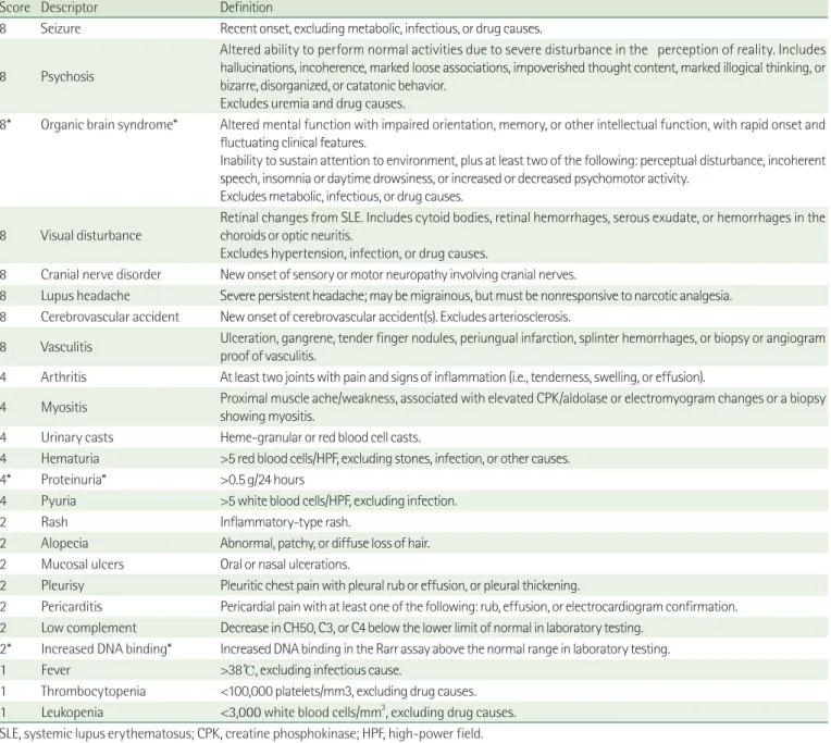
pletely free from symptoms on the 24th day. The SLE Disease Activity Index 2000 (SLEDAI2K) score12) was 14 (Table 1).
Sjögren’s syndrome was diagnosed based on an ophthalmologic evaluation for dryeye syndrome, delayed passage on a parotid gland scan, and consistent pathologic findings in a biopsy of the
Table 1. Systemic Lupus Erythematosus Disease Activity Index 2000 (SLEDAI-2K)12)
Score Descriptor Definition
8 Seizure Recent onset, excluding metabolic, infectious, or drug causes.
8 Psychosis
Altered ability to perform normal activities due to severe disturbance in the perception of reality. Includes hallucinations, incoherence, marked loose associations, impoverished thought content, marked illogical thinking, or bizarre, disorganized, or catatonic behavior.
Excludes uremia and drug causes.
8* Organic brain syndrome* Altered mental function with impaired orientation, memory, or other intellectual function, with rapid onset and fluctuating clinical features.
Inability to sustain attention to environment, plus at least two of the following: perceptual disturbance, incoherent speech, insomnia or daytime drowsiness, or increased or decreased psychomotor activity.
Excludes metabolic, infectious, or drug causes.
8 Visual disturbance Retinal changes from SLE. Includes cytoid bodies, retinal hemorrhages, serous exudate, or hemorrhages in the choroids or optic neuritis.
Excludes hypertension, infection, or drug causes.
8 Cranial nerve disorder New onset of sensory or motor neuropathy involving cranial nerves.
8 Lupus headache Severe persistent headache; may be migrainous, but must be nonresponsive to narcotic analgesia.
8 Cerebrovascular accident New onset of cerebrovascular accident(s). Excludes arteriosclerosis.
8 Vasculitis Ulceration, gangrene, tender finger nodules, periungual infarction, splinter hemorrhages, or biopsy or angiogram proof of vasculitis.
4 Arthritis At least two joints with pain and signs of inflammation (i.e., tenderness, swelling, or effusion).
4 Myositis Proximal muscle ache/weakness, associated with elevated CPK/aldolase or electromyogram changes or a biopsy showing myositis.
4 Urinary casts Heme-granular or red blood cell casts.
4 Hematuria >5 red blood cells/HPF, excluding stones, infection, or other causes.
4* Proteinuria* >0.5 g/24 hours
4 Pyuria >5 white blood cells/HPF, excluding infection.
2 Rash Inflammatory-type rash.
2 Alopecia Abnormal, patchy, or diffuse loss of hair.
2 Mucosal ulcers Oral or nasal ulcerations.
2 Pleurisy Pleuritic chest pain with pleural rub or effusion, or pleural thickening.
2 Pericarditis Pericardial pain with at least one of the following: rub, effusion, or electrocardiogram confirmation.
2 Low complement Decrease in CH50, C3, or C4 below the lower limit of normal in laboratory testing.
2* Increased DNA binding* Increased DNA binding in the Rarr assay above the normal range in laboratory testing.
1 Fever >38℃, excluding infectious cause.
1 Thrombocytopenia <100,000 platelets/mm3, excluding drug causes.
1 Leukopenia <3,000 white blood cells/mm3, excluding drug causes.
SLE, systemic lupus erythematosus; CPK, creatine phosphokinase; HPF, high-power field.
*The present patient had an SLEDAI-2K score of 14 (positive for organic brain syndrome, proteinuria, and increased DNA binding).
minor salivary gland.
Discussion
NPSLE may occur during any phases of SLE, whose symptoms vary among patients3). Although both the central nervous system (CNS) and PNS may be involved, CNS symptoms such as headache, cognitive dysfunction, mood disorder, seizure, and psychosis are more common7). GBS, plexopathy, and peripheral neuropathy have been reported in less than 1% of SLE cases7). While brain MRI is the modality of choice for diagnosing NPSLE, negative findings do not completely exclude NPSLE; for example, Toledano et al. reported normal brain MRI findings in 25 of 43 patients (58.1%) with NPSLE13).
Neuropsychiatric events are the most severe manifestations of SLE and are commonly associated with a worse prognosis.
Zirkzee et al. reported that Dutch patients with NPSLE had an almost tenfold higher mortality rate compared to the general Dutch population14). Among NPSLE patients, less than 40% are directly associated with the active disease itself, while more than 60% are linked to treatment complications4). The most common causes of death in patients with NPSLE are the disease activity of NPSLE itself and several miscellaneous types of infection14). The SLEDAI2K scoring system reflects the status of disease activity and consists of 24 items that include rash, mucosal ulcers, alopecia, and proteinuria12). An appropriate cutoff value for defining active disease has been reported as 3 or 415); the score in our patient was 14.
In cases of mild NPSLE, conservative therapies including analgesics, anticonvulsants, antidepressants, or antipsychotics are effective for symptoms of headache, seizures, or mood disorders, whereas more intensive treatments are required in severe NPSLE cases with high SLEDAI2K scores5). It is also known that highdose corticosteroid, cyclophosphamide, plasmapheresis, or intravenous immunoglobulin is needed in the acute phase, while lowdose corticosteroid or immunosup
pressants should be considered in the chronic phase for maintenance5).
Sjögren’s syndrome is characterized by dry eye and dry mouth due to chronic inflammation of the exocrine glands16). Sjögren’s syndrome involves both the CNS and PNS, as does SLE, which also more frequently involves the CNS than the PNS16). PNS disease in Sjögren’s syndrome presents as sensory neuropathy, demyelinating neuropathy, or autonomic neuropathy16). However, BBE has not been reported previously in Sjögren’s syndrome. The coexistence of Sjögren’s syndrome with SLE is
not rare, with an estimated incidence rate of between 8% and 30% based on published studies17). Manoussakis et al. published a paper about adult cases of SLE associated with Sjögren’s syndrome17). Neuropsychiatric event can occur simultaneously along with those two disease17). But childhood case of SLE accompanied with Sjögren’s syndrome have not been reported in Korea yet.
Neuropsychiatric manifestations are common in SLE patients7). However, NPSLE with PNS conditions such as GBS or BBE have been rarely reported. Our patient showed the rapid onset of motor weakness in her low extremities, bulbar palsy, and drowsiness with normal deep tendon reflexes, which was suggestive of brainstem encephalitis despite her negative findings for brain MRI, serum antiganglioside antibodies, and CSF albuminocytologic dissociation. Although initial immunoglobulin treatment failed, early treatment with high
dose steroid after the diagnosis of NPSLE based on her dermatologic manifestation and positive serologic results was very effective. Sjögren’s syndrome was also diagnosed, which could have contributed to her neurologic manifestations in association with SLE.
요약
신경정신성 전신 홍반성 루푸스는 소아 루푸스 환자에서 빈번하게 발생하며, 예후 및 사망률과 밀접한 관련이 있다. 전신홍반성루푸스는 대부분 중추신경계를 침범하며 말초신경계 침범은 드문 것으로 알려 져 있다. 저자들은 하지 및 안면의 근력 저하와 기면으로 내원하였던 12세 여아에서 감염성 수막뇌염 혹은 자가면역성 뇌줄기 뇌염을 의심 하였다. 환자는 구강 궤양과 건조증, 사지 망상형 홍반을 보였고, 뇌자 기공명영상 및 뇌자기공명혈관촬영술은 정상이었으나 뇌척수액 검사 에서 백혈구 증가증을 보였다. 내원 직후 항생제, 항바이러스제, 저용 량 덱사메타손과 면역글로불린을 투여하였으나 증상 호전이 없다가, 내원 3일 째 안면 홍조와 함께 혈청 항핵항체 및 antidsDNA 양성 소견을 보여 신경정신성 전신 홍반성 루푸스로 진단하고 고용량 스테 로이드 투여한 후 환자의 임상경과는 호전되었으며, 안과검진, 침샘 스캔검사 및 소타액샘 조직검사에서 쇼그렌 증후군으로 추가 진단되 었다. 이에 저자들은 Bickerstaff 뇌줄기 뇌염 양상으로 발현된, 쇼그 렌 증후군을 동반한 소아 신경정신성 전신 홍반성 루푸스 환아에서 고용량 스테로이드 사용 후 성공적으로 치료된 증례를 보고하는 바이 다.
References
1) Zhou HQ, Zhang FC, Tian XP, Leng XM, Lu JJ, Zhao Y, et al. Clini
cal features and outcome of neuropsychiatric lupus in Chinese:
analysis of 240 hospitalized patients. Lupus 2008;17:939.
2) Kampylafka EI, Alexopoulos H, Kosmidis ML, Panagiotakos DB, Vlachoyiannopoulos PG, Dalakas MC, et al. Incidence and pre
valence of major central nervous system involvement in syste
mic lupus erythematosus: a 3year prospective study of 370 patients. PLoS One 2013;8:e55843.
3) Kim SY, Yoon TS, Suh JH. Concomitant occurrence of cervical myelopathy, cerebral infarction, and peripheral neuropathy in systemic lupus erythematosus: a case report. Ann Rehabil Med 2014;38:2638.
4) Bertsias GK, Boumpas DT. Pathogenesis, diagnosis and mana
gement of neuropsychiatric SLE manifestations. Nat Rev Rheumatol 2010;6:35867.
5) Sanna G, Bertolaccini ML, Mathieu A. Central nervous system lupus: a clinical approach to therapy. Lupus 2003;12:93542.
6) Steinlin MI, Blaser SI, Gilday DL, Eddy AA, Logan WJ, Laxer RM, et al. Neurologic manifestations of pediatric systemic lupus erythematosus. Pediatr Neurol 1995;13:1917.
7) Unterman A, Nolte JE, Boaz M, Abady M, Shoenfeld Y, Zand
manGoddard G. Neuropsychiatric syndromes in systemic lupus erythematosus: a metaanalysis. Semin Arthritis Rheum 2011;41:111.
8) Benseler SM, Silverman ED. Neuropsychiatric involvement in pediatric systemic lupus erythematosus. Lupus 2007;16:56471.
9) Kang SH, Yum MS, Lee EH, Ko TS. Concurrent GullainBarre syndrome and acute transverse myelitis as an initial presenta
tion of systemic lupus erythematosus. J Korean Child Neurol Soc 2012;20:1218.
10) Shahrizaila N, Yuki N. Bickerstaff brainstem encephalitis and Fisher syndrome: antiGQ1b antibody syndrome. J Neurol Neurosurg Psychiatry 2013;84:57683.
11) Overell JR, Hsieh ST, Odaka M, Yuki N, Willison HJ. Treatment for Fisher syndrome, Bickerstaff's brainstem encephalitis and related disorders. Cochrane Database Syst Rev 2007:CD004761.
12) Gladman DD, Ibanez D, Urowitz MB. Systemic lupus erythema
tosus disease activity index 2000. J Rheumatol 2002;29:28891.
13) Toledano P, Sarbu N, Espinosa G, Bargallo N, Cervera R. Neuro
psychiatric systemic lupus erythematosus: magnetic resonance imaging findings and correlation with clinical and immuno
logical features. Autoimmun Rev 2013;12:116670.
14) Zirkzee EJ, Huizinga TW, Bollen EL, van Buchem MA, Middel
koop HA, van der Wee NJ, et al. Mortality in neuropsychiatric systemic lupus erythematosus (NPSLE). Lupus 2014;23:318.
15) Yee CS, Farewell VT, Isenberg DA, Griffiths B, Teh LS, Bruce IN, et al. The use of Systemic Lupus Erythematosus Disease Activity Index2000 to define active disease and minimal clinically meaningful change based on data from a large cohort of sys
temic lupus erythematosus patients. Rheumatology (Oxford) 2011;50:9828.
16) Bhattacharyya S, Helfgott SM. Neurologic complications of systemic lupus erythematosus, sjogren syndrome, and rheuma
toid arthritis. Semin Neurol 2014;34:42536.
17) Manoussakis MN, Georgopoulou C, Zintzaras E, Spyropoulou M, Stavropoulou A, Skopouli FN, et al. Sjogren's syndrome associated with systemic lupus erythematosus: clinical and laboratory profiles and comparison with primary Sjogren's syndrome. Arthritis Rheum 2004;50:88291.