三子化痰煎이 천식모델 생쥐의 CD4, CD8 세포에 미치는 영향
남태흥․박양춘*
대전대학교 한의과대학 폐계내과학교실
Effect of Samjahwadam-jeon on CD4, CD8 Cells in OVA-induced Asthmatic Mice
Tae Heung Nam, Yang Chun Park*
Division of Respiratory System, Depatment of Internal Medicine, College of Oriental Medicine, Daejeon University
This study aimed to examine the effects of Samjahwadam-jeon (SJHDJ) on CD4+ T cells and CD8+ T cells in ovalbumin (OVA)-induced asthmatic mice. C57BL/6 mice were injected, inhaled and sprayed with OVA for 12 weeks (four a week) for asthma induction. Two experimental groups were treated with different concentrations of SJHDJ (400 mg/kg and 200 mg/kg) extract and cyclosporin A (10 mg/kg) for the later 8 weeks. At the end of the experiment, the mice lung, peripheral lymph node (PLN), and spleen were removed and CD4+ T cells and CD8+ T cells for analyzed by flow cytometer. Number of CD4+ T cells in lung, PLN and spleen of the SJHDJ group (400 ㎎/㎏ and 200 ㎎/㎏) were significantly decreased compared with that of control group. Number of CD8+ T cells in PLN and spleen of the SJHDJ group (400 ㎎/㎏, 200 ㎎/㎏) were significantly decreased compared with that of control group. Conclusion : These results suggest that SJHDJ alleviated asthmatic hyperreactivity through CD4+ and CD8+ T cells. Further study of relative cytokines is expected.
Key words : Samjahwadam-jeon (Sanzihuatan-jian), asthma, CD4+ T cells, CD8+ T cells
* 교신저자 : 박양춘, 청주시 상당구 용담동 173-9 대전대학교 청주한방병원
․E-mail : [email protected], ․Tel : 043-229-3704
․접수 : 2008/08/21 ․수정 : 2008/09/29 ․채택 : 2008/10/01
서 론
기관지천식(이하 천식)은 전 세계적으로 유병률이 증가하고 있으며 이에 따라 사회경제적 부담이 증가하고 있는 질환이다1). 우리나라의 경우 2006년도 국민건강보험 통계지표에 의하면 천 식환자가 지난 2001년보다 26.9% 증가하여 488,977명에 이르는 것으로 조사되었다2).
천식은 韓醫學에서 呼吸急促하며 喉中有聲響한 것을 특징으 로 하는 哮喘證의 범주에 해당한다. 哮喘의 발생에 대한 素因說에 의하면 특수하게 내재된 素因인 夙根을 가진 사람이 寒冷을 만나 거나, 疲勞 혹은 勞力할 때 哮喘이 발생하는 것으로 설명하고 있 는데 이는 알레르기에 의한 천식의 발생과 유사하다고 할 수 있다
3). 천식의 병리학적 기전에는 다양한 면역세포, 화학매체, 사이토 카인 및 유착분자가 관여하는데 이중 T세포는 염증세포를 기도내 로 끌어 모으고 활성화시켜 천식 병인에 중요한 역할을 한다.
CD4+인 보조T세포(이하 Th세포)는 사이토카인 분비양상에 따라 Th1세포와 Th2세포로 나뉘는데 Th2세포는 IL-3, IL-4, IL-5 등의 사이토카인들을 분비하여 천식의 면역학적 반응에서 중요 한 역할을 한다4,5). CD8+ 세포는 세포독성T세포(이하 Tc세포)로 Th세포와 마찬가지로 사이토카인 분비 양상에 따라 IL-2, IFN-γ 를 분비하는 Tc1세포와 IL-4, IL-5 등을 분비하는 Tc2세포로 분류 하게 되었고6), 이후 천식과 같은 알레르기성 기도염증에서 CD8+ T세포의 역할에 대한 관심이 높아졌다. 따라서 CD4+ T세포와 CD8+ T세포에 대하여 한약 처방이 미치는 영향을 관찰하는 것은 천식 치료 약물의 효과 평가에서 매우 중요하다고 할 수 있다.
최근 천식과 관련하여 다양한 單味 및 處方을 대상으로 동물 천식 모델을 이용한 연구7-10)가 이루어지고 있다. 三子化痰煎은 蘇子導痰降氣湯에 가감한 처방으로 痰涎이 氣道에 壅塞하여 短 氣 喘促하며 咳喘 面浮하는 오래된 천식에 사용하는 처방이다11). 이에 호흡기질환에 다용하고 있는 三子化痰煎을 대상으로 면역세포에 미치는 영향을 규명하고자 기관지천식 생쥐모델을 통해 폐, 말초림프절, 비장 조직의 CD4+세포와 CD8+세포의 수의 변화를 측정하여 유의한 결과를 얻었기에 보고하는 바이다.
재료 및 방법
1. 재료 1) 동물
체중 18-25 g의 C57BL/6(샘타코, Korea) 생쥐를 실험 당일 까지 고형사료(조단백질 22.1% 이상, 조지방 8.0% 이하, 조섬유 5.0% 이하, 조회분 8.0% 이하, 칼슘 0.6% 이상, 인 0.4%이상 ; 삼 양사, Korea)와 물을 충분히 공급하고, 실온 22±2℃, 상대습도 50±10%, 조명시간 12시간(07:00-19:00), 조도 150-300 Lux로 설정 하여 2주일간 실험실 환경에 적응시킨 후 체중 변화가 일정하고 건강한 동물만을 선별하여 실험에 사용하였다.
2) 약재
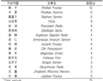
본 실험에 사용한 三子化痰煎(Samjahwadam-jeon, SJHDJ)의 구성 약물은 대전대학교 부속한방병원에서 구입한 후 정선하여 사용하였고, 처방 1첩의 내용과 분량은 다음과 같다(Table 1).
Table 1. Herb Composition of Samjahwadam-jeon(SJHDJ)
구성약물 生藥名 용량(g)
蘇 子 Perillae Fructus 12
半 夏 Pinelliae rhizoma 6
蘿葍子 Raphani Semen 6
白茯苓 Poria 6
前 胡 Peucedani Radix 4
皂角刺 Gleditsiae Spina 4
當 歸 Angelicae Gigantis Radix 4 杏 仁 Armeniacae Amarum Semen 4
枳 殼 Aurantii Fructus 4
陳 皮 Citri Pericarpium 4
厚 朴 Magnoliae Cortex 4
款冬花 Farfarae Flos 3
白芥子 Sinapis Semen 3
甘 草 Glycyrrhizae Radix 3
生 薑 Zingiberis Rhizoma Recens 3
大 棗 Jujubae Fructus 3
Total amount 73
2. 시약 및 기기 1) 시약
Chicken egg albumin(ovalbumin : OVA), aluminum potassium sulfate (Alum), trichloroacetic acid, SRB, amphotericin, antibiotics, DNase, collagenase, chloroform, collagenase, RPMI-1640 배양액, isopropanol, 적혈구 용혈액 (RBC lysis solution), ethidium bromide (EtBr), dulbecco's phosphate buffered saline (D-PBS), formaldehyde, magnesium chloride (㎎Cl2)는 Sigma사(USA) 제품을, 우태아혈청(fetal bovine serum, FBS)는 Hyclone사(USA) 제품을 사용하였으며 기 타 일반 시약은 특급 시약을 사용하였다.
2) 기기
기기는 열탕추출기(대웅 Co., Korea), rotary vaccum evaporator (Büchi, Switzerland), freeze dryer (EYELA, Japan), CO2 incubator (Forma Scientific, USA), clean bench (Vision Scientific, Korea), autoclave(Sanyo, Japan), micro-pipet(Gilson, France), water bath(Vision Scientific, Korea), vortex
mixer(Vision Scientific, Korea), spectrophotometer(Shimazue, Japan), centrifuge(Sigma, USA), deep-freezer(Sanyo, Japan), flow cytometometer (Becton Dickinson, USA), thermocycler system(MWG Biotech, Germany), ice-maker(Vision Scientific, Korea), homogenizer(OMNI, USA), plate shaker (Lab-Line, USA) 등을 사용하였다.
3. 방법
1) 三子化痰煎 추출물 분리
SJHDJ 4첩 분량을 증류수 2,000 ㎖를 가하여 열탕 추출기에서 3 시간 추출하였다. 추출액을 여과한 후 감압 증류장치(B-480, BUCHI, Switzerland)로 농축하고, 다시 동결 건조기(FDU-540, EYELA, Japan)를 이용하여 18.5 g의 분말을 얻었다. 완전 건조한 SJHDJ을 냉 동(-84℃) 보관하면서 적당한 농도로 희석하여 사용하였다.
2) 기관지 천식 생쥐 모델
500 ㎍/㎕의 난알부민(OVA, chicken egg ovalbumin; Grade IV)과 10% (w/v) aluminum potassium sulfate(Alum)를 PBS로 용해한 후 혼합하였다. 이 혼합물을 10 N NaOH로 pH를 6.5로 조정하여 상온에서 1시간 동안 방치하고 750 × g 에서 5분 동안 원심분리 하였다. 원심분리한 OVA/ Alum 침전물을 증류수를 가하여 원래의 양으로 용해한 후, 100 ㎍ OVA를 0.2 ㎖로 조정 하여 복강 내로 주사하여 전신 감작시켰다. 이 후 4주째에 생쥐 를 마취한 후, 난알부민(500 ㎍/㎖) 100 ㎕를 기관으로 주사하여 직접 투여(I.T. : intra trachea)하였다. 5주째부터 분무기를 이용 하여 2.5 ㎎/㎖ 난알부민 용액을 하루에 30분씩 일주일에 3회씩 8주 동안 비강 및 기도내로 흡입시켰다. 이 때 음성대조군(wild type)은 PBS 또는 Alum 만을 복강과 기관에 주사하고, 분무기로 흡입시켰다(Scheme 1).
Scheme 1. Asthma OVA-induced mouse model.
3) 경구 투여
OVA/Alum로 전신 감작 시킨 후 4주째부터 SJHDJ (400, 200 ㎎/㎏)을 일주일에 5회 경구 투여 하였다. 대조군에는 증류 수를 동량 경구 투여하였다.
4) 폐장, 말초림프절 및 비장의 면역세포수 측정
실험 종료 후 OVA 천식 생쥐를 ethyl ether로 마취시킨 후 폐장, 말초림프절(lung, peripheral lymph node; PLN), 그리고 비장을 분리하여 buffered ammonium chloride(ACK) 용액을 37oC에서 5분 동안 처리하여 적혈구를 용해시켰다. 이를 다시 배 지로 세척한 후 0.04% trypan blue로 염색하여 총 면역세포수를 측정하였다.
5) 유세포 분석
실험 종료 후 비장, 말초림프절, 폐를 각각 적출하여 100 mesh로 세포를 분리하여 D-PBS로 5분간 원심분리(1,700 rpm)하 여 2회 세척한 후 cell strainer에 통과시켜 세포 이외의 분해되지 않은 조직이나 불순물을 제거하였다. 그리고 이를 잘게 chopping한 후, collagenase 1 ㎎/㎖ (in 2% FBS + RPMI 1640) 을 넣고 37℃ shaker (180 rpm, 20 min) 배양기에서 배양한 후 상층액을 회수하는 방법으로 4회 반복하였다. 이들 세포들을 ACK 용액(8.3 g NH4Cl, 1 g KHCO3, in 1 L of demineralized water + 0.1 mM EDTA)을 실온에서 5분 동안 처리하여 적혈구 를 용해시키고 다시 D-PBS로 2회 세척한 후 0.04% trypan blue 로 염색한 후 총 세포수를 측정하였다. 측정한 비장, 말초림프절, 폐의 세포를 5×10⁵ 세포로 조정한 후 4℃에서 면역 형광염색 (immunofluorescence staining)을 실시하였다. 각각에 FITC-anti-CD4, FITC-anti-CD8을 넣고 30분간 얼음에서 반응시 켰다. 반응 후 3회 이상 인산완충 생리식염수로 수세한 후 flow cytometer의 Cell Quest 프로그램을 이용하여 CD4+, CD8+ 세포 수를 백분율(%)로 분석한 후 총세포수를 적용하여 각 조직에서 의 절대세포수(absolute number)를 산출하였다.
6) 통계처리
실험으로부터 얻은 결과는 mean ± standard error로 기록하 였고, 유의성 검증은 Student's T-test 분석 방법을 이용하여 결정 하였다.
결 과
1. CD4+ 세포 수에 미치는 영향 1) Lung
Lung에서 분리한 세포에서 CD4+ 세포 수를 측정한 결과는 다음과 같다. 정상군은 1.4 ± 0.1 (×105), 대조군은 13.0 ± 0.8 (×105), 양성대조군 (CsA)은 4.4 ± 0.0 (×105), SJHDJ 400 mg/kg 투여군은 6.6 ± 1.0 (×105), 200 mg/kg 투여군은 8.5 ± 0.7 (×105) 로 나타나, 대조군에 비하여 SJHDJ 모든 실험 농도에서 유의성 있는 (***p<0.001, ***p<0.001) 감소 효과를 나타내었다(Fig. 1).
2) PLN
PLN에서 분리한 세포에서 CD4+ 세포 수를 측정한 결과, 정 상군은 24.8 ± 0.3 (×105), 대조군은 57.9 ± 3.2 (×105), 양성대조군 (CsA)은 35.0 ± 4.5 (×105), SJHDJ 400 mg/kg 투여군은 31.6 ± 12.0 (×105), 200 mg/kg 투여군은 35.3 ± 0.3 (×105)으로 나타나, 대조군에 비하여 SJHDJ 모든 실험 농도에서 유의성 있는 (**p<0.01, ***p<0.001) 감소 효과를 나타내었다(Fig. 2).
3) Spleen
Spleen에서 분리한 세포에서 CD4+ 세포 수를 측정한 결과, 정상군은 5.2 ± 1.5 (×106), 대조군은 17.8 ± 4.2 (×106), 양성대조 군 (CsA)은 4.6 ± 0.9 (×106), SJHDJ 400 mg/kg 투여군은 6.7 ± 1.2 (×106), 200 mg/kg 투여군은 7.4 ± 1.2 (×106)로 나타나, 대조 군에 비하여 SJHDJ 모든 실험 농도에서 유의성 있는 (**p<0.01,
**p<0.01) 감소 효과를 나타내었다(Fig. 3).
Fig. 1. Effect of SJHDJ extract on CD4+ absolute cell number in lung of OVA-induced mice. C57BL/6 mice were injected, inhaled and sprayed with OVA for 12 weeks (four a week) for asthma induction. Two experimental groups were treated with different concentrations of SJHDJ (400 mg/kg and 200 mg/kg) extract and cyclosporin A (10 mg/kg) for the later 8 weeks. At the end of the experiment, the mice lung was removed and CD4+ T cells for analyzed by flow cytometer. The results are expressed the mean
± S.E (N=5). Statistically significant value compared with control group data by T test (***p<0.001).
Fig. 2. Effect of SJHDJ extract on CD4+ absolute cell number in peripheral lymph node (PNL) of OVA-induced mice. C57BL/6 mice were injected, inhaled and sprayed with OVA for 12 weeks (four a week) for asthma induction. Two experimental groups were treated with different concentrations of SJHDJ (400 mg/kg and 200 mg/kg) extract and cyclosporin A (10 mg/kg) for the later 8 weeks. At the end of the experiment, the mice PLN were removed and CD4+ T cells for analyzed by flow cytometer. The results are expressed the mean
± S.E (N=5). Statistically significant value compared with control group data by T test (**p<0.01, ***p<0.001).
Fig. 3. Effect of SJHDJ extract on CD4+ absolute cell number in spleen of OVA-induced mice. C57BL/6 mice were injected, inhaled and sprayed with OVA for 12 weeks (four a week) for asthma induction. Two experimental groups were treated with different concentrations of SJHDJ (400 mg/kg and 200 mg/kg) extract and cyclosporin A (10 mg/kg) for the later 8 weeks.
At the end of the experiment, the mice spleen was removed and CD4+ T cells for analyzed by flow cytometer. The results are expressed the mean ± S.E (N=5).
Statistically significant value compared with control group data by T test (**p<0.01).
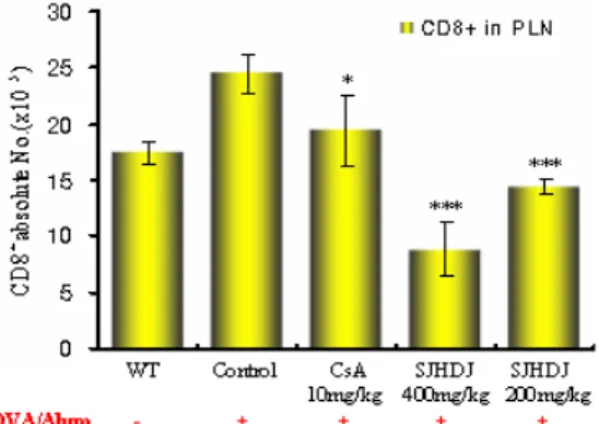
2. CD8+ 세포 수에 미치는 영향 1) Lung
Lung에서 분리한 CD8+ 세포 수를 측정한 결과, 정상군은 0.7 ± 0.1 (×105), 대조군은 4.9 ± 0.6 (×105), 양성대조군 (CsA)은 2.7 ± 0.5 (×105), SJHDJ 400 mg/kg 투여군은 3.5 ± 1.2 (×105), SJHDJ 200 mg/kg 투여군은 3.6 ± 0.9 (×105)로 나타났다(Fig. 4).
Fig. 4. Effect of SJHDJ extract on CD8+ absolute cell number in lung of OVA-induced mice. C57BL/6 mice were injected, inhaled and sprayed with OVA for 12 weeks (four a week) for asthma induction. Two experimental groups were treated with different concentrations of SJHDJ (400 mg/kg and 200 mg/kg) extract and cyclosporin A (10 mg/kg) for the later 8 weeks. At the end of the experiment, the mice lung was removed and CD8+ T cells for analyzed by flow cytometer. The results are expressed the mean ± S.E (N=5). Statistically significant value compared with control group data by T test.
2) PLN
PLN에서 분리한 CD8+ 세포 수를 측정한 결과, 정상군은 17.5 ± 1.0 (×105), 대조군은 24.5 ± 1.7 (×105), 양성대조군 (CsA) 은 19.5 ± 3.1 (×105), SJHDJ 400 mg/kg 투여군은 8.8 ± 2.4 (×105), 200 mg/kg 투여군은 14.3 ± 0.7 (×105)으로 나타나, 대조 군에 비하여 SJHDJ 투여군 모든 실험 농도에서 유의성 있는 (***p<0.001, ***p<0.001) 감소 효과를 나타내었다(Fig. 5).
Fig. 5. Effect of SJHDJ extract on CD8+ absolute cell number in peripheral lymph node (PNL) of OVA-induced mice. C57BL/6 mice were injected, inhaled and sprayed with OVA for 12 weeks (four a week) for asthma induction. Two experimental groups were treated with different concentrations of SJHDJ (400 mg/kg and 200 mg/kg) extract and cyclosporin A (10 mg/kg) for the later 8 weeks. At the end of the experiment, the mice PLN were removed and CD8+ T cells for analyzed by flow cytometer. The results are expressed the mean
± S.E (N=5). Statistically significant value compared with control group data by T test (*p<0.05, ***p<0.001).
3) Spleen
Spleen에서 분리한 CD8+ 세포 수를 측정한 결과, 정상군은 21.3 ± 1.0 (×106), 대조군은 31.2 ± 2.3 (×106), 양성대조군 (CsA) 은 19.4 ± 0.9 (×106), SJHDJ 400 mg/kg 투여군은 14.9 ± 4.1 (×106), 200 mg/kg 투여군은 19.5 ± 0.5 (×106)으로 나타나, 대조 군에 비하여 SJHDJ 모든 실험 농도에서 유의성 있는 (**p<0.01,
**p<0.001) 감소 효과를 나타내었다(Fig. 6).
Fig. 6. Effect of SJHDJ extract on CD8+ absolute cell number in spleen of OVA-induced mice. C57BL/6 mice were injected, inhaled and sprayed with OVA for 12 weeks (four a week) for asthma induction. Two experimental groups were treated with different concentrations of SJHDJ (400 mg/kg and 200 mg/kg) extract and cyclosporin A (10 mg/kg) for the later 8 weeks.
At the end of the experiment, the mice spleen was removed and CD8+ T cells for analyzed by flow cytometer. The results are expressed the mean ± S.E (N=5).
Statistically significant value compared with control group data by T test (**p<0.01,
***p<0.001).
고 찰
천식은 가역적 기도폐색과 기관지과민성이라는 임상적 특징 을 갖는 질환이면서 다양한 면역관련 세포가 증가하며 이들을 통해 화학매체, 사이토카인, 유착분자, 성장인자 등의 분비를 일 으키는 만성 염증성 질환으로 인식되고 있다12). 따라서 천식의 병리에 관여하는 면역세포들의 역할과 이에 대한 조절 가능성은 천식 치료 방법의 개발에서 중요한 부분이라고 할 수 있다.
천식의 병리에 중요하게 관여하는 면역세포의 하나인 T세포 는 가슴샘 유래의 림프구로 표면 항원 수용체(T cell receptor, 이 하 TCR)를 갖는데 TCR은 TCR1과 TCR2로 구별되고 그 중 TCR2를 가진 T세포는 CD4+인 보조T세포와 CD8+인 세포독성T 세포로 나뉜다13). 보조T세포(이하 Th세포)는 사이토카인 분비양 상에 따라 서로 길항작용을 나타내는 Th1세포와 Th2세포로 나 뉜다. 주로 IFN-γ, TNF-β, IL-2를 생산하는 Th1세포는 바이러스 같은 세포내 병원체를 공격하고, 지연형 과민반응을 일으키고, 종양에 대한 숙주반응에 관여하는데 Th1 경로가 지나치게 활성 화되면 류마티스 관절염, 다발성 경화증, 1형 당뇨병과 같은 자 가면역질환을 발생시킨다. IL-4, IL-5, IL-6, IL-10 등을 생산하는 Th2세포는 기생충감염에 대한 방어작용에 관여하는데 Th2 경로 는 기관지천식과 같은 알레르기성 질환에서 활발하게 작용하여 천식환자의 기관지 폐포세척액에서 Th2세포 기능의 활성화가 관
찰되고 있다14-16).
CD8+ 세포는 세포독성T세포로 감염시 적세포를 죽이고 억 제하는 기능인 방어세포로서의 기능만 주로 알려져 있었으나
17,18)
, 항원에 의한 자극에서 CD8+ 세포에서도 CD4+ 세포와 같이 IL-4의 생성이 많고 IFN-γ의 생산이 적다고 알려지면서19,20), Th 세포와 마찬가지로 사이토카인 분비 양상에 따라 IL-2, IFN-γ를 분비하는 T cytotoxic 1(Tc1) 세포와 IL-4, IL-5 등을 분비하는 T cytotoxic 2(Tc2) 세포로 분류하게 되었다6). 또한 기관지천식환자 에서 기관지폐포세척액내 CD8+ 분율은 정상 대조군에 비해 증 가되어 있고, 기관지폐포세척액내 호산구 분율 및 기도폐쇄 정도 와 상관관계가 있는 것으로 보고되고 있다21). 따라서 CD4+ 세포 뿐만 아니라 CD8+ 세포도 기관지 천식의 병인에서 중요한 역할 을 담당함을 알 수 있다.
三子化痰煎은 蘇子導痰降氣湯에 溫肺祛痰, 下氣定喘하는 白 芥子, 蘿菔子, 宣肺化痰, 止咳平喘하는 杏仁, 款冬花, 消腫排膿하 는 皁角子, 破氣行痰하는 枳殼, 利水滲濕하는 白茯苓을 加味한22) 처방으로 痰涎이 氣道에 壅塞하여 短氣 喘促하며 咳喘 面浮하는 오래된 천식에 사용하는 처방이다11). 이에 기관지천식 모델을 대 상으로 CD4+ 세포와 CD8+ 세포에 대한 영향을 평가해 보고자 하였다.
실험 결과 천식을 유발한 대조군은 폐, 말초림프절, 비장 모 두에서 정상군보다 CD4+ 세포와 CD8+ 세포의 수가 증가하였으 며, 三子化痰煎을 투여하였을 때 CD4+ 세포가 폐, 말초림프절, 비장 모두에서 대조군보다 유의하게 감소하였고(Fig. 1-3), CD8+ 세포는 폐에서 대조군보다 감소하였으나 유의성은 없었으며(Fig.
4), 말초림프절, 비장에서는 대조군보다 유의하게 감소하였다 (Fig. 5, 6). 항원으로 유발한 특이 기관지유발검사의 시행 전후에 측정한 기관지폐포세척액에서 CD4+ 세포가 증가한다고 보고되
었고23-25), 이후 CD8+ 세포 또한 증가하여 기관지천식의 병인에
관여한다는 결과들이 보고되고 있다26-28). 특히 호흡기 바이러스 감염은 기관지천식 악화의 가장 중요한 원인으로 알려져 있는데 호흡기 바이러스 감염으로 CD8+ 세포가 활성화되는 것이 type2 사이토카인의 과잉 분비를 유발하며 이로 인해 증가된 알레르기 염증 반응이 기관지천식 악화의 기전으로 설명되고 있다29-30). 따라서 三子化痰煎이 CD4+ 세포와 CD8+ 세포의 증가를 모 두 억제하는 것은 항원에 의한 기관지천식의 알레르기 염증반응 뿐만 아니라 주요악화 요인인 호흡기 바이러스 감염에 의한 염 증반응을 포함한 좀 더 다양한 병리기전에 작용할 가능성을 보 여주는 것으로 생각된다. 이는 기존의 천식모델을 대상으로 한 연구에서 神秘湯과 神秘湯加味31), 定喘湯32), 加味淸金降火湯33)은 CD4+ 세포의 증가를 억제하고 CD8+ 세포의 증가에는 유의한 효 과를 나타내지 않았으나, 麥門冬湯34)과 加味地黃湯35)이 CD4+ 세 포와 CD8+ 세포의 증가를 모두 유의하게 억제한 것으로 나타난 결과와 유사하였다. 향후 각 처방의 약물구성과 결과와의 연관성 을 분석하는 연구도 필요하리라 생각된다.
이상의 결과는 三子化痰煎이 기관지천식의 병리에서 T 세포 와 관련된 염증반응에 일정하게 작용한다는 근거를 제시하는 것 으로 생각되며, 향후 三子化痰煎을 대상으로 기관지천식에 관련
되는 다양한 세포들과 사이토카인 및 케모카인에 미치는 영향에 대한 추가 연구가 필요할 것으로 사료된다.
결 론
천식을 유발시킨 생쥐에 三子化痰煎(200, 400 ㎎/㎏)을 투여 하여 폐, 말초림프절, 비장의 CD4+ 세포의 수를 측정한 결과 두 농도 모두에서 대조군에 비하여 유의성있게 감소하였고, 폐, 말 초림프절, 비장의 CD8+ 세포의 수를 측정한 결과 말초림프절과 비장에서 두 농도 모두 대조군에 비하여 유의성있게 감소함을 알 수 있었다. 따라서 三子化痰煎이 기관지천식으로 인한 과민반 응에 영향을 나타내는 것으로 생각된다.
참고문헌
1. Braman, S.S. The global burden of asthma. Chest 130(1 Suppl):4S-12S, 2006.
2. 건강보험심사평가원. 건강보험통계지표.
http://www.hira.or.kr/cms/rg/rgc/pds_02/1186060_
1278.html, 2006년.
3. Busse, W.W., Lemanske, R.F. Asthma, N Engl J Med, 344(5):350-362, 2001.
4. Constant, S.L., Bottomly, K. Induction Th1 and Th2 CD4+
T cell responses. The alternative approaches. Annu Rev Immunol, 15: 97-322, 1997.
5. Sad, S., Marcotte, R., Mosmann, T.R. Cytokine-induced differentiation of precursor mouse CD8+ T cells into cytotoxic CD8+ T cells secreting Th1 or Th2 cytokines.
Immunity, 2(3):271-279, 1995.
6. 전국한의과대학폐계내과학교실. 동의폐계내과학. 서울, 한문 화사, pp 329-331, 2002.
7. Ko, E., Rho, S., Cho, C., Choi, H., Ko, S., Lee, Y., Hong, M.C., Shin, M.K., Jung, S.G., Bae, H. SoCheongRyongTang, traditional Korean medicine, suppresses Th2 lineage development. Biol Pharm Bull, 27(5):739-743, 2004.
8. 한영주, 박양춘. 감초(Glycyrrhiza uralensis Fisch, GLU)가 천 식모델 생쥐의 BALF내 면역세포 및 Cytokine에 미치는 영 향. 대한한방내과학회지 25(3):408-417, 2004.
9. 곽상교, 최선미, 박양춘. 관동화가 천식모델 생쥐의 BALF내 면역세포 및 사이토카인에 미치는 영향. 동의생리병리학회 지 19(3):716-721, 2005.
10. 조철준, 임도희, 황지호, 양수영, 박양춘. 황금이 천식모델 생쥐의 면역세포 및 사이토카인에 미치는 영향. 대한한방내 과학회지 27(1):114-125, 2006.
11. 김운길, 박양춘. 가미지황탕이 천식모델 생쥐의 CD4, CD8 세 포에 미치는 영향. 동의생리병리학회지 21(2):438-443, 2007.
12. 최인선. 천식의 병리. 대한 천식 및 알레르기학회. 천식과 알 레르기 질환. 서울, 군자출판사, pp 257-264, 2002.
13. 김세종. 면역학. 서울, 고려의학, pp 150-153, 1994.
14. Kidd, P. Th1/Th2 balance: the hypothesis, its limitations, and implications for health and disease. Altern Med Rev, 8(3):223-246, 2003.
15. Mazzarella, G., Bianco, A., Catena, E., De, Palma. R., Abbate, G.F. Th1/Th2 lymphocyte polarization in asthma.
Allergy, 55(Suppl 61):6-9, 2000.
16. Brightling, C.E., Symon, F.A., Birring, S.S., Bradding, P., Pavord, I.D., Wardlaw, A.J. TH2 cytokine expression in bronchoalveolar lavage fluid T lymphocytes and bronchial submucosa is a feature of asthma and eosinophilic bronchitis. J Allergy Clin Immunol, 110(6):899-905, 2002.
17. Mehrotra, P.T., Wu, D., Crim, J.A., Mostowski, H.S., Siegel, J.P. Effects of IL-12 on the generation of cytotoxic activity in human CD8+ T lymphocytes. J Immunol.
151(5):2444-2452, 1993.
18. Horvat, B., Loukides, J.A., Anandan, L., Brewer, E., Flood, P.M. Production of interleukin 2 and interleukin 4 by immune CD4-CD8+ and their role in the generation of antigen-specific cytotoxic T cells. Eur J Immunol.
21(8):1863-1871, 1991.
19. Seder, R.A., Boulay, J.L., Finkelman, F., Barbier, S., Ben-Sasson, S.Z., Le Gros, G., Paul, W.E. CD8+ T cells can be primed in vitro to produce IL-4. J Immunol.
148(6):1652-1656, 1992.
20. Croft, M., Carter, L., Swain, S.L., Dutton, R.W. Generation of polarized antigen-specific CD8 effector populations:
reciprocal action of interleukin (IL)-4 and IL-12 in promoting type 2 versus type 1 cytokine profiles. J Exp Med. 180(5):1715-1728, 1994.
21. 이숙영, 윤형규, 신윤, 이상학, 김석찬, 김관형, 문화식, 송정 섭, 박성학. 기관지천식 환자의 기관지폐포세척액내 T 세포 아형과 임상양상과의 관계. 천식 및 알레르기 19(6):904-911, 1999.
22. 전국한의과대학본초학교수 공편. 본초학. 서울, 영림사, pp 121-123, 136-137, 178-179, 369-370, 440-441, 450-451, 478-479, 481-485, 540-541, 587-588, 1991.
23. Yurovsky, V.V., Weersink, E.J., Meltzer, S.S., Moore, W.C., ostma, D.S., Bleecker, E.R., White, B. T-Cell repertoire in the blood and lungs of atopic asthmatics before and after ragweed challenge. Am J Respir Cell Mol Biol.
18(3):370-383, 1998.
24. Gerblich, A.A., Salik, H., Schuyler, M.R. Dynamic T-cell
changes in peripheral blood and bronchoalveolar lavage after antigen bronchoprovocation in asthmatics. Am Rev Respir Dis. 143(3):533-537, 1991.
25. Corrigan, C.J., Haczku, A., Gemou-Engesaeth, V., Doi, S., Kikuchi, Y., Takatsu, K., Durham, S.R., Kay, A.B. CD4 T-lymphocyte activation in asthma is accompanied by increased serum concentrations of interleukin-5. Effect of glucocorticoid therapy. Am Rev Respir Dis. 147(3):540-547, 1993.
26. 이상엽, 이승룡, 김제형, 신철, 심재정, 강경호, 유세화, 인광 호, 이지혜, 정운용, 김한겸. 천식과 호산구성 기관지염에서 CD4, CD8 림프구 침윤. 결핵 및 호흡기질환 55(5):459-466, 2003.
27. 박수영, 조영주. 내인성 천식 및 외인성 천식 환자의 CD8 양 성 세포에서 interleukin 4 및 interferon gamma 생산. 천식 및 알레르기 21(1):65-72, 2001.
28. Cho, S.H., Stanciu, L.A., Holgate, S.T., Johnston, S.L.
Increased interleukin-4, interleukin-5, and interferon- gamma in airway CD4+ and CD8+ T cells in atopic asthma. Am J Respir Crit Care Med. 171(3):224-230, 2005.
29. O'Sullivan, S., Cormican, L., Faul, J.L., Ichinohe, S., Johnston, S.L., Burke, C.M., Poulter, L.W. Activated, cytotoxic CD8(+) T lymphocytes contribute to the pathology of asthma death. Am J Respir Crit Care Med.
164(4):560-564, 2001.
30. Stanciu, L.A., Roberts, K., Papadopoulos, N.G., Cho, S.H., Holgate, S.T., Coyle, A.J., Johnston, S.L. IL-4 increases type 2, but not type 1, cytokine production in CD8+ T cells from mild atopic asthmatics. Respir Res. 6: 67, 2005.
31. 염종훈, 정희재, 정승기, 이형구. 定喘湯과 定喘湯加減方이 알 레르기 천식모델 흰쥐의 BALF내 면역세포 및 혈청 IgE에 미 치는 영향. 대한한의학회지 24(1):169-180, 2003.
32. 조영민, 정희재, 정승기, 이형구. 加味淸金降火湯 및 加味六味 地黃湯이 Allergy 천식 모델 흰쥐의 BALF내 면역세포 및 혈 청 IgE에 미치는 영향. 대한한의학회지 24(3):1-10, 2003.
33. 김승수, 정희재, 정승기, 이형구. 신비탕 및 신비탕가미방이 Allergy 천식 모델 흰쥐의 BALF내 면역세포 및 혈청 IgE에 미치는 영향에 관한 연구. 대한의학회지 23(2):198-210, 2002.
34. 김진주, 정희재, 정승기, 이형구. 맥문동탕 및 정천화담강기탕 이 Allergy 천식 모델 흰쥐의 BALF내 면역세포 및 혈청 IgE 에 미치는 영향에 관한 연구. 대한의학회지 23(1):37-49, 2002.
35. 김운길, 박양춘. 가미지황탕이 천식모델 생쥐의 CD4, CD8 세 포에 미치는 영향. 동의생리병리학회지 21(2):438-443, 2007.