* Corresponding author: Sang Ku Park
Department of Neurology, Institute of Neuroscience, Samsung Medical Center, 81 Irwon-ro, Gangnam-gu, Seoul 06351, Korea
E-mail: [email protected]
* ORCID: https://orcid.org/0000-0002-0546-9014
†
The first two authors contributed equally to this work.
ORIGINAL ARTICLE
Principles of Intraoperative Neurophysiological
Monitoring with Insertion and Removal of Electrodes
Sung Hyuk Lim 1,† , Soon Bu Park 2,† , Dae Young Moon 3 , Jong Sik Kim 4 , Young Doo Choi 5 , Sang Ku Park 1
1
Department of Neurology, Institute of Neuroscience Center, Samsung Medical Center, Seoul, Korea
2
Physiologic Diagnostic Laboratory, Seoul National University Bundang Hospital, Seongnam, Korea
3
Department of Neurosurgery, Ajou University Hospital, Suwon, Korea
4
Department of Neurology, Asan Medical Center, Seoul, Korea
5
Department of Neurosurgery, Seoul National University College of Medicine, Seoul, Korea
수술 중 신경계감시검사에서 검사에 따른 전극의 삽입 및 제거방법
임성혁 1,† , 박순부 2,† , 문대영 3 , 김종식 4 , 최영두 5 , 박상구 1
1
삼성서울병원 뇌신경센터 신경과,
2분당서울대학교병원 특수검사부,
3아주대학교병원 신경외과,
4아산병원 신경과,
5서울대학교병원 신경외과
ARTICLE INFO ABSTRACT
Received September 3, 2019 Revised 1
stSeptember 22, 2019 Revised 2
ndSeptember 29, 2019 Revised 3
rdOctober 7, 2019 Accepted October 8, 2019
Intraoperative neurophysiological monitoring (INM) examination identifies the damage caused to the nervous system during surgery. This method is applied in various surgeries to validate the procedure being performed, and proceed with confidence. The assessment is conducted in an operating room, using subdermal needle electrodes to optimize the examination. There are no textbooks or guides for the correct stimuli and recording areas for the surgical laboratory test. This article provides a detailed description of the correct stimuli and recording parts in motor evoked potential (MEP), soma- tosensory evoked potential (SSEP), brainstem auditory evoked potentials (BAEP) and visual evoked potentials (VEP). Free-running Electromyography (EMG) is an observation of the EMG that occurs in the muscle, wherein the functional state of most cranial nerves and spinal nerve roots is determined.
In order to help understand the test, an image depicting the inserting subdermal needle electrodes into each of the muscles, is attached. Furthermore, considering both the patient and the examiner, a safe method is suggested for removal of electrodes after conclusion of the test.
Copyright © 2019 The Korean Society for Clinical Laboratory Science. All rights reserved.
Key words
Brainstem auditory evoked potential Electromyography
Motor evoked potential Somatosensory evoked potential Visual evoked potential
서 론
수술 중 발생하는 신경계 손상 여부를 감별하는 검사인 수술 중 신경계 감시검사(Intraoperative neurophysiological monitoring, INM)는 신경외과, 정형외과 그리고 흉부외과 등 신경계 수술에서 이용되어 수술 중 신경계의 손상을 조기에 찾
아서 수술 후의 합병증을 줄여주어 집도의가 수술을 안전하게 진행할 수 있도록 도움을 주는 매우 중요한 검사다[1-4]. 검사실 에서 보통 시행하는 유발전위(evoked potential), 뇌파(elec- troencephalography), 신경전도(nerve conduction velocity) 검사 등은 표면전극(surface electrode) 또는 컵전극(cup electrode)을 사용하고 있다. 저항값을 줄이기 위해 피부표면 을 깨끗하게 닦아야 하는 번거로움이 있지만 각성(alert)상태의 환자에게 많은 침 전극(subdermal needle electrode)을 사용 할 수는 없기 때문이다. INM은 환자가 마취된 이후에 검사 셋팅 을 하므로 표면전극보다 침 전극을 사용 할 수 있다. 또한 자극 및 측정부위에 전극을 정확하게 삽입하여야 문턱위자극(supra–
Korean Society for Clinical Laboratory Science
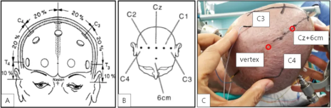
Figure 1. The current headset design implements the 10-20 system the internationally recognized method for placing electrodes on the human scalp in the context of EEG.
threshold stimulus)에서 다극성파형(polyphasic wave)과 좀 더 큰 진폭(amplitude)을 얻을 수 있다[4]. 만약 정확한 부위 에 삽입되지 않으면 검사가 위음성 또는 위양성이 되어 오히려 수술에 방해만 되는 검사가 될 수도 있다. 임상병리사는 비침습 적인 검사를 담당하고 있으나 INM은 예외로 침습적인 방법을 이용하고 있다.
다양한 수술 중 신경계 모니터링 검사들 마다 정확한 검사가 이루어지도록 구체적인 전극 삽입 방법과 부위, 매개변수 그리 고 전극제거 방법을 제시하여 안전하고 확실한 검사가 이루어 지는데 도움을 주고자 한다.
재료 및 방법
수술 중 신경계 감시 검사는 운동유발전위검사(motor evoked potential, MEP), 체성감각유발전위검사(somato- sensory evoked potential, SSEP), 청각유발전위검사 (brainstem auditory evoked potential, BAEP), 시각유발전 위검사(visual evoked potential, VEP), 자유진행 및 유발근 전도검사(free-running and triggered EMG)로 크게 분류할 수 있다[4, 5]. 이 검사 방법들은 수술의 종류 및 부위에 따라 적 용이 달라질 수 있다. 현재 우리나라에 수입된 장비중 the Xltek_ Protektor IOM system (Natus Medical Incorporated, Pleasanton, CA, USA)을 이용하여 각 검사마다 자극부위 (stimulation site)와 기록부위(recording site)에 대하여 해부 학적으로 알아보고 실제환자에 침 전극(Xi'an Friendship Medical Electronics Co., No. 9 Gao Xin 1st Road, Hi-Tech Development Zone, Xi'an, Shaanxi, China)을 삽입하는 과정도 자세히 언급하였다. 본 연구의 환자들은 수술 전 의료진에게 수술동의서를 작성하였으며, 수술동의서에 따라 수술 중 유발전위검사를 시행하였다. 신경계 검사자는 수술장 에서 파형 측정이 원활하지 않은 경우, 의사의 지도 하에 유발전 위의 검사 술기를 효율적으로 제안 및 개선하여 수행하였다.
결 과
1. 운동유발전위 검사(motor evoked potential, MEP) 1) 자극부위(stimulation site)
Limb MEP는 10-20 system의 C1-C2 또는 C3-C4 부위에 설치하고 facial MEP는 C3/C4-Cz 부위에 설치한다. C1-C2 부위에 자극을 주면 주로 하지 쪽 파형이 잘 측정되고, C3-C4 부위에 자극을 주면 상지의 파형이 잘 측정된다(Figure 1). 특별 히 다리 부위의 파형을 집중적으로 잘 관찰하기 위한 방법으로 는 vertex- Cz+6 cm 부위에 설치하여 자극을 주면 매우 잘 관 찰된다) [4-7]. 이러한 이유는 1차운동영역(primary motor cortex)으로 볼 때 뇌도(homunculus) 부위 중 C1-C2 (vertex- Cz+6 cm) 부위는 다리 쪽 영역이고, C3-C4 부위는 팔 쪽 영역으로 자극 시 해당 부위가 더 잘 흥분되기 때문이다 (Figure 2). 자극은 항전압(constant voltage) 방식 또는 항전 류(constant current) 방식으로 연속펄스(train)를 3∼7회 반복 하여 주며, 펄스간격(interpulse interval)은 1∼5 ms (250~1,000 Hz)으로 하고, 펄스기간(pulse duration)은 50∼
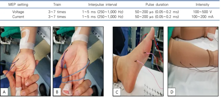
200 s (0.05~0.2 ms)로 한다. 민감도(sensitivity)는 70 V ~5 mV로 환자의 근이완 정도에 따라 범위를 선택할 수 있고, 자극 강도(intensity)는 파형이 형성되는 최저 자극으로 100∼500 voltage (또는 100~200 mA)로 검사한다(Table 1).
2) 기록부위(recording site)
좌, 우측 반구의 운동영역 부위의 두피에 전기자극을(anodal stimulation) 가하면 운동중추가 흥분을 하여 얼굴 및 상지와 하지의 근육으로 흥분된 전위가 전달된다. 상지에서는 승모근 (trapezius), 삼각근(deltoid), 이두근(biceps), 삼두근(triceps), 짧은엄지벌림근(abductor pollicis brevis, APB), 새끼벌림근 (abductor digitis quinti, ADQ) 중 하지에서는 대퇴근 (vastus), 앞정간근(tibialis anterior, TA), 엄지벌림근(abductor hallucis, AH) 중 얼굴에서는 눈둘레근(orbicularis oculi), 입
Table 1. Stimulation setting in motor evoked potential
MEP setting Train Interpulse interval Pulse duration Intensity
Voltage 3∼7 times 1∼5 ms (250∼1,000 Hz) 50∼200 s (0.05∼0.2 ms) 100∼500 V
Current 3∼7 times 1∼5 ms (250∼1,000 Hz) 50∼200 s (0.05∼0.2 ms) 100∼200 mA
Figure 3. Muscle for motor evoked potential, (A) insert electrode APB muscle, (B) insert electrode ADQ muscle, (C) insert electrode AH muscle, (D) insert electrode TA muscle.
Abbreviations: APB, abductor pollicis brevis; ADQ, abductor digitis quinti; AH, abductor halluces; TA, tibialis anterior.
Figure 2. Homunculus of penfield.
둘레근(orbicularis oris), 턱끝근(mentails), 저작근(masseter) 중 수술종류 및 부위에 따라 기록부위를 선택(필요에 따라 muscle 추가)하여 흥분된 근육의 파형을 기록한다. 하나의 근 육에는 2~4 cm정도 떨어진 두 바늘을 삽입하여 복합근활동전 위(compound muscle action potential, CMAP)를 기록한 다[4-7]. 하나의 근육에 전극간 간격차이가 있거나 근위축 (muscle atrophy)이 있을 경우 파형이 작거나 관찰되지 않을 수 있다.
실제로 환자의 얼굴 및 상지와 하지에 전극 삽입하는 과정은
알코올 솜으로 전극 삽입부위를 잘 닦아서 소독한 후 전극을 해 당 근육에 삽입하는데, 침전극의 끝부분이 몸의 안쪽으로 향하 도록 하여 삽입한다. 전극을 삽입할 때는 수술이 끝난 후 제거할 때도 미리 생각하여 삽입방향을 정해야 한다. 아무렇게나 전극 삽입을 하게 되면 전극 제거 시 침 전극의 바늘이 꺾이거나 휘어 지게 되어서 환자의 피부 및 근육이 손상될 수 있다. 그러므로 수 술 환자의 자세를 잘 파악하고, 전극의 제거까지 염두 해 두고 전 극을 삽입해야 한다(Figure 3).
2. 체성감각유발전위 검사(somatosensory evoked potential, SSEP)
1) 자극부위(stimulation site)
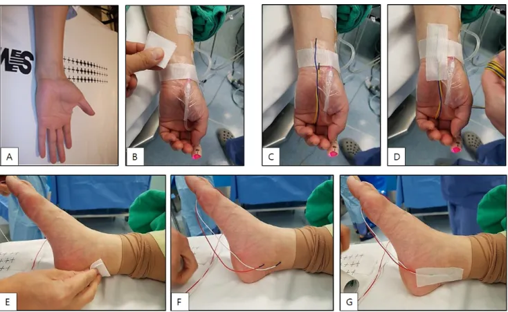
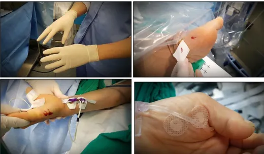
상지에서는 정중신경(median nerve), 하지에서는 뒤정강 신경(posterior tibial nerve)의 원위부를 자극하는 것이 일반 적이다. 자극은 항전류(constant current) 방식으로 5~30 mA 로 하고, 자극폭은 100∼300 s (또는 0.1~0.3 ms), 자극빈도 는 1.95∼5 Hz, 민감도는 1~10 V, 50~100번의 평균화 (averaging)로 검사를 한다(Figure 4, Table 2) [8, 9]. 검사 진 행은 좌상지, 우상지와 좌하지, 우하지 네 곳을 자동으로 장비에 서 순차적으로 자극하는 방식으로 동시에 사지의 파형을 기록하 며 분석한다.
Figure 4. Insert stimulator electrode for median nerve somatosensory evoked potential, (A) patient’s hand, (B) disinfection with alcohol cotton, (C) insert electrode SSEP stimulator, (D) tapping for fixing. Insert stimulator electrode for posterior tibialis nerve somatosensory evoked potential, (E) disinfection with alcohol cotton, (F) insert electrode SSEP stimulator, (G) tapping for fixing.
Abbreviation: SSEP, somatosensory evoked potential.
Table 2. Stimulation setting in somatosensory evoked potential
SSEP setting Electrical stimulation Stimulation rate Stimulation duration Intensity
Constant current 1.95∼5 Hz 100∼300 s (0.1∼0.3 ms) 5∼30 mA
Figure 5. Recording site in somato- sensory evoked potential, (A) insert the reference electrode into the frontal region of the Fpz, (B) insert the active electrode into the parietal region of the C3′, Cz′, C4′, (C) insert the active electrode into the cervical region of the C5s.
2) 기록부위(recording site)
기록전극은 10-20 system을 기준으로 하여 피질전위 (cortical potential)인 C3′ (C3보다 2 cm posterior 부위), Cz′ (Cz보다 2 cm posterior 부위), C4′ (C4보다 2 cm posterior 부위)와 피질하전위(subcortical potential)인 cervical 5 level (C5s)을 활동전극으로 하고 Fpz를 기준전극 으로 하여 파형을 기록한다. 피질하전위에서 기록한 파형은 마
취의 영향을 배제하기에 유용하다. 좌측상지 또는 우측상지를 자극하면 반대편 부위에서 파형이 우세하게 측정된다. 하지에 서는 Cz′-Fpz 채널에서 파형이 우세하게 측정된다. 상지와 하 지의 말초신경자극에 의해 측정하는 파형은 위에서 언급한 활동 전극 4개의 채널을 동시에 기록하게 되는데 자극 부위의 동측에 서 측정된 파형은 검사에 대한 대조군으로 의미를 갖는다 (Figure 5) [5].
Table 4. Stimulation setting in visual evoked potential
VEP setting Visual stimulation Stimulation rate Averaging time Timebase
Flash ≤1 Hz 50∼200 50 ms/div
Figure 6. Recording site in brainstem auditory evoked potential, (A) patient’s ear, (B) insert BAEP stimulator, (C) insert the active electrode into the earlobe, (D) tapping for fixing.
Table 3. Stimulation setting in brainstem auditory evoked potential
BAEP setting Sound stimulation Stimulation rate Averaging time Intensity
Click Sound 10∼45 Hz 300∼1,500 120∼140 dB SPL (90∼110 dB nHL)
3. 청각유발전위 검사(brainstem auditory evoked potentials, BAEP)
1) 자극부위(stimulation site)
일반적으로 소리자극은 주로 클릭 음(click sound)으로 왼 쪽, 오른쪽 각각 한 쪽씩 검사한다. 보통 1 kHz의 가청주파수를 이용하고 자극은 10 Hz의 빈도로 1,000번 이상 기록한다[10, 11]. 하지만 수술 중 청신경 손상여부는 빠르게 감지하는 것이 중요하므로 40 Hz 이상의 빠른 빈도와 400번 정도의 평균화로 10초 이내로 검사하는 새로운 검사방법도 있다. 민감도는 0.1~0.3 V로 하고, 자극의 극성은 교환전위(alternating polarity), 희박전위(rarefaction polarity), 압축전위(con- densation polarity) 중 파형 형성이 가장 좋은 것으로 선택 하 며, 일반적으로 교환전위(alternating polarity)가 artifact없 이 I파의 모양을 온전하게 관찰할 수 있고, 희박전위(rare- faction polarity)는 I, V파의 절대 지연(absolute latency)이 다른 전위 보다 짧아 이 두 가지 방법을 가장 많이 사용한다. 수술 중 신경손상으로 인하여 발생하는 파형 변화의 재현성을 높이기 위해 파형이 형성되는 최소자극 즉, 역치자극(Threshold
stimulation)으로 검사를 하지 않고 소리의 자극 세기는 120 dB SPL (sound pressure level) 또는 90 dB nHL (normal hearing level)로 자극을 준다[12, 13]. 수술중 파형의 재현성 이 낮아 질 경우 140 dB SPL (sound pressure level) 또는 110 dB nHL (normal hearing level)로 자극의 세기를 높여서 확인 하기도 한다. 하지만 이렇게 높여서 자극을 주면 고막에 소상이 발생할 수 도 있기 때문에 매우 일시적으로 잠시 확인하는 경우 에만 적용한다. 반대쪽 귀는 차폐음(masking sound)을 준다 (Table 3).
2) 기록부위(recording site)
양쪽 귓볼에 설치한 A1, A2 전극을 활성 전극으로 하고, 두정 부(vertex) 부위에 설치한 Cz 전극을 기준 전극으로 한다 (Figure 6) [5, 10-13].
4. 시각유발전위 검사(visual evoked potentials, VEP) 1) 자극부위(stimulation site)
고글안경을 통해 플래쉬(flash) 형태의 빛 자극을 반복해서 주며, 기록 방식은 1 Hz 이하의 빛 자극을 50~200회 주고 각각
Figure 8. A photograph with a direct precipitator inserted for observation about around the eyeballs. (A) Trochlear nerve (ICN IV), abducens nerve (ICN VI), (B) oculomotor nerve (ICN III), abducens nerve (ICN VI).
Figure 9. Special electrode attached to the endotracheal tube to observe vagus nerve (ICN X).
Figure 7. The back of the electrodes site in visual evoked potential. (A) Insert the active electrodes into the occipital region of the LO, MO, RO, (B) insert the reference electrodes into the mastoid region of the A1 and A2.
기록된 파형들을 평균화하여 민감도 5 V, 50 ms 척도로 기록 한다(Table 4).
2) 기록부위(recording site)
검사는 International 10-20 system보다 좀더 파형 형성이 원활하고 진폭도 큰 Queen square 전극 부착법을 이용하여, 뒤통수점(inion)에서 5 cm 상방에 middle occipital (MO), 좌측으로 5 cm, 우측으로 5 cm 지점에 left occipital (LO), right occipital (RO)에 부착을 하고, A1, A2를 기준전극으로 하여 측정한다(Figure 7) [5, 14-16].
5. 자유진행 및 유발근전도 검사(Free-running and Triggered EMG)
신경과 연관되어 있는 근육에서 발생하는 근전도의 파형은 대부분 뇌신경(intra cranial nerve, ICN)및 척수신경근 (spinal nerve root)의 상태파악에 활용된다. 즉, 해당 근육에 침전극을 삽입하여 자유진행 및 유발근전도 검사를 하며, 근전 도 검사에서 이상 파형이 관찰되면 수술로 인하여 신경이 영향 을 받고 있다는 것을 감지하게 되는 것이다[17-19].
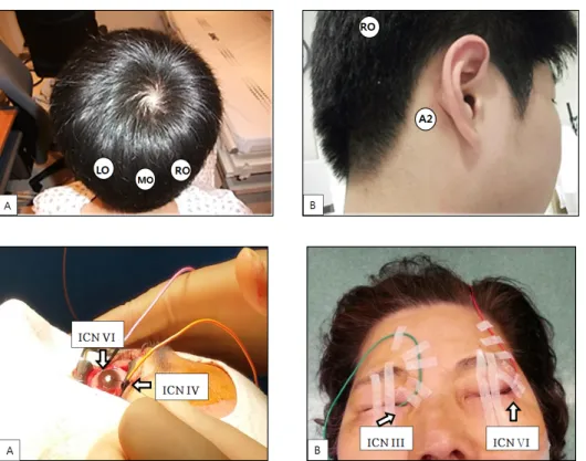
각각의 뇌신경과 관련된 근육들을 살펴보자면, 눈동자 움직 임과 관련된 신경으로 동안신경(ICN III, oculomotor nerve), 활차신경(ICN IV, trochlear nerve), 외전신경(ICN VI, abducens nerve)이 있고 안구 주위의 근육에 wire 또는 침전
극(needle electrode)을 삽입하거나 눈꺼풀 밖에 전극을 부착 하여 눈동자 움직임을 관찰하기도 한다(Figure 8) [20, 21].
삼차신경(ICN V, trigeminal nerve)은 저작과 관련된 근육 으로 temporalis, masseter 근육이 관여하며, 얼굴신경(ICN VII, facial nerve)은 frontalis, oculi, oris, mentalis 근육이 관여한다[5, 22].
미주신경(ICN X, vagus nerve)은 자율신경기능(autonomic nerve function)과 관련되어 있고, 기도 삽관에 부착되어 있는 특수 전극으로 관찰하며(Figure 9), 더부신경(ICN XI, accessory nerve)은 어깨의 승모근(trapezius)에 전극을 부착하여 관찰하
Figure 10. Electrodes attached to the back muscle to observe accessory nerve (ICN XI).
Figure 11. (A) Insert directly into the tongue to observe hypoglossal nerve (ICN XII), after electrodes are removed, there’s a lot of blood. (B) Insert a precipitate in a place corresponding to the root of the tongue below the chin without attaching it to the tongue to observe the hypoglossal nerve (easy to remove electric poles and little blood after removal).
Table 5. Muscle for electromyography in nerve root myotomal distribution
Spinal level Nerve root Muscle for electromyography
Cervical C2, 3, 4 Trapezius, SCM
C5, 6 Biceps, Deltoid
C6, 7 FCU, Triceps
C8, T1 APB, ADQ
Thoracic T5, 6 Upper rectus abdominis T7, 8 Middle rectus abdominis T9, 10, 11 Lower rectus abdominis Lumbar L2, 3, 4 Vastus lateralis/medialis
L4, 5 Tibialis anterior Sacral S1, 2 Gastrocnemius, Abductor hallucis
S3, 4, 5 External anal sphincter
고(Figure 10), 혀밑신경(ICN XII, hypoglossal nerve)은 혀 (tongue)에 침전극을 삽입하여 파형을 관찰한다(Figure 11) [23, 24].
뇌신경과 유사하게 척수신경근(spinal nerve root)과 관련 된 근육들을 살펴보자(Table 5).
위와 같이 분류되며 파형의 진폭은 적게는 5 V에서 크게는 100 mV 까지 다양하게 형성된다. 척수신경근과 관련한 근육은 더 자세하고 많이 구분되나, 일반적으로 수술장에서 사용되는 대표적인 근육만을 기술하였다[25, 26].
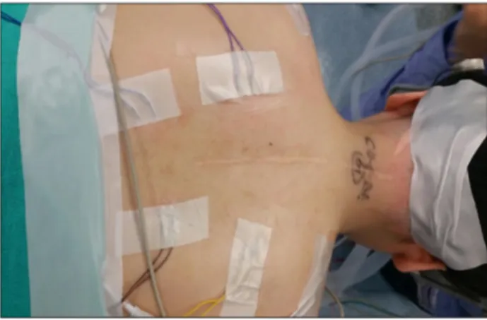
6. 검사 후 전극제거 방법
검사를 위하여 삽입하였던 침 전극들은 수술 이후 제거를 하 여야 한다. 전극이 바늘로 되어있기 때문에 제거하는 과정에서 피가 조금 날 수 밖에 없다. 그래서 전극제거 하기 전에 반드시 장갑을 착용하여 검사자의 감염관리에 문제가 발생하지 않도록 주의해야 한다. 경우에 따라서 혈관근처에 침전극이 설치되었 던 경우라면 전극제거 후 많은 양의 피가 날 수도 있지만 피부나 근육이 손상된 경우는 없었다. 이럴 경우에는 채혈 후 붙이는 지 혈용 밴드를 미리 준비하여 피가 나는 곳에 부착하면 지혈시키
는 시간도 단축할 수 있고, 피로 인한 환자 주변침대를 오염시키 는 것도 방지할 수 있어 매우 좋다(Figure 12).
고 찰
수술 중 신경계 감시는 수술 부위, 수술 방법에 따라 청각을 자 극하여 뇌간에서 발생하는 유발전위를 기록하는 검사로서 뇌간 의 기능을 간접적으로 평가할 수 있으며 진폭 등을 분석하여 청 신경의 이상유무를 감별할 수 있는 청각유발전위검사, 안구에 Goggle을 이용하여 빛 자극을 주어 뇌의 후두부에서 기록하는 방법으로 안구병변을 포함한 시신경교차전과 시신경교차후의 병변을 감별하고 그 병변(종양)을 제거할 때의 시신경 이상 유무 를 감별 할 수 있는 시각유발전위검사, 말초감각신경(median nerve, posterior tibial nerve)에 자극을 가하여 자극이 감각 로(sensory pathway)를 따라 전달되어 대뇌피질에서 파형을 기록하여 전기자극–말초신경–척수-대뇌피질로 전달되는 중추 신경계와 말초신경계의 기능을 모두 파악할 수 있는 체성감각유 발전위검사, 대뇌 좌우측 반구의 운동영역부위를 두피에서 자 극을 가하고 상지와 하지의 원위부 근육에서 파형을 기록하는
Figure 12. Make sure to wear gloves and attach a blood control band after removing the electrodes.
방법을 이용하여 운동신경경로의 이상을 파악할 수 있는 운동유 발전위검사, 상하지 근육 및 뇌신경에 의해 지배를 받는 근육(안 면신경 등)들에서 시행하여 수술 중 신경손상에 의하여 발생하 는 운동단위전위(motor unit potentials, MUP)를 기록하는 방법(free-running EMG)과 직접 신경근을 전기 자극하여 신 경에 이상이 없는지 확인하는 방법(triggered EMG)을 적극 활 용하여 보다 안전한 수술이 되도록 노력하고 있다[4, 5]. 예전보 다 수술의 기술적인 부분이 발달하면서 수술 중 신경계 감시 또 한 많은 발전을 하였다. 수술 중 얼굴 운동유발전위와 평균화 (averaging)의 단점을 최소화 한 청각유발전위검사는 얼굴신 경과 청각신경의 보존율을 높이는데 기여가 크다[6, 7, 12, 13].
그러나 시각유발전위검사는 현재까지도 만족할만한 결과를 얻 지 못하고 있어 많은 연구가 필요하다[14-16]. 기도 삽관에 부 착하는 방식이거나 일체형인 특수전극의 개발로 미주신경을 추 적감시 할 수 있어 목소리가 중요한 직업(가수, 성우, 성악가 등) 을 가진 환자들에게 도움을 주고 있다[23, 24]. 수술장 검사에서 는 환자가 마취되어 있어서 근육잡파(muscle artifact)의 혼입 을 최소화하기 위해 평균화 과정을 오래해야 할 필요는 없다. 하 지만 유발전위 검사를 원활하게 하도록 몇 분씩 수술을 하지 않 고 기다려 주지는 못한다. 특히 수술실에서는 마취 및 수술과 관 련하여 많은 장비들이 환자 주변에 배치되기 때문에 전기적 영 향을 쉽게 받게 된다. 전기소작기(bovie)를 매우 자주 사용하며 식염수를 지속적으로 뿌리고 흡입기로 흡입하는 일들이 너무나 빈번하게 이루어지므로 이러한 영향을 검사 파형은 받을 수 밖 에 없다[5]. 그러므로 수술실에서의 검사는 이러한 영향을 최소 화 하기 위한 방법으로 침전극(subdermal needle electrode) 을 사용한다. 그러나 수술실에서 이루어지는 검사라는 특수성
때문에 우리는 수술실 검사에 대하여 자세히 배우는 것이 힘들 고 실습을 하기도 매우 어려운 점이 있다. 또한 수술실에서 시행 하는 검사에 대하여 올바른 방법을 제시한 책자나 교재가 없는 실정이다. 본 논문이 여러 병원에서 시행하고 있는 수술 중 모니 터링 검사자에게 확실한 지침서 역할을 하길 바란다.
요 약
수술 중 발생하는 신경계 손상 여부를 감별하는 검사인 수술 중 신경계 모니터링(intraoperative neurophysiological monitoring, INM) 검사는 다양한 수술에서 안정적으로 수술 이 잘 진행되고 있음을 확신하며 수술을 진행할 수 있도록 도움 을 주는 매우 중요한 검사다. 수술실이라는 특수한 환경에서 검 사의 최적화를 위하여 침 전극을 사용하여 검사를 진행하며, 수 술실검사에 대하여 정확한 자극부위와 측정부위에 대한 교재나 안내책자가 없는 것이 실정이다. 그래서 이번 논문에서 운동유 발전위검사, 체성감각유발전위검사, 청각유발전위검사, 시각 유발전위검사에서 올바른 자극부위와 측정부위에 대하여 자세 하게 설명을 하였다. 그리고 자유진행 및 유발근전도검사 (free-running and triggered EMG)는 근육에서 발생하는 근 전도의 관찰로 대부분의 뇌신경(cranial nerve)과 척수신경근 (spinal nerve root)의 기능상태 파악을 한다. 검사의 이해를 돕기 위해 각각의 해당 근육에 전극을 삽입하는 사진을 첨부하 였고, 척수신경근에 따른 해당근육도 표로 제시하였다. 검사 후 전극제거를 할 때에도 환자와 검사자 모두 안전한 방법을 제시 하여 보다 완벽한 검사가 되었으면 한다.
Acknowledgements: None Conflict of interest: None
Author’s information (Position): Lim SH
1
, M.T.; Park SB2
, M.T.; Moon DY3
, M.T.; Kim JS4
, M.T.; Choi YD5
, M.T.;Park SK
1
, M.T.REFERENCES