BRCA1 Plays a Role in the Hypoxic Response by Regulating HIF-1 ␣ Stability and by Modulating Vascular Endothelial Growth Factor Expression
*Received for publication, December 6, 2005, and in revised form, March 8, 2006Published, JBC Papers in Press, March 16, 2006, DOI 10.1074/jbc.M513033200
Hyo Jin Kang‡, Hee Jeong Kim‡, Jeong-Keun Rih‡, Thomas L. Mattson‡, Kyu Won Kim§, Chi-Heum Cho¶, Jennifer S. Isaacs储, and Insoo Bae‡1
From the‡Department of Oncology, Lombardi Comprehensive Cancer Center, Georgetown University, Washington, D. C. 20057, the§Division of Pharmaceutical Bioscience, College of Pharmacy, Research Institute of Pharmaceutical Sciences, Seoul National University, Seoul 135-725, Republic of Korea,the¶Department of Obstetrics and Gynecology, Keimyung University School of Medicine, Daegu 700-712, Republic of Korea, and the储Department of Cell and Molecular Pharmacology, Medical University of South Carolina, Charleston, South Carolina 29425
A recent study of breast cancer patients with and without BRCA1 gene mutations found significantly lower levels of VEGF in serum from patients with BRCA1 mutations (Tarnowski, B., Chudecka-Glaz, A., Gorski, B., and Rzepka-Gorska, I. (2004) Breast Cancer Res. Treat. 88, 287–288). Here, we describe a pos- sible mechanistic explanation for this correlation. Because hypoxia in tumors stimulates VEGF expression and secretion we hypothesized that altered BRCA1 protein levels in breast tumors could affect hypoxia-stimulated VEGF promoter activity. This possibility was tested in cells transfected with various combina- tions of expression plasmids for BRCA1, BRCA1 specific inhibi- tory RNAs (BRCA1-siRNAs), HIF-1␣, and a VEGF promoter-re- porter and then incubated in normoxia (21%, O2) or hypoxia (0.1%, O2). As predicted, increased BRCA1 levels enhanced both hypoxia-stimulated VEGF promoter activity and the amounts of VEGF mRNA, as determined by semiquantitative RT-PCR and quantitative real time PCR. Using the ChIP assay, we discovered that BRCA1 could be recruited to the endogenous human VEGF promoter along with HIF-1␣ following hypoxia. An interaction between BRCA1 and HIF-1␣ was found in human breast cancer cells. We also found that hypoxia-stimulated VEGF promoter activity and secretion was reduced in cells containing reduced amounts of endogenous BRCA1 protein (obtained by transfect- ing with BRCA1 siRNAs). A mechanistic explanation for these effects is provided by our finding a reduced half-life and reduced accumulation of HIF-1␣ in hypoxic cells transfected with BRCA1-siR- NAs and that proteasome inhibitors blocked these effects of BRCA1- siRNAs. Thus, our results suggest that normal amounts of BRCA1 function in hypoxia to regulate HIF-1␣ stability, probably by inter- acting with HIF-1␣.
The gene encoding the breast cancer susceptibility gene 1 (BRCA1),2 which is frequently mutated in the germline of women with a genetic
predisposition to breast and ovarian cancers, was cloned in 1994 (1).
Although somatic mutations inactivating BRCA1 are rare in sporadic breast cancers, BRCA1 levels are often decreased or undetectable in cells of these cancers. These reduced BRCA1 levels, whether reflecting promoter methylation or other causes, suggests a role for normal levels of BRCA1 in protecting against non-hereditary tumors (2, 3). Although the specific functions of BRCA1 that contribute to tumor suppression remain unknown, functions related to its roles in regulating the cell cycle, transcription and DNA damage repair have been suggested (reviewed in Ref. 4).
Reports describing new roles for BRCA1 in regulating transcription activity continue to increase. The earliest evidence showed that a con- served acidic domain of BRCA1 can activate transcription in both yeast and mammalian cells when fused to the GAL4 DNA binding domain (5). Although BRCA1 is not known to bind directly to specific DNA sequences, it may regulate transcription through several indirect mech- anisms; by interacting with components of the basal transcription factor (e.g. RNA helicase A and RNA pol II), by interacting with transcription coactivators and corepressors (e.g. p300 and its functional homolog CBP (the CREB-binding protein), retinoblastoma 1 (RB1), by interacting with RB-associated proteins (RbAp46/48), by interacting with several histone deacetylases (HDAC-1/2), and/or by interacting with sequence- specific transcription factors (e.g. p53, c-Myc, estrogen receptor, and other proteins)) (6 –11).
Increased VEGF levels are considered to be an important marker of the aggressiveness of breast cancer. VEGF presents about 7-fold higher in tumor regions compared with normal adjacent breast tissue (12).
Multiple studies have shown that VEGF secretion by tumor cells is required for the early stages of breast tumor growth (13, 14). Consistent with these observations, other experiments have shown that inhibition of angiogenesis, by anti-angiogenic agents such as angiostatin and endostatin, resulted in reduced tumorigenesis and/or some regression of established tumors (15–18). In addition, VEGF expression was found to be essential for the survival of metastatic breast carcinoma cells (19).
VEGF expression is regulated by estradiol. A recent study shows that BRCA1 interacts with ER-␣ to inhibit estrogen-induced VEGF expres- sion and secretion (20).
In response to hypoxia, HIF-1␣, a major hypoxia-inducible transcrip- tion factor, becomes stabilized, translocates into the nucleus, binds to
*This work was supported by Grant E014440-01 (to I. B.) from the NIEHS, National Insti- tutes of Health and by the United States Department of Defense Army Idea Award (DAMD17-02-1-0525) (to I. B.) and the American Cancer Society (IRG 97-152-13) (to I. B.). The costs of publication of this article were defrayed in part by the payment of page charges. This article must therefore be hereby marked “advertisement” in accordance with 18 U.S.C. Section 1734 solely to indicate this fact.
1To whom correspondence should be addressed: Dept. of Oncology, Lombardi Compre- hensive Cancer Center, Georgetown University, 3970 Reservoir Rd., NW, Washington, D. C. 20057-1469. Tel.: 202-687-5267; Fax: 202-687-7256; E-mail: [email protected].
2The abbreviations used are: BRCA1, breast cancer susceptibility gene-1; ARNT, aryl hydro- carbon receptor nuclear translocator; ChIP, chromatin immunoprecipitation; qRT, quan- titative reverse transcriptase; HBC, hereditary breast cancer; HIF-1␣, hypoxia-
inducible transcription factor 1␣; IP, immunoprecipitation; VEGF, vascular endothelial growth factor; VHL, von Hippel-Lindau gene; WB, Western blotting assays; CHX, cyclohex- imide; GFP, green fluorescent protein; HRE, hypoxia responsive element; Luc, luciferase;
wt, wild type.
at KEIMYUNG UNIV MED LIB on January 7, 2016http://www.jbc.org/Downloaded from
ARNT, forming HIF-1␣䡠ARNT heterodimers (21). The heterodimeric complex acts as a transcription factor, regulating a variety of hypoxia- inducible target genes, including VEGF (22). In this study, we investi- gated whether and how BRCA1 affects hypoxia-induced VEGF expres- sion or secretion.
EXPERIMENTAL PROCEDURES
Cell Culture and Reagents—The human breast cell lines (MCF-7, MCF-10A, and T47D) were purchased from the American Type Cul- ture Collection, and the human embryonic kidney cell line (HEK293T) was obtained from the Tissue Culture Shared Resource, at Georgetown University (Washington, D. C.). All cell lines were grown in Dulbecco’s modified Eagle’s medium, fetal bovine serum (10%),L-glutamine (5 mM), non-essential amino acids (5 mM), penicillin (100 units per ml), and streptomycin (100 mg per ml). All cell culture reagents were obtained from Bio Whittaker (Walkersville, MD). Several proteasome inhibitors, including ALLN, MG115 and epoxomycin were purchased from Peptides International (Louisville, KY).
Subcellular Fractionation—Biochemical fractionation of the cells was done using the Nuclear Extract kit (Panomics) following the man- ufacturer’s protocol. MCF-7 cells grown in 100-mm tissue culture plates were transfected with siRNAs (control versus BRCA1) for 72 h and then incubated in hypoxia for 6 h, when the cells were washed twice with ice-cold phosphate-buffered saline and lysed on ice for 10 min in Buffer A mix (10 mMHEPES, pH 7.9, 10 mMKCl, 10 mMEDTA, 4% IGEPAL, 10 mMdithiothreitol, protease inhibitor mixture) with shaking. Cells were harvested and centrifuged at 15,000 rpm for 3 min. The superna- tant (cytoplasmic fraction) was collected in a tube; the nuclear pellet remained. The nuclear pellet was resuspended in Buffer B mix (20 mM HEPES, pH 7.9, 0.4MNaCl, 1 mMEDTA, 10% glycerol, 10 mMdithio- threitol, protease inhibitor mixture) was incubated on a rocking plat- form at 4 °C for 2 h. The mixture was centrifuged at 15,000 rpm for 5 min, and the supernatant (nuclear extract) was collected. 25g of the cell fractions were resolved on a 7.5% SDS-PAGE gel and probed with antibodies against BRCA1 and ARNT.
Quantitative Real Time PCR (qRT-PCR)— qRT-PCR was performed using ABI PRISM 7900HT Sequence Detection System (Applied Bio- systems, Foster City, CA) with a standard temperature protocol and 2⫻
SYBR Green PCR Master Mix reagent (Applied Biosystems) in a 20-l volume in triplicate as previously described (23). As a control, the mRNA level of-actin was determined in the real time PCR assay for each RNA sample and was used to correct for experimental variations.
The following primer sequences were used: VEGF121 forward primer was 5⬘-ccc tga tga gat cga gta cat ctt-3⬘, and VEGF121 backward primer was 5⬘-gcc tcg gct tgt cac att tt-3⬘. The VEGF121 forward primer was also used as the VEGF165 forward primer. The VEGF165 backward primer was 5⬘-agc aag gcc cac agg gat tt-3⬘. VEGF121 and VEGF165 are two most abundant VEGF mRNA isoforms.
Semiquantitative RT-PCR—Semiquantitative reverse transcription- PCR assays were performed as described previously (24). -Actin, whose expression is unaffected by BRCA1 overexpression or hypoxia, was used as a loading control. The PCR products were loaded on a 1%
agarose gel, stained with ethidium bromide (0.1 mg per ml), and photo- graphed with a digital camera under UV illumination.
Transient Transfection of DNA and Luciferase Assays—The HIF-1␣
expression vector (pCEP4-HIF-1␣), the VEGF promoter-reporter plas- mid (pVEGF-kpnI-Luc containing the 2.6-kb VEGF promoter in pGL2), p11wt (the VEGF promoter containing the wild-type HRE in the pGL2 vector), and p11mt (the VEGF promoter containing a mutated HRE in the pGL2 vector) were purchased from ATCC. For assaying promoter
activity, subconfluent proliferating cells in 24-well plates were trans- fected with various expression vector(s) (125 ng per well) and luciferase reporter plasmid (125 ng per well), unless indicated otherwise in the figure legends, using Lipofectamine Plus (Invitrogen). Luciferase activ- ity values (minus background) were normalized to the control (reporter only) and expressed as means⫾ S.E. of quadruplicate wells. We repeated these promoter reporter experiments at least three times. Measured lucif- erase activity levels were normalized to relative transfection efficiencies, which were monitored using the control plasmid pCMV--gal as previ- ously described (24).
Transfection of siRNAs—The BRCA1-siRNA-1, BRCA1-siRNA-3, and control (scrambled)-siRNA were previously described (24, 25).
These siRNAs were chemically synthesized and provided by Dharma- con (Boulder, CO). For siRNA treatments, subconfluent proliferating cells were transfected with each siRNA (50 nM), using Lipofectamine 2000. The cells were incubated with siRNA for 48 or 72 h prior to the start of the experiment (cotransfection with fresh siRNA and a reporter
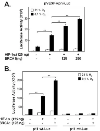
FIGURE 1. Effects of BRCA1 and HIF-1␣ on hypoxia-induced VEGF promoter activity.
A, effect of exogenous BRCA1 and HIF-1␣ on VEGF reporter activity. Exponentially pro- liferating cells (MCF-7) were transfected with various combinations of pVEGF-kpnI-Luc (125 ng), HIF-1␣ (125 ng), and either of two doses of a BRCA1 expression vector (125 ng or 250 ng), incubated for 24 h and then exposed to hypoxia (0.1%, O2) for 6 h, harvested, and assayed for luciferase activity as described under “Experimental Procedures.” Trans- fection of HIF-1␣ alone into MCF-7 cells induced statistically significant pVEGF-kpnI-Luc activity under hypoxic condition ( p⬍ 0.005 for comparisons of cells transfected with or without the HIF-1␣ expression vector in 0.1% O2) and co-transfections with both the BRCA1 and HIF-1␣ expression vectors further enhanced this expression ( p ⬍ 0.005 for comparisons between cells transfected with and without the BRCA1 expression vector, in the presence of exogenous HIF-1␣). B, role of the HRE in BRCA1-enhanced, HIF-1␣- induced VEGF promoter activity during hypoxia. To determine if the ability of BRCA1 to affect VEGF promoter activity during hypoxia might require interaction between the HRE and HIF-1␣, cells were transfected with either p11wt-Luc, p11mt-Luc, or empty vector (125 ng) plus HIF-1␣ and BRCA1 expression vectors (125 ng of each) as indicated in the figure, incubated for 24 h, and then exposed to hypoxia (0.1%, O2) for 6 h before meas- uring luciferase activities. As in A, BRCA1 significantly enhanced HIF-1␣-induced Luc activity under hypoxic conditions when the VEGF promoter contained a wild-type HRE ( p⬍ 0.005 for comparisons of cells transfected with and without the BRCA1 expression vector). In contrast, neither HIF-1␣ alone nor HIF-1␣ plus BRCA1 induced statistically significant increased amounts of reporter activity in cells transfected with the HRE mutant reporter plasmid (p11mt-Luc).
at KEIMYUNG UNIV MED LIB on January 7, 2016http://www.jbc.org/Downloaded from
plasmid as described in the figure legends). The control siRNA had no effect on BRCA1 levels and siRNAs (BRCA1 or control) were not toxic to the cells under these experimental conditions, as determined using 3-(4,5-dimethylthiazol-2-yl)-2,5-diphenyltetrazolium bromide (MTT) assays (data not shown). Alternatively, we used DNA-based siRNA (pKD-negative control (NC) empty vector and pKD-BRCA1 siRNA expression vector) (Upstate Biotechnology).
Immunoprecipitation (IP) and Western Blotting (WB)—All IP proce- dures were carried out at 4 °C. Cells grown on 100-mm dishes were washed twice with 1⫻ PBS before lysis. Then, cells were lysed in a buffer (50 mMTris, pH 8.0, 150 mMNaCl, 1% Nonidet P-40) and immunopre- cipitated with either anti-HIF-1␣ rabbit polyclonal antibody (H-206, Santa Cruz Biotechnology) or a combination of anti-BRCA1 mouse monoclonal antibodies against N- and C-terminal epitopes on BRCA1 (Ab-1⫹ Ab-2 ⫹ Ab-3, Oncogene Research Products) as previously described (11). Immunoprecipitated proteins were run on SDS-PAGE gels, and Western blots were analyzed using anti-HIF-1␣ mouse mono- clonal antibody (Transduction Laboratories) and anti-BRCA1 rabbit polyclonal antibody (C-20, Santa Cruz Biotechnology) as the primary antibodies. Anti-GFP mouse monoclonal antibody (BD Sciences, Inc) was also used for IP and WB to detect transfected GFP-HIF-1␣. The membranes were then washed and incubated with horseradish peroxi- dase-conjugated secondary antibodies for 1 h at room temperature. The
bound secondary antibodies were visualized by enhanced chemilumi- nescence (ECL) (Santa Cruz Biotechnology) detection kits using Fuji x-ray films.
Chromatin Immunoprecipitation (ChIP) Assay—The ChIP assay was performed according to the protocol provided with the ChIP assay kit (Upstate Biotechnology). MCF-7 and HEK293T cells exposed to hypoxic gas (0.1%, O2) for the times indicated in the figure legends were cross-linked by adding formaldehyde to a final concentration of 1% and incubating at 37 °C for 10 min. Then, cells were washed with 1⫻ phos- phate-buffered saline and sonicated to yield DNA fragments ranging in size from 200 to 1000 base pairs. Sonicated samples were centrifuged for 10 min at 4 °C (13,000 rpm), supernatants recovered, diluted 10-fold into ChIP dilution buffer, and then precleared by incubating with 75l of salmon sperm DNA/protein A-agarose-50% slurry for 30 min at 4 °C with agitation to reduce the nonspecific interaction of chromatin DNA with the agarose. Supernatants were immunoprecipitated with each antibody (ARNT, HIF-1␣, BRCA1) or control IgG. The anti-ARNT rab-
FIGURE 2. Effects of BRCA1 on the endogenous levels of hypoxia-induced VEGF121 and VEGF165 mRNA. A, agarose gel analysis. Exponentially proliferating HEK293T cells were transiently transfected with empty vector (pCDNA3) or the BRCA1 expression vec- tor, incubated for 24 h and then exposed to hypoxia (0.1%, O2) for 6 h when total RNA was extracted used for semiquantitative RT-PCR. The gel image presented is representative of three independent experiments. B, quantitative analysis of agarose gel densitometry data. Each PCR band on photographs of the three agarose gels was quantitated using densitometry, and means⫾ S.E. of these values were calculated. BRCA1 overexpression significantly enhanced hypoxia-induced VEGF121 and VEGF165 mRNA expression ( p⬍
0.005 for comparisons of cells transfected with or without BRCA1 in 0.1% O2). C, confir- mation of PCR results. Real time PCR were performed in quadruplicate in three inde- pendent experiments to confirm the findings shown in A and B.-Actin was used as the loading control for semiquantitative RT-PCR and to normalize the quantitative real time PCR results. The results were normalized to the control (reporter only) in 21% O2and are presented as bar graphs showing the means⫾ S.E. The effect of overexpressing BRCA1 on hypoxia-induced VEGF expression is statistically significant for both VEGF mRNAs ( p⬍ 0.005).
FIGURE 3. The effect of hypoxia on ChIP assays for genomic VEGF promoter DNA. A, agarose gel analysis of PCR products. Exponentially proliferating cells exposed to hypoxia for the times indicated were harvested for ChIP assays as described under
“Experimental Procedures.” (Protein-DNA cross-linked samples were immunoprecipi- tated with the indicated antibodies or control IgG, processed, and used as templates for PCR reactions to measure relative amounts of the HRE element-containing region of the genomic VEGF promoter DNA that had been immunoprecipitated.) B, densitometer trac- ings of the results in A. The amount of each PCR product band was quantitated by densitometry for three independent experiments, and means⫾ S.E. were calculated.
Statistically significant increases ( p⬍ 0.005) of VEGF promoter DNA were observed for chromatin immunoprecipitated with the BRCA1, ARNT, and HIF-1␣ antibodies in hypoxic versus normoxic conditions. C, negative controls: as in A except that the primers were for genomic VEGF promoter sequences lacking known HRE sites.
at KEIMYUNG UNIV MED LIB on January 7, 2016http://www.jbc.org/Downloaded from
bit polyclonal antibody was H-172 (Santa Cruz Biotechnology). The antibodies for HIF-1␣ and BRCA1 were the same as used for IPs (see above). Immunoprecipitated complexes were eluted from the beads (with 1% SDS, 0.1MNaHCO3), the DNA was separated from protein (as described in the ChIP kit), and used as templates for PCR reactions.
The genomic primer sequences, designed to amplify the HRE element- containing region of the VEGF promoter, used as previously described (27), were: VEGF forward primer (5⬘-aca gac gtt cct tag tgt tgg-3⬘) and VEGF reverse primer (5⬘-agc tga gaa cgg gaa gct gtg-3⬘). As negative controls, we used two sets of primers for regions of the genomic VEGF promoter that do not have any known HRE sites, as follows: for NC-1 (negative control primer set 1): forward primer (5⬘-gga cac cat acc gat gga ac-3⬘) and reverse primer (5⬘-ccc ctt ttc ctc caa ctc tc-3⬘) and for NC-2 (negative control primer set 2): forward primer (5⬘-gaa ttc tgt gcc ctc act cc-3⬘) and reverse primer (5⬘-gta gac atc ttg ggg cag ga-3⬘).
VEGF Secretion Assay—Cells were transiently transfected with either control (scrambled)-siRNA or BRCA1-siRNA as previously described (24). The next day the medium was changed to serum-free Dulbecco’s modified Eagle’s medium. After 48 h, cells were exposed to hypoxic gas (0.1%, O2) for an additional 16 h when the supernatants were collected for a VEGF secretion assay, which was measured using a Quantikine human VEGF ELISA immunoassay (R&D Diagnostics, Minneapolis,
MN). The values obtained were normalized to the total protein concen- tration in the total cell extracts prepared from each dish.
Hypoxic Treatment—Human cells were dispensed into a 100-mm culture dishes. The dishes were placed in a sealed hypoxia chamber (Billups-Rothenberg, Del Mar, CA) equilibrated with a humidified 5%
CO2atmosphere or with certified gas containing 0.1% O2, 5% CO2, and 94% N2(28).
BRCA1 Mutants—The panel of BRCA1 mutant constructs are shown in Fig. 4A. The 5382insC mutant (Q1756term), which is commonly found in Ashkenazi Jews, a population with a significantly increased risk for BRCA1 mutant breast cancers (29 –31), encodes a truncated protein missing part of the C-terminal transcriptional activation domain (TAD).
The C5365G mutant, (p1749R) encodes a full-length BRCA1 protein con- taining a point mutation in the C-terminal TAD region that abolishes TAD activity (5). The 5677insA mutant, (Y1853term), encodes a near full-length BRCA1 protein that is missing only the last 11 amino acids. This muta- tion reduces TAD activity. The T300G mutant (61Cys3 Gly), encodes a full-length protein whose single point mutation inactivates the N-ter- minal RING finger domain function (11).
Statistical Methods—Statistical comparisons (of densitometer trac- ings of digital images (of either agarose gels or x-ray films) or of graphically determined half-lives from each of three independent experiments) were
FIGURE 4. Effect of BRCA1 mutants on hypoxia- stimulated VEGF promoter activity. A, genetic maps. Diagrams of wtBRCA1 and mutant BRCA1 (5382insC, C5365G, 5677insA, and T300G) expres- sion vector constructs. B, effect of BRCA mutants on reporter activity. MCF-7 cells were co-trans- fected with pVEGF-kpnI-Luc, HIF-1␣, and either wtBRCA1 or one the mutant BRCA1s as indicated.
24 h later, cells were treated with or without 0.1%
hypoxic gas for 6 h. pCMV--gal and pCDNA3 vec- tors were included as controls for data normaliza- tion. The data are presented as bar graphs of the means⫾ S.E. of quadruplicate wells of three inde- pendent experiments. C, Western blot of the rela- tive wild-type and mutant BRCA1 protein levels in the cells analyzed in B.
at KEIMYUNG UNIV MED LIB on January 7, 2016http://www.jbc.org/Downloaded from
made using the two-tailed Student’s t test where appropriate. The symbols used in the figures (*) and (**), indicate p⬍ 0.05 and p ⬍ 0.005, respectively.
RESULTS
Effects of BRCA1 on HIF-1␣ Transcriptional Activity—In this study, a concentration of 0.1% O2was used to induce hypoxia. This hypoxic condition is physiologically relevant, for some breast cancer tissues have extremely low O2concentrations (0 to 0.3% O2) (32–34). To determine if BRCA1 affects hypoxia-induced VEGF expression, we used a VEGF promoter-reporter system. Following transient co-transfection with HIF-1␣ and pVEGF-kpnI-Luc, reporter plasmid activity was measured after 6 h of hypoxia. Higher reporter activity under our hypoxic condi- tions was observed in extracts of HIF-1␣-transfected cells than in extracts of cells lacking the HIF-1␣ expression vector. Co-transfection of MCF-7 cells with BRCA1 and the HIF-1␣ expression vector further enhanced activity from the VEGF promoter construct (Fig. 1A). The amount of this effect depends on the amount of BRCA1 plasmid DNA (Fig. 1A). Thus, under hypoxia, a statistically significant effect on reporter activity was observed when only HIF-1␣ was overexpressed, and this effect was significantly more enhanced when BRCA1 was also overexpressed. To determine if this ability of BRCA1 to enhance HIF- 1␣-induced VEGF promoter activity requires the presence of a func- tional HRE (hypoxia response element) in the VEGF promoter, cells were co-transfected with three plasmids, BRCA1, HIF-1␣, and a VEGF reporter plasmid containing either a wild-type HRE (p11wt) or an HRE containing a point mutation (p11mt) and then incubated under hypoxic conditions and assayed for luciferase reporter activity. The results show that exogenous BRCA1 has no independent ability to enhance activity from a VEGF promoter under hypoxic conditions when this promoter contains a mutation blocking its responsiveness to exogenous HIF-1␣
(Fig. 1B). This finding suggests that BRCA1 may alter VEGF promoter activity via protein-protein interactions rather than by protein-DNA interactions.TodirectlydeterminewhethertheBRCA1-enhanced,HRE- dependent VEGF reporter activity reflects, at least in part, increased transcription, we measured the effect of overexpressed BRCA1 on endogenous VEGF mRNA levels. For this test, HEK293T cells were transfected with BRCA1 or empty vector and exposed to hypoxic con- ditions for 6 h when total RNA was isolated and used for semiquantita- tive RT-PCR (Fig. 2, A and B) and quantitative real time PCR (Fig. 2C).
As expected, we found that BRCA1 increased the amounts of the two most abundant endogenous isoforms of VEGF mRNA (VEGF121 and VEGF 165) in cells exposed to hypoxic conditions (Fig. 2) ( p⬍ 0.005 for comparisons of cells transfected with pCDNA3 versus BRCA1 under hypoxia).
BRCA1 Is Recruited to VEGF Promoter Regions—ChIP was used to identify and quantitate interactions of proteins with specific genomic regions. Because BRCA1 is known to regulate transcriptional activity by directly interacting with various transcription factors, it is possible that BRCA1 (and other factors) are recruited to the endogenous VEGF pro- moter during hypoxia. Indeed, significantly increased binding of HIF- 1␣, ARNT, and BRCA1 to the promoter region of the endogenous (wild type, HRE-containing) VEGF promoter was found in both MCF-7 and HEK293T cells after either 6 or 16 h of hypoxia (Fig. 3, A and B) ( p⬍ 0.005 for each protein in cells incubated in hypoxia versus normoxia).
The negative control, rabbit IgG, could not precipitate detectable amounts of chromatin containing VEGF promoter DNA. Combined, these results suggest that the observed enrichment of VEGF promoter sequences in ChIPs precipitated by specific antibodies reflect increased interaction between the targeted proteins and genomic VEGF promoter DNA. Our two sets of negative control primers (VEGF-NC1 and VEGF-
NC2), from regions of the genomic VEGF promoter that do not have any known or putative HRE binding sites, did not yield detectable amounts of VEGF promoter DNA under any of the tested conditions (Fig. 3C).
BRCA1 Sequences Important for Regulating the Enhanced VEGF Pro- moter Activity—To begin identifying regions(s) of BRCA1 important for regulating VEGF-reporter plasmid activity under hypoxic conditions we used the panel of BRCA1 mutant constructs shown in Fig. 4A. Two of the BRCA1 mutants (5677insA and C5365G) significantly (but only partially) impaired BRCA1 ability to enhance hypoxia-mediated VEGF expression ( p⬍ 0.005 or 0.05), one (T300G) was completely defective ( p⬍ 0.005) and one (5382insC) had no detectable defect in stimulating hypoxia-induced VEGF (Fig. 4B). These differences cannot be explained simply as differences in BRCA1 protein levels produced by the different expression vectors (Fig. 4C).
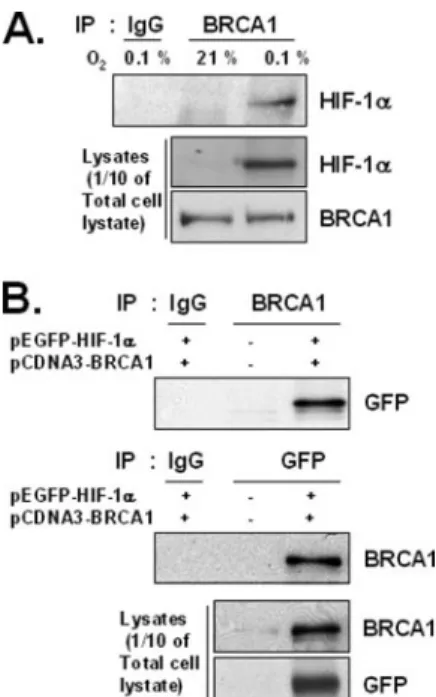
BRCA1 and HIF-1␣ Interact—Because BRCA1 can regulate tran- scription activity via physically interacting with various transcription factors, we looked for potential interactions between endogenous BRCA1 and endogenous HIF-1␣ by using standard IP-Western blot (WB) analysis. Following IP performed with anti-BRCA1 antibodies the precipitated proteins were analyzed by WB. HIF-1␣ protein was detected in the total lysates only in samples from cells incubated in the hypoxic condition (Fig. 5A). This hypoxia-dependent interaction did not significantly affect total BRCA1 protein levels (although as expected, hypoxia did increase HIF-1␣ levels in this experiment). As a positive control, to be sure that our IP assay could detect BRCA1- HIF-1␣ interactions under normoxic conditions, if they occurred, the BRCA1 expression vector was co-transfected with a GFP-HIF-1␣
expression vector into HEK293T cells. Cell lysates were prepared and precipitated with either an anti-GFP antibody or anti-BRCA1 antibod- ies. Western blot analysis revealed that BRCA1 was present in IP com- plexes precipitated with the anti-GFP antibody and that GFP-HIF-1␣
FIGURE 5. Interactions between BRCA1 and HIF-1␣ under hypoxic and normoxic conditions. A, interactions between endogenous proteins. Total extracts of MCF-7 cells incubated in either normoxia (21%, O2) or hypoxia (0.1%, O2) for 6 h were immunopre- cipitated using anti-BRCA1 antibodies. The presence of HIF-1␣ or BRCA1 in the immuno- precipitated complexes was determined by Western blot analysis. B, interactions between exogenous proteins. Total normoxic extracts of HEK293T cells were prepared 24 h after co-transfection with GFP-HIF-1␣ and BRCA1 expression vectors and immuno- precipitated with either anti-GFP or anti-BRCA1 antibodies. The immunoprecipitates were analyzed by our standard Western blot method. at KEIMYUNG UNIV MED LIB on January 7, 2016http://www.jbc.org/Downloaded from
was present in IP complex precipitated with the anti-BRCA1 antibodies (Fig. 5B). However, neither protein was detected in samples precipi- tated with control antibodies (mouse IgG) or in samples transfected with empty vector (GFP), suggesting that normoxic conditions, per se, do not block BRCA1 and HIF-1␣ from being able to interact, directly or indirectly.
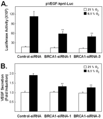
Endogenous BRCA1 Levels Affect Hypoxia-induced VEGF Promoter Activity and Endogenous VEGF Protein Secretion—We employed small inhibitory RNA (siRNA) to evaluate whether endogenous BRCA1 protein levels affect hypoxia-induced VEGF gene expression and secretion (Fig. 6).
We designed two BRCA1-siRNAs (BRCA1-siRNA-1 and BRCA1-siRNA- 3), complementary to two different regions of the BRCA1 gene, to avoid potential artifacts resulting from inappropriate siRNA sequences (35). Cells transfected with siRNA for 48 h were then co-transfected with the pVEGF- kpnI-Luc reporter and fresh siRNA, incubated for 24 h and then exposed to our standard hypoxic condition for an additional 6 h, when luciferase activ- ity was measured. Both BRCA1-siRNA constructs significantly reduced hypoxia-induced VEGF-Luc reporter activity when compared with the control (Fig. 6A; p⬍ 0.005). In addition, reducing endogenous BRCA1 levels by either siRNA construct also reduced secretion of endogenous VEGF protein following hypoxia (Fig. 6B; (p⬍ 0.005). However, this reduced VEGF secretion may reflect, at least partially, reduced VEGF induction in these BRCA1-depeleted hypoxic cells. Note that BRCA1 siRNA treatments did not significantly reduce either total or secreted levels of VEGF (or VEGF promoter activity) in the absence of hypoxia. The molecular basis for the apparent BRCA1 level, independence of normoxic VEGF levels, has not been investigated.
BRCA1 Regulates HIF-1␣ Stability—Proteasome-mediated degrada- tion is known to regulate HIF-1␣ protein stability (36). In particular, the rapid degradation of HIF-1␣ that occurs under normoxia is blocked under hypoxic conditions (36). These observations suggest a mecha- nism for how BRCA1 levels could affect HIF-1␣-dependent transcrip- tional activity during hypoxia: perhaps BRCA1 influences HIF-1␣ sta- bility under hypoxic conditions. To test this hypothesis, we first examined the effect of BRCA1-siRNAs on the steady state levels of endogenous HIF-1␣ protein. In the absence of hypoxia, BRCA1 siRNA
FIGURE 6. The effect of endogenous BRCA1 levels on hypoxia-induced VEGF pro- moter activity and VEGF secretion. A, effect of BRCA1 siRNAs on VEGF promoter activ- ity. HEK293T cells were transiently transfected with control-siRNA, BRCA1-siRNA-1 or BRCA1-siRNA-3 for 48 h when the remaining siRNA was removed. The cells were then immediately co-transfected with the pVEGF-kpnI-Luc reporter construct and the same siRNAs, (freshly prepared), incubated for 24 h, and then exposed to hypoxia for 6 h before luciferase activity was measured. The activity levels, relative to the control luciferase activity (control-siRNA in normoxia), are presented as bar graphs of the means⫾ S.E. of four wells from each of three independent experiments. The effects of both BRCA1 siR- NAs on hypoxia-induced VEGF-Luc reporter activity was statistically significant ( p⬍ 0.005). B, effect of BRCA1 siRNAs on hypoxia-induced secretion of endogenous VEGF.
VEGF secretion into the medium was measured with a VEGF ELISA kit (see “Experimental Procedures”). Cells (T47D) transiently transfected with control-siRNA, BRCA1-siRNA-1, or BRCA1-siRNA-3 for 48 h were exposed to hypoxia or normoxia for an additional 16 h (without removing the siRNAs) when the amount of VEGF secreted into the medium was measured. The values presented as bar graphs give the means⫾ S.E. of quadruplicate wells from three independent experiments. The effects of both BRCA1 siRNAs on the secretion of endogenous VEGF protein are statistically significant under hypoxic condi- tions ( p⬍ 0.005) but not under normoxic conditions.
FIGURE 7. The effect of endogenous BRCA1 pro- tein levels on HIF-1␣ inducibility under hypoxic conditions. A, exponentially proliferating MCF-7 cells transiently transfected with 50 nMof either con- trol-siRNA or BRCA1-siRNA-1 for 72 h were exposed to hypoxic stress (and fresh siRNAs) for an additional 16 h, when nuclear (NE) and cytosol extracts (CE) were prepared as described under “Experimental Procedures” and analyzed by Western blots. B, results from A and two additional independent experiments were quantitated by densitometry and are presented as bar graphs of means⫾ S.E. C, expo- nentially proliferating MCF-10A cells were trans- fected with siRNA (BRCA1 versus control) for 72 h and then exposed to hypoxia for 0, 8, or 24 h before total cell extracts were prepared and used for Western blot analysis with anti-BRCA1 and anti-HIF-1␣ anti- bodies.-actin antibody was used for the transfer and loading control. D, results from C and two addi- tional independent experiments were quantitated and are presented as bar graphs of the means⫾ S.E.
The Western blots shown in A and C are representa- tive of triplicate experiments. Reduced HIF-1␣ inducibility following hypoxic stress in BRCA1-de- pleted cells, compared with the control, was statisti- cally significant ( p⬍ 0.005).
at KEIMYUNG UNIV MED LIB on January 7, 2016http://www.jbc.org/Downloaded from
treatment had little or no effect on basal HIF-1␣ levels. However, under hypoxic conditions, and as expected, BRCA1-siRNA pretreatments, of either MCF-7 (Fig. 7, A and B) or MCF-10A (Fig. 7, C and D) cells, significantly reduced subsequent hypoxia-induced HIF-1␣ accumula- tion, either in nuclear extracts (Fig. 7B) or in total cell extracts (Fig. 7D;
p⬍ 0.005 for comparisons of cells transfected with control-siRNA ver- susBRCA1-siRNA).
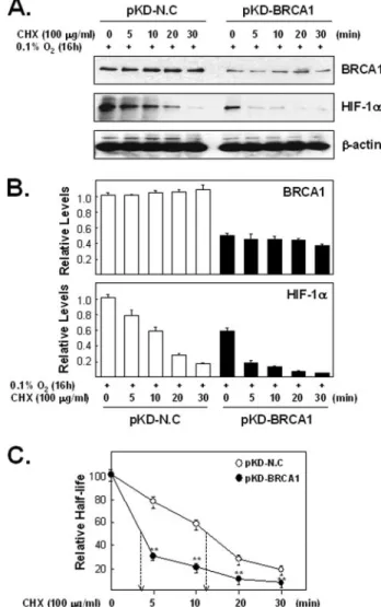
We then tested whether the effect of BRCA1 on HIF-1␣ accumula- tion under hypoxic conditions could reflect altered HIF-1␣ stability.
Cells transfected with BRCA1-siRNAs were exposed to hypoxia for 20 h and then treated with various proteasome inhibitors for an additional 4 h under hypoxic conditions (Fig. 8). Each proteosome inhibitor treat- ment significantly increased HIF-1␣ accumulation (in comparison to Me2SO alone) only in cells containing reduced endogenous BRCA1 levels (Fig. 8; p⬍ 0.005). This finding suggests that reducing BRCA1 levels under hypoxic conditions contributes to a more rapid degradation of HIF-1␣. To directly test whether reduced BRCA1 levels in hypoxic cells is associated with decreased HIF-1␣ stability, we measured HIF-1␣ half-lives
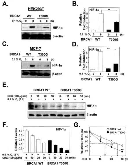
(Fig. 9). For this test cells were transfected with expression plasmids encod- ing either control-siRNA (pKD-NC) or BRCA1-siRNAs (pKD-BRCA1), then exposed to hypoxia and then treated, under hypoxia, with cyclohexi- mide for up to 30 min. Indeed, reducing the endogenous BRCA1 level had a rapid and significant effect on the stability of HIF-1␣ under hypoxic con- ditions (p⬍ 0.005 for the difference between hypoxic cells with and with- out BRCA1 knockdown at each time point after adding 100g/ml cyclo- heximide). Consequently, reducing endogenous BRCA1 levels also significantly shortened the HIF-1␣ half-life under hypoxic conditions (p ⬍ 0.005). Finally, since reduced levels of BRCA1 reduce HIF-1␣ half-lives under hypoxic conditions, we also tested whether increasing the level of BRCA1, by transient transfection, would increase the stability of endoge- nous HIF-1␣. For this test (Fig. 10) we transfected cells with expression vectors for either wtBRCA1 or the T300G BRCA1 mutant and measured HIF-1␣ half-lives under hypoxic conditions as described for the Fig. 9 experiment. The T300G mutant did not increase VEGF promoter activity
FIGURE 8. The effect of proteasome inhibitors on endogenous HIF-1␣ protein levels in hypoxic cells containing reduced levels of endogenous BRCA1 protein. A, MCF-7 cells transiently transfected with control-siRNA or BRCA1-siRNA-1 for 72 h, were exposed to hypoxia for 20 h. when various proteasome inhibitors, including ALLN (50M), MG115 (10M), and epoxomycin (1M), were added, and the hypoxic condition continued for an additional 4 h. The accumulation of endogenous HIF-1␣ protein with or without various proteasome inhibitors was determined by using an anti-HIF-1␣ antibody in Western blot analysis. The effects of the BRCA1-siRNA treatments on BRCA1 protein levels were mon- itored on Western blots with an anti-BRCA1 antibody (C-20). A-actin antibody was used to normalize densitometer tracing values for differences in loading and transfer efficien- cies. This experiment was repeated three times with similar results. B, bar graphs pre- senting calculated means⫾ S.E. of densitometer tracings values from three independent experiments of A. Each proteasome inhibitor (in comparison to Me2SO alone) signifi- cantly increased HIF-1␣ accumulation in cells transfected with BRCA1 siRNA but not in cells transfected with control siRNA ( p⬍ 0.005).
FIGURE 9. The effect of endogenous BRCA1 levels on the half-life of HIF-1␣ in hypoxic cells. A, lysates of cells transfected with DNA-based siRNA expression vectors (pKD-NC versus pKD-BRCA1) for 72 h, then exposed to hypoxia for 16 h, and then treated with CHX (100g/ml) for the indicated time periods under hypoxic were used for West- ern blotting analysis with anti-HIF-1␣ and anti-BRCA1 antibodies. B, mean intensities (⫾ S.E.) of the HIF-1␣ signals from triplicate experiments was calculated from densitom- eter tracings, normalized to-actin and plotted relative to a control sample (pKD-NC transfected but not CHX treated) that was set at 1. C, relative amounts of the HIF-1␣
signals, normalized to the values obtained in the absence of CHX, were plotted and used to calculate half-lives from each of three independent experiments (data not shown) and from the combined data (shown here). BRCA1 depletion significantly reduced the HIF-1␣
half-life ( p⬍ 0.005). In addition, the amounts of HIF-1␣ in CHX-treated cells, relative to the 0 time points, is reduced significantly more at each time point in the BRCA1-depleted hypoxic cells than in non-depleted BRCA1 cells ( p⬍ 0.005).
at KEIMYUNG UNIV MED LIB on January 7, 2016http://www.jbc.org/Downloaded from
under hypoxic conditions as much as wt BRCA1 (as also seen in Fig. 4).
Most importantly, the increased level of wtBRCA1 is associated with a statistically significant increased half-life of the endogenous HIF-1␣ (p ⬍ 0.005; Fig. 10G) when compared with cells over-expressing the T300G mutant BRCA1. This half-life is also increased about 2-fold when compared with cells having a normal wtBRCA1 level (compare with Fig. 9).
DISCUSSION
The data presented here suggest a critical role for BRCA1 in the transcriptional regulation of VEGF expression in hypoxia. BRCA1 forms complexes with HIF-1␣ under hypoxic conditions (Fig. 5), and increased BRCA1 protein levels can increase the response of the VEGF promoter to hypoxia in a HIF-1␣ dependent fashion (Fig. 1). Further- more, under hypoxic conditions functional wtBRCA1 is necessary for maintaining the stability of HIF-1␣ and for inducing normal (hypoxic) amounts of VEGF expression and secretion (Figs. 6 –10). Thus, our data strongly suggest that BRCA1 plays an important role in hypoxia-in- duced expression of VEGF and that, most likely, BRCA1 might also be involved in the transcriptional induction of other hypoxia-responsive genes.
Our studies revealed that C-terminal mutants of BRCA1 (5382insC, C5365G, and 5677insA) retained partial ability to stimulate reporter activity whereas a point mutation in the N-terminal RING domain (T300G) completely abrogated the ability of BRCA1 to stimulate tran- scriptional activity in human breast cancer cells (Fig. 4). These results
suggest that both the N-terminal and C-terminal domains of BRCA1 are necessary to fully enhance hypoxia-induced VEGF expression. More complete mapping of the sites necessary for the interaction between BRCA1 and HIF-1␣ may increase our understanding of how these widely separated BRCA1 domains modulate hypoxia-induced gene expression. The results of our siRNA studies (Figs. 6 and 7), (showing that reduced endogenous BRCA1 protein levels reduce the ability of hypoxia to induce VEGF expression) suggest that our data may be rel- evant to sporadic breast cancers that also contain reduced amounts of BRCA1 protein. However, the nature of this connection is not clear.
That is, because reducing BRCA1 levels reduces both VEGF expression and potential angiogenic capacity, it is hard to understand why cancer- promoting BRCA1 point mutants, that presumably reduce both BRCA1 activity and angiogenesis, would nevertheless, promote tumorigenesis.
One interesting possibility for the cancer-promoting effects of reduced BRCA1 levels is suggested by recent reports showing that the survival of certain stem cells and/or precursor cells, or the inhibition of differenti- ation is promoted by hypoxia (37). A recent article suggested that one key function of BRCA1 may be as a stem cell regulator, a suggestion based on clinicopathological features of breast cancer related to BRCA1 (38). Thus, a major effect of BRCA1 mutants on breast cancer progres- sion may be via cancer stem cells, by intensifying hypoxia and the hypoxic response.
Previous studies have demonstrated that HIF-1␣ mediated transacti- vation requires a coactivator, such as, CBP/p300 (39, 40), SRC-1 (41), or
FIGURE 10. The effect of exogenous BRCA1 on the stability of endogenous HIF-1␣ in hypoxic cells. A, accumulation of HIF-1␣ protein in lysates of HEK293T cells transfected with wt BRCA1 or the T300G mutant for 24 h and then exposed to hypoxia for 8 h was monitored on Western blots.
B, mean (⫾S.E.) amounts of HIF-1␣ (relative to
-actin and then normalized to the amounts present just before hypoxia) from three inde- pendent experiments were calculated from densi- tometer tracings and plotted as bar graphs, C, as in A except that MCF-7 cells were used. D, as in B except that the data are from three independent experiments with MCF-7 cells. E and F, three inde- pendent experiments with MCF-7 cells trans- fected with wt BRCA1 or the T300G mutant and exposed to hypoxia as in A and C were then incu- bated with CHX (100g/ml) for the indicated times, analyzed by Western blot (E), and quanti- fied (F) as in B and plotted (G) as in Fig. 9 to graph- ically calculate half-lives. BRCA1 overexpression significantly increased the HIF-1␣ half-life ( p ⬍ 0.005). In addition, the amounts of HIF-1␣ in CHX- treated cells, relative to the time 0 points, are reduced significantly more at each time point in the T300G-transfected hypoxic cells than in wild- type BRCA1-transfected cells ( p⬍ 0.005).
at KEIMYUNG UNIV MED LIB on January 7, 2016http://www.jbc.org/Downloaded from
TRIP230 (27). It has been reported that overexpression of BRCA1 down-regulates p300 in a subset of human cancer cell lines (42). How- ever, ectopic expression of BRCA1 did not affect p300 protein levels in either of the two cell types used here, MCF-7 and HEK293T (data not shown).
Here, using the ChIP assay, we demonstrated a new role for BRCA1 in HIF-1␣ mediated transactivation. BRCA1 is recruited to chromosomal sites where it is presumably part of activated transcriptional complexes containing HIF-1␣ and ARNT, at least in vivo at the endogenous VEGF promoter (Fig. 3). These data suggest that BRCA1 is a physiologically important co-activator of HIF-1␣ under hypoxic stress.
Our data showed that significantly less HIF-1␣ accumulated in hypoxic conditions if cells were transfected with either of two BRCA1- siRNAs. Because BRCA1 depletion did not significantly alter HIF-1␣
mRNA levels in either normoxia or hypoxia (data not shown), decreased accumulation of HIF-1␣ protein during hypoxia is likely not to caused by decreased HIF-1␣ gene transcription. Under normoxic conditions, the low, barely detectable amount of HIF-1␣ protein was not detectably affected by BRCA1-siRNA. Our finding showing that proteasome inhib- itor treatments lead to an increased accumulation of HIF-1␣ protein in BRCA1-siRNA transfected hypoxic cells (Fig. 8), suggests that normal amounts of BRCA1 block proteasome-mediated degradation of HIF-1␣
during hypoxia. This result suggests that BRCA1 may have a role in vivo in regulating HIF-1␣ ubiquitination and/or other steps of the proteaso- mal degradation pathway. However, because BRCA1 and HIF-1␣
detectably interact only under hypoxic conditions, it is more likely that the BRCA1 effect on HIF-1␣ accumulation is a protein-specific effect rather than a general effect on proteasomal-mediated degradation. Because the von Hippel-Lindau tumor suppressor protein (VHL) degrades HIF-1␣ via the proteasome-mediated degradation system, we investigated whether BRCA1 affects VHL-mediated HIF-1␣ degradation (43) and found that BRCA1 does not affect either VHL expression or VHL-mediated HIF-1␣
degradation (data not shown). Two other human proteins, Jab1 and Nur77, have also been reported to interact with HIF-1␣ and regulate its stability (44, 45) during hypoxia. BRCA1 levels greater than those found in the con- trol hypoxic cells studied here appear to further increase HIF-1␣ protein stability (compare Figs. 9 and 10). The physiological significance of this effect, if any, has not been determined.
BRCA1 contains a RING finger domain that functions as an E3 ubiq- uitine ligase in vitro. This activity is greatly increased in a complex with its heterodimeric partner molecule, BARD1 (46, 47). Candidate in vivo substrates for poly- and monoubiquitination by BRCA1/BARD1 have been identified from in vitro studies. These include BRCA1, histones H2A and H2AX, FANCD2, p53,␥-tubulin, and nucleophosmin (NPM)/
B23 (48 –52). Unlike the standard ubiquitination pathway (i.e. ubiquiti- nation preceeds and automatically leads to degradation), BRCA1/
BARD1 activity appears to stabilize NPM/B23, which might be due, in this instance, to ubiquitination of NPM/B23. In any case, the role, if any, of BRCA1 in regulating ubiquitination of HIF-1␣ can be determined by additional studies.
Considering that the primary function of BRCA1 is to act as a tumor suppressor, the BRCA1 function identified here, to positively regulate hypoxia-mediated VEGF expression, is somewhat paradoxical: reduced BRCA1 levels (and thus presumably reduced BRCA1 activity) would appear to both decrease tumor expansion (via reducing VEGF levels and thus reducing angiogenic potential (53)) and to promote tumor expan- sion (via multiple possible mechanisms). Nevertheless, our results may help explain why there are significantly lower levels of VEGF expression in tumors of breast cancer patients with BRCA1 mutations than in breast cancer patient tumors without these mutations (54). A study
comparing tumors from patients with hereditary breast cancer (HBC) (mutation in either BRCA1 or BRCA2) and tumor from patients with non-HBC, showed that tumors from patients with HBC showed decreased angiogenesis compared with controls (55). In human mela- noma cells, the expression of VEGF was strongly inhibited after irradi- ation of BRCA1 knock-down cells compared with control cells (26).
Each of these studies is consistent with our data suggesting that the presence of a normal amount of BRCA1 protein may have a positive regulatory effect on the accumulation of VEGF protein and angiogene- sis. Together with published articles (26, 54, 55), our findings suggest that the down-regulation of BRCA1 in many breast cancer tumor cells may not be responsible for the angiogenesis-promoting effects gener- ated by cancer cells isolated from such tumor.
REFERENCES
1. Miki, Y., Swensen, J., Shattuck-Eidens, D., Futreal, P. A., Harshman, K., Tavtigian, S., Liu, Q., Cochran, C., Bennett, L. M., and Ding, W. (1994) Science 266, 66 –71 2. Wilson, C. A., Ramos, L., Villasenor, M. R., Anders, K. H., Press, M. F., Clarke, K.,
Karlan, B., Chen, J. J., Scully, R., Livingston, D., Zuch, R. H., Kanter, M. H., Cohen, S., Calzone, F. J., and Slamon, D. J. (1999) Nat. Genet. 21, 236 –240
3. Esteller, M., Silva, J. M., Dominguez, G., Bonilla, F., Matias-Guiu, X., Lerma, E., Bussaglia, E., Prat, J., Harkes, I. C., Repasky, E. A., Gabrielson, E., Schutte, M., Baylin, S. B., and Herman, J. G. (2000) J. Natl. Cancer Inst. 92, 564 –569
4. Rosen, E. M., Fan, S., Pestell, R. G., and Goldberg, I. D. (2003) J. Cell. Physiol. 196, 19 – 41
5. Monteiro, A. N., August, A., and Hanafusa, H. (1996) Proc. Natl. Acad. Sci. U. S. A. 93, 13595–13599
6. Anderson, S. F., Schlegel, B. P., Nakajima, T., Wolpin, E. S., and Parvin, J. D. (1998) Nat. Genet. 19,254 –256
7. Pao, G. M., Janknecht, R., Ruffner, H., Hunter, T., and Verma, I. M. (2000) Proc. Natl.
Acad. Sci. U. S. A. 97,1020 –1025
8. Yarden, R. I., and Brody, L. C. (1996) Proc. Natl. Acad. Sci. U. S. A. 96, 4983– 4988 9. Wang, Q., Zhang, H., Kajino, K., and Greene, M. I. (1998) Oncogene 17, 1939 –1948 10. Ouichi, T., Monteiro, A. N., August, A., Aaronson, S. A., and Hanafusa, H. (1998)
Proc. Natl. Acad. Sci. U. S. A. 95,2302–2306
11. Fan, S., Wang, J. A., Ma, Y. X., Yuan, R. Q., Meng, Q., Erdos, M. R., Pestell, R. G., Goldberg, I. D., and Rosen, E. M. (2001) Oncogene 20, 77– 87
12. Yoshiji, H., Gomez, D. E., Shibuya, M., and Thorgeirsson, U. P. (1996) Cancer Res. 56, 2013–2016
13. Yoshiji, H., Harris, S. R., and Thorgeirsson, U. P. (1997) Cancer Res. 57, 3924 –3928 14. Kranz, A., Mattfeldt, T., and Waltenberger, J. (1999) Int. J. Cancer. 84, 293–298 15. O’Reilly, M. S., Holmgren, L., Shing, Y., Chen, C., Rosenthal, R. A., Moses, M., Lane,
W. S., Cao, Y., Sage, E. H., and Folkman, J. (1994) Cell 79, 315–328
16. O’Reilly, M. S., Boehm, T., Shing, Y., Fukai, N., Vasios, G., Lane, W. S., Flynn, E., Birkhead, J. R., Olsen, B. R., and Folkman, J. (1997) Cell 88, 277–285
17. Boehm, T., Folkman, J., Browder, T., and O’Reilly, M. S. (1997) Nature 390, 404 – 407 18. Bergers, G., Javaherian, K., Lo, K. M., Folkman, J., and Hanahan, D. (1999) Science
284,808 – 812
19. Bachelder, R. E., Crago, A., Chung, J., Wendt, M. A., Shaw, L. M., Robinson, G., and Mercurio, A. M. (2001) Cancer Res. 61, 5736 –5740
20. Kawai, H., Li, H., Chun, P., Avraham, S., and Avraham, H. K. (2002) Oncogene 21, 7730 –7739
21. Semenza, G. L. (1999) Annu. Rev. Cell Dev. Biol. 15, 551–578
22. Forsythe, J. A., Jiang, B. H., Iyer, N. V., Agani, F., Leung, S. W., Koos, R. D., and Semenza, G. L. (1996) Mol. Cell. Biol. 16, 4604 – 4613
23. Xu, J., Fan, S., and Rosen, E. M. (2005) Endocrinology. 146, 2031–2047
24. Bae, I., Fan, S., Meng, Q., Rih, J. K., Kim, H. J., Kang, H. J., Xu, J., Goldberg, I. D., Jaiswal, A. K., and Rosen, E. M. (2004) Cancer Res. 64, 7893–7909
25. Xiong, J., Fan, S., Meng, Q., Schramm, L., Wang, C., Bouzahza, B., Zhou, J., Zafonte, B., Goldberg, I. D., Haddad, B. R., Pestell, R. G., and Rosen, E. M. (2003) Mol. Cell. Biol. 23, 8668 – 8690
26. Hesling, C., D’Incan, M., D’Incan, C., Souteyrand, P., Monboisse, J. C., Pasco, S., Madelmont, J. C., and Bignon, Y. J. (2004) J. Invest. Dermatol. 122, 369 –380 27. Beischlag, T. V., Taylor, R. T., Rose, D. W., Yoon, D., Chen, Y., Lee, W. H., Rosenfeld,
M. G., and Hankinson, O. (2004) J. Biol. Chem. 279, 54620 –54628
28. Yudoh, K., Nakamura, H., Masuko-Hongo, K., Kato, T., and Nishiok, K. (2005) Arthritis Res. Ther. 7,R904 –R914
29. Fodor, F. H., Weston, A., Bleiweiss, I. J., McCurdy, L. D., Walsh, M. M., Tartter, P. I., Brower, S. T., and Eng, C. M. (1998) Am. J. Hum. Genet. 63, 45–51
30. Abeliovich, D., Kaduri, L., Lerer, I., Weinberg, N., Amir, G., Sagi, M., Zlotogora, J., Heching, N., and Peretz, T. (1997) Am. J. Hum. Genet. 60, 505–514
31. Struewing, J. P., Hartge, P., Wacholder, S., Baker, S. M., Berlin, M., McAdams, M.,
at KEIMYUNG UNIV MED LIB on January 7, 2016http://www.jbc.org/Downloaded from
Timmerman, M. M., Brody, L. C., and Tucker, M. A. (1997) N. Engl. J. Med. 336, 1401–1408
32. Vaupel, P., Schlenger, K., Knoop, C., and Hockel, M. (1991) Cancer Res. 51, 3316 –3322
33. Vujaskovic, Z., Rosen, E. L., Blackwell, K. L., Jones, E. L., Brizel, D. M., Prosnitz, L. R., Samulski, T. V., and Dewhirst, M. W. (2003) Int. J. Hyperthermia. 19, 498 –506 34. Dachs, G. U., Greco, O., and Tozer, G. M. (2004) Methods Mol. Med. 90, 371–387 35. Editorial (2003) Nat. Cell. Biol. 5, 489 – 490
36. Salceda, S., and Caro, J. (1997) J. Biol. Chem. 272, 22642–22647
37. Gustafsson, M. V., Zheng, X., Pereira, T., Gradin, K., Jin, S., Lundkvist, J., Ruas, J. L., Poellinger, L., Lendahl, U., and Bondesson, M. (2005) Dev. Cell. 9, 617– 628 38. Foulkes, W. D. (2004) J. Med. Genet. 41, 1–5 (review)
39. Arany, Z., Huang, L. E., Eckner, R., Bhattacharya, S., Jiang, C., Goldberg, M. A., Bunn, H. F., and Livingston, D. M. (1996) Proc. Natl. Acad. Sci. U. S. A. 93, 12969 –12973 40. Kobayashi, A., Numayama-Tsuruta, K., Sogawa, K., and Fujii-Kuriyama, Y. (1997)
J. Biochem.(Tokyo) 122, 703–710
41. Carrero, P., Okamoto, K., Coumailleau, P., O’Brien, S., Tanaka, H., and Poellinger, L.
(2000) Mol. Cell. Biol. 20, 402– 415
42. Fan, S., Ma, Y. X., Wang, C., Yuan, R. Q., Meng, Q., Wang, J. A., Erdos, M., Goldberg, I. D., Webb, P., Kushner, P. J., Pestell, R. G., and Rosen, E. M. (2002) Cancer Res. 62, 141–151
43. Maxwell, P. H., Wiesener, M. S., Chang, G. W., Clifford, S. C., Vaux, E. C., Cockman, M. E., Wykoff, C. C., Pugh, C. W., Maher, E. R., and Ratcliffe, P. J. (1999) Nature 399,
271–275
44. Bae, M. K., Ahn, M. Y., Jeong, J. W., Bae, M. H., Lee, Y. M., Bae, S. K., Park, J. W., Kim, K. R., and Kim, K. W. (2002) J. Biol. Chem. 277, 9 –12
45. Yoo, Y. G., Yeo, M. G., Kim, D. K., Park, H., and Lee, M. O. (2004) J. Biol. Chem. 279, 53365–53373
46. Lorick, K. L., Jensen, J. P., Fang, S., Ong, A. M., Hatakeyama, S., and Weissman, A. M.
(1999) Proc. Natl. Acad. Sci. U. S. A. 96, 11364 –11369
47. Baer, R., and Ludwig, T. (2002) Curr. Opin. Genet. Dev. 12, 86 –91 (review) 48. Vandenberg, C. J., Gergely, F., Ong, C. Y., Pace, P., Mallery, D. L., Hiom, K., and Patel,
K. J. (2003) Mol. Cell. 12, 247–254
49. Mallery, D. L., Vandenberg, C. J., and Hiom, K. (2002) EMBO J. 21, 6755– 6762 50. Chen, A., Kleiman, F. E., Manley, J. L., Ouchi, T., and Pan, Z. Q. (2002) J. Biol. Chem.
277,22085–22092
51. Sato, K., Hayami, R., Wu, W., Nishikawa, T., Nishikawa, H., Okuda, Y., Ogata, H., Fukuda, M., and Ohta, T. (2004) J. Biol. Chem. 279, 30919 –30922
52. Starita, L. M., Machida, Y., Sankaran, S., Elias, J. E., Griffin, K., Schlegel, B. P., Gygi, S. P., and Parvin, J. D. (2004) Mol. Cell. Biol. 24, 8457– 8466
53. Plate, K. H., Breier, G., Weich, H. A., and Risau, W. (1992) Nature 359, 845– 848 54. Tarnowski, B., Chudecka-Glaz, A., Gorski, B., and Rzepka-Gorska, I. (2004) Breast
Cancer Res. Treat. 88,287–288
55. Lynch, B. J., Holden, J. A., Buys, S. S., Neuhausen, S. L., and Gaffney, D. K. (1998) Hum.
Pathol. 29,1140 –1144
at KEIMYUNG UNIV MED LIB on January 7, 2016http://www.jbc.org/Downloaded from
Bae
Chi-Heum Cho, Jennifer S. Isaacs and Insoo Rih, Thomas L. Mattson, Kyu Won Kim, Hyo Jin Kang, Hee Jeong Kim, Jeong-Keun
Growth Factor Expression
and by Modulating Vascular Endothelial Stability α
Response by Regulating HIF-1 BRCA1 Plays a Role in the Hypoxic
doi: 10.1074/jbc.M513033200 originally published online March 16, 2006 2006, 281:13047-13056.
J. Biol. Chem.
10.1074/jbc.M513033200 Access the most updated version of this article at doi:
. JBC Affinity Sites Find articles, minireviews, Reflections and Classics on similar topics on the
Alerts:
When a correction for this article is posted
•
When this article is cited
•
to choose from all of JBC's e-mail alerts Click here
http://www.jbc.org/content/281/19/13047.full.html#ref-list-1
This article cites 55 references, 27 of which can be accessed free at
at KEIMYUNG UNIV MED LIB on January 7, 2016http://www.jbc.org/Downloaded from