online©ML Comm
707
Korean J Otorhinolaryngol-Head Neck Surg 2007;50:707-9전이성 다형성 선종 1예
계명대학교 의과대학 이비인후과학교실
조유미·이준엽·구민본·여창기
Metastasizing Pleomorphic Adenoma:Report of a Case
Yu Mi Cho, MD, Jun Yeop Lee, MD, Min Bon Koo, MD and Chang Ki Yeo, MD Department of Otolaryngology, Keimyung University School of Medicine, Daegu, Korea
ABSTRACT
Benign metastasizing pleomorphic adenoma is an extremely rare neoplasm, although the pleomorphic adenoma is the most com- mon neoplasm affecting the salivary glands. Metastasis commonly develops many years after the excision of the primary tumor and is usually proceeded by local recurrences. This is a case of pleomorphic adenoma in the parotid gland without a local re- currence. We describe a case of metastasizing pleomorphic adenoma in a 69 year-old woman. The literature concerning the subject is reviewed. The treatment of choice for the metastatic pleomorphic adenoma appears to be complete surgical excision. (Korean J Otorhinolaryngol-HeadNeck Surg2007;50:707-9)
KEY WORDS:Pleomorphic adenoma·Metastasis.
서 론
다형성 선종은 타액선에서 발생하는 양성 종양 중 가장 빈도가 높은 종양으로 주로 이하선과 악하선에서 발생하며 약 10% 정도만 소타액선에서 발생하는 것으로 알려져 있 다.1) 이 종양은 병리 조직학적으로 양성 종양이지만 종양 의 표면에 위족을 형성하여 수술 시 불완전 절제가 되기 쉽 고, 재발률이 높아 치료에 어려움이 많다.2) 치료는 다른 양 성 종물들의 경우와 마찬가지로 피막을 포함한 완전한 외과 적 절제이며 일반적으로 불완전한 절제 이후 약 20~45%에 서 재발이 관찰되고, 악성 전환은 2~9%에서 관찰된다.3-5) 다형성 선종의 악성 형태인 악성 혼합종에는 다형선종유래 암종, 암육종, 전이성 다형성 선종이 있다.6) 그 중 조직학적 으로는 양성이나 전이가 발생하는 전이성 다형성 선종은 극히 드물다.5-7) 이에 저자는 이하선에 발생한 다형성 선종 의 완전 절제 22년 후 국소 재발 없이 경부로 전이된 전이 성 다형성 선종 1예를 치험하였기에 문헌고찰과 함께 보고 하는 바이다.
증 례
69세 여자 환자가 약 3년 전부터 좌측 경부 세 군데에 촉 지되는 종물들을 주소로 2006년 7월 본원 이비인후과에 내 원하였다. 환자는 과거력상 내원 22년 전 본원에서 좌측 이 하선 다형성 선종으로 이하선 천엽 절제술을 시행 받았고 (Fig. 1), 가족력에서 특이소견이 없었으며 신체검사에서 좌측 경부 level II, III, IV에 각각 약 1×1 cm, 1×1 cm , 0.5×0.5 cm 크기의 무통성이며 부드러운 종괴가 촉지되 었고 그 외 특이 소견이 없었다.
임상 검사 소견상 혈액, 뇨, 간 기능 및 신 기능 검사, 심전 도 검사 등은 정상이었다. 경부 전산화단층촬영에서 좌측 이 하선에는 종괴가 없었으며 좌측 경부 Level II, III, IV 에 각각 강하게 조영되는 둥근 표재성 결절들이 관찰되었 다(Fig. 2). 세침 흡인 세포 검사상 좌측 경부 다형성 선종 으로 확인되었다. 과거 수술 받은 좌측 이하선 부위에 세 침 흡입 검사를 시행하여 원발 부위에 국소 재발이 없음을 확인하였다. 2006년 9월 전신 마취하에 좌측 경부 종양 절 제술을 시행하였다. 각각 크기가 1 cm 정도 되는 세 개의 둥 근 종괴들로 대 이개 신경(Greater Auricular Nerve)과 유 착되어 있었으며 비교적 피막이 잘 형성되어 있어서 피막 이 손상되지 않도록 주의하면서 종괴를 완전히 제거하였고 논문접수일:2006년 10월 20일 / 심사완료일:2007년 2월 26일
교신저자:여창기, 700-712 대구광역시 중구 동산동 194 계명대학교 의과대학 이비인후과학교실
전화:(053) 250-7711·전송:(053) 256-0325 E-mail:ckyeo@dsmc.or.kr
전이성 다형성 선종
Korean J Otorhinolaryngol-Head Neck Sung 2007;50:707-9
708
절개 부위는 단순 봉합술을 시행하였다(Fig. 3). 적출된 종양 은 각각 다수의 소엽으로 구성되어 있었으며 회색의 매끄러 운 소견을 보였다. 병리 조직학적 검사에서 종양의 외부 표 면은 불규칙하지만 섬유성 피막으로 둘러싸여 경계가 명확
하게 관찰되었고 풍부한 점액질 및 연골양 간질 사이로 상피 성 성분의 이원적인 종양세포들로 전형적인 다형성 선종의 소견을 보였다(Fig. 4).
환자는 특이한 합병증 없이 술 후 2일째 퇴원하였으며 현 재 술 후 4주째로 외래 추적 관찰 중이다.
고 찰
다형성 선종은 주로 주타액선에서 발생하는 양성 종양으 로 타액선에서 발생하는 모든 양성 종양의 약 70%를 차지 하고 병리 조직학적으로 피막이 형성된 회색의 종양이다. 상 피세포 성분과 중배엽성 조직이 혼재되어 있으며 위족을 형 성하므로 일반적으로 불완전한 절제 이후 흔한 재발이 관찰 되고, 악성 전환은 2~9%에서 관찰된다.3-5) 따라서 피막을 포함한 완전한 국소 절제가 반드시 필요하다. 다형성 선종의 악성 형태인 악성 혼합종에는 다형선종유래암종, 암육종, 전 이성 다형성 선종이 있다.6) 이 중 조직학적으로는 양성이나 전이가 발생하는 전이성 다형성 선종은 극히 드물다.
대부분의 전이성 다형성 선종은 임상증상이 없는 경우가
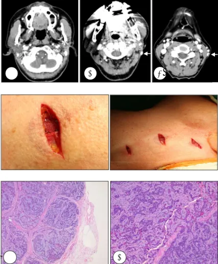
Fig. 1. Pathologic finding of the parotid is compatible with benign pleomorphic adenoma showing benign epithelial cells in the myx- oid stroma (H&E ×40).
Fig. 3. Intraoperative photographs show three ma- sses adherent to the greater auricular nerve.
A B
Fig. 4. Microscopic finding of the masses is com- patible with pleomorphic adenoma. The cellular smear is composed of epithelial cells admixed with fi-bromyxoid background. A:H&E ×40, B:H&E × 200.
Fig. 2. A:Postcontrast neck CTs show no mass le- sion on Lt. Parotidectomy area. B, C:There are ovoid superficial tumors with strong enhancement
in the left posterolateral neck. A B C
조유미 외
709
많으며 대개 진단은 전이가 확인된 이후 후향적으로 이루어지고 경부로 전이될 경우 경부 종물이 주 증상이다. 전 이 병소와 원발 병소의 양성 소견과 악성화가 없는 것이 특징이다.7,8) 전이된 병소의 종양은 양성 상피와 기질 성분 으로 이루어져 있으며 조직학적으로 원발 부위와 동일하다.
병리 조직학적 감별 진단으로는 Myxoid MFH, Dediffer- entiated Chondrosarcoma가 있다.
전이성 다형성 선종을 보고한 문헌을 살펴보면 전이성 다형성 선종에서 원발성 다형성 선종의 진단 이후 전이 병 소의 발견까지는 3~52년의 상당 기간이 있었으며 평균 35 년이었다.5) 성별에는 유의한 차이가 없었으며 나이는 8~
72세이었고, 전이 병소 발견의 평균 나이는 60세였다.5,9) 원발성 병소는 대부분 이하선이었으며 악하선과 소타액선들 도 있었다. 대부분의 전이 병소는 뼈와 폐였으나 간혹 림 프절, 구강, 인두, 피부, 간, 후복막, 신장, 중추 신경 조직도 있었다.
치료는 서서히 성장하고 오랜 기간 동안에도 주위 조직 과 구분되는 종양의 특징 때문에 적절한 변연을 포함한 완 벽한 외과적 절제가 필요하며 방사선 치료는 전이 병소가 감수성이 적기 때문에 제한적으로 사용되어진다.5,9) 대부분 의 전이성 다형성 선종은 원발성 다형성 선종의 국소 재발 이후 전이가 발생한다. 그러므로 피막의 파괴나 파종에 의 한 원발성 다형성 선종의 국소 재발을 낮추기 위해 주위의 정상 조직을 포함한 완전한 외과적 절제가 전이성 다형성 선종을 예방하는 가장 좋은 방법으로 보인다.7)
전이성 다형성 선종의 전이 기전은 더 많은 연구가 필요 하지만, 대부분의 증례에서 수술적 치료 후 발생했다는 점 에서 혈관 착상에 의한 혈행성 전이 가설이 유력하며 본
예에서는 대 이개 신경을 통한 전이가 의심되었다.5,7,8) 본 증례는 좌측 이하선 종양 절제술 후 국소 재발 없이 경부로 전이된 전이성 다형성 선종을 치험한 매우 드문 경 우로 과거력에 다형성 선종이 있었던 환자에게는 전이성 병변의 감별 진단에 전이성 다형성 선종이 포함되어야 하 며 향후 재발에 대한 추적 관찰이 필요하리라 생각된다.
중심 단어:다형성 선종·전이성.
REFERENCES
1) Waldron CA, el-Mofty SK, Gnepp DR. Tumors of the intraoral minor salivary glands: A demographic and histologic study of 426 cases.
Oral Surg Oral Med Oral Pathol 1988;66 (6):323-33.
2) Henriksson G, Westrin KM, Carlsoo B, Silfversward C. Recurrent pri- mary pleomorphic adenomas of salivary gland origin: Intrasurgical rupture, histopathologic features, and pseudopodia. Cancer 1998;82 (4) :617-20.
3) Stennert E, Guntinas-Lichius O, Klussmann JP, Arnold G. Histopathol- ogy of pleomorphic adenoma in the parotid gland: A prospective un- selected series of 100 cases. Laryngoscope 2001;111 (12):2195-200.
4) Chen KT. Metastasizing pleomorphic adenoma of the salivary gland.
Cancer 1978;42 (5):2407-11.
5) Marioni G, Marino F, Stramare R, Marchese-Ragona R, Staffieri A. Be- nign metastasizing pleomorphic adenoma of the parotid gland: A clino- copathologic puzzle. Head Neck 2003;25 (12):1071-6.
6) Hwang JH, Park IS, Kim YB, Yang JM. A case of adenoid cystic car- cinoma Ex pleomorphic adenoma of submandibular gland. Korean J Otolaryngol-Head Neck Surg 2004;47 (6):587-9.
7) Raja V, China C, Masaki KH, Nakano G. Unusual presentations of un- common tumors: Case 1. benign metastasizing pleomorphic adenoma.
J Clin Oncol 2002;20(9):2400-3.
8) Goodisson DW, Burr RG, Creedon AJ, Stirling RW, Morgan PR, Odell EW. A case of metastasizing pleomorphic adenoma. Oral Surg Oral Med Oral Pathol Oral Radiol Endod 1999;87 (3):341-5.
9) Takeuchi E, Shimizu E, Sano N, Yamaguchi T, Yanagawa H, Sone S.
A case of pleomorphic adenoma of the lung with multiple distant metas- tasis observations on its oncogene and tumor suppressor gene expres- sion. Anticancer Res 1998;18 (3B):2015-20.