Original Article
Notch signaling affects biliary fibrosis via transcriptional regulation of RBP-jκ in an animal model of chronic
liver disease
Sun-Jae Lee1, Kyung-Hyun Kim1, Sok Cheon Pak2, Yu-Na Kang3, Ghil-Suk Yoon4, Kwan-Kyu Park1
1Department of Pathology, Catholic University of Daegu School of Medicine, 3056-6 Daemyung-4 Dong, Nam- Gu, Daegu, Republic of Korea; 2School of Biomedical Sciences, Charles Sturt University, Bathurst, NSW 2795, Australia; 3Department of Pathology, Keimyung University School of Medicine, 2800, Dalgubeoldaero, Dalseo-Gu, Daegu, Republic of Korea; 4Department of Pathology, Kyungpook National University School of Medicine, 680 Gukchaebosang-Ro, Jung-Gu, Daegu, 700-842, Republic of Korea
Received August 6, 2015; Accepted September 25, 2015; Epub October 1, 2015; Published October 15, 2015 Abstract: Liver repair in patients with a chronic liver disease requires the orchestrated action of epithelial, mesen- chymal, and inflammatory cells. Notch components are expressed in both the epithelial and mesenchymal compart- ments of the adult liver and are differentially regulated after injury. However, the functional role of Notch signaling in regulating epithelial/mesenchymal cross-talk during fibrogenic pathologic repair remains unknown. The aim of this study was to investigate how proliferation of the bile duct influences biliary fibrosis and to recognize the effect of inhibiting Notch signaling in biliary fibrotic tissue of the injured liver. We designed a synthetic decoy oligodeoxynu- cleotide (ODN) for recombination signal binding protein immunoglobulin kappa J (RBP-jκ), which is a common DNA- binding partner of Notch receptors. The effect of blocking RBP-jκ on fibrogenesis was assessed in the 3,5-Diethoxy- carbonyl-1,4-dihydrocollidine (DDC) diet mouse model. We observed the reduced fibrosis and decreased expression of associated signaling molecules after the RBP-jκ decoy ODN treatment. These data demonstrate that Notch signal- ing may play an important role in progression of ductular reaction and fibrosis. Further studies are required to unveil how ductular cells interact with other liver cell types, such as hepatic stellate cells or Kupffer cells,in patients with cholestatic liver diseases based on Notch signaling. These results suggest that controlling the ductular reaction us- ing a synthetic ring type decoy RBP-jκ ODN will help develop a novel therapeutic approach targeting biliary fibrosis in patients with chronic liver diseases.
Keywords: Liver fibrosis, Notch, RBP-jκ, decoy ODN
Introduction
Chronic liver injury, including chronic hepatitis and cirrhosis, triggers a repair response, and fibrosis occurs with atypical proliferation of the dysfunctional bile duct when repair becomes deregulated. This biliary fibrous tissue gradual- ly replaces the hepatic parenchyma and causes a gradual decrease in the number of mature hepatocytes in the hepatic lobules. The bile duct basement membranes undergo degrada- tion in patients with fibrogenic liver diseases and cholangiocytes assume fibroblast-like, non-cuboidal shapes. It is well established that proliferating cholangiocytes detectable in all types of chronic liver disease express a variety
of pro-fibrogenic growth factors and cytokines that likely contribute to fibrosis and inflamma- tion by promoting activation, proliferation, and collagen synthesis in the surrounding pro-fibro- genic cells [1-7]. However, the distinct patho- genesis of intrahepatic bile duct regeneration in patients with chronic liver diseases is poorly understood.
Transforming growth factor (TGF)-β1 is a major profibrogenic cytokine and a potent inducer of collagen production [8]. Activation of the TGF- β1 pathway results in nuclear translocation of phosphorylated Smad2 and Smad3, which form hetero-oligomers with Smad4, where they regulate transcription of target genes [9].
Overexpression of TGF-β1 is linked to liver fibro- sis in diverse animal models [10] and in human patients with chronic liver diseases [11]. TGF- β1 regulates deposition of the extracellular matrix by controlling expression of vimentin, α-smooth muscle actin (α-SMA), and fibronec- tin. Participation of TGF-β1 in the regulation of Notch signaling during fibrogenesis has been reported previously in mammary glands, kidney tubules, and epidermis [12].
Several studies have indicated that the Notch pathway plays a key role maintaining liver homeostasis during post-natal life and is involved in the reparative reaction to biliary damage, as well as in liver carcinogenic, meta- bolic and inflammatory responses [13]. Ligand- activated Notch receptors are cleaved at the Notch intracellular domain (NICD). The NICD translocates to the nucleus where it binds the recombination signal binding protein immuno- globulin kappa J (RBP-jκ) transcription factor [14-17] and initiates transcription of Notch tar- get genes. Liver repair in patients with chronic liver diseases requires the orchestrated action of epithelial, mesenchymal, and inflammatory cells. Notch components are expressed in both the epithelial and mesenchymal compartments in adult liver and are differentially regulated after injury [13]. However, the functional role of Notch signaling in regulating epithelial/mesen- chymal cross-talk during fibrogenic pathologic repair remains unknown [13].
Studies to control the proliferation of reactive cholangiocytes associated with fibrosis at the molecular level remain limited in adult patients with chronic liver disease. Therefore, we attempted to suppress biliary fibrosis by regu- lating bile duct proliferation at the transcription level via blocking the transcription factor RBP- jκ. Several attempts have been made previ- ously to inhibit specific targeted gene expres- sion to control the pathologic condition by mod- ulating transcription factor function. Decoy technology uses synthetic double-stranded oli- godeoxynucleotide (ODN) containing the con- sensus binding site sequence of a targeted transcription factor. The decoy ODN blocks the activity of specific transcription factor and inhibits specific gene expression at the DNA level [18, 19]. This decoy ODN strategy is an effective approach to suppress specific gene expression in vitro and in vivo [20, 21]. It has
been proposed as an effective therapeutic tool for inhibiting specific gene expression to treat several disorders [22-24]. Several studies have reported that introducing a decoy ODN with high affinity for target transcription factors into specific cells leads to selective down regulation and expression of the genes regulated by those transcription factors [25-27]. In addition, we previously demonstrated that several decoy ODNs effectively suppressed the expression of fibrosis-related genes in vivo and in vitro [28, 29].
The aim of this study was to investigate how proliferation of the bile duct influences biliary fibrosis and to identify the effect of inhibiting Notch signaling in biliary fibrotic tissue of the injured liver. The current data demonstrate that liver cell plasticity could contribute to regenera- tion of the intrahepatic bile duct in mice by dis- rupting Notch signaling and regulating tran- scription factor RBP-jκ activity. These results suggest that Notch signaling may play an important role in biliary fibrosis seen in chronic diseases. Novel approaches to treat chronic liver diseases could be developed using the synthetic decoy ODN strategy with transcription factor RBP-jκ to regulate biliary fibrosis.
Materials and methods
ODN synthesis (ring type decoy)
We designed a synthetic decoy ODN for RBP-jκ, the common DNA-binding partner of Notch receptors, as described previously [18, 30].
The sequence of each decoy ODN was as fol- lows (consensus sequence for RBP-jκ is underlined):
RBP-jκODN: 5’-GAAATTTCATTCCCACTTCAAGA- AAACTTGAAGTGGGAAT-3’; Scrambled ODN: 5’- GAATTCAATTCAGGGTACGGCAAAAAATTGCCGT- ACCCTGAATT-3’.
The decoy ODN was purchased as a high per- formance liquid chromatography-purified prod- uct from Bionics (Seoul, Korea). These ODNs were annealed for 8 h, as temperature was decreased from 80 to 25°C. Both ODNs were predicted to form a stem-ring-type. Following the addition of T4 DNA ligase (1 U; Takara Bio, Shiga, Japan), the mixture was incubated for 16 h at 16°C to obtain covalently ligated ring-type decoy ODN molecules.
Experimental animal model and transfer of decoy ODN
Experiments were performed with mice to induce chronic liver injuries using a 3,5-Die- thoxycarbonyl-1,4-dihydrocollidine (DDC) con- taining diet. Male C57BL/6 mice (6 weeks old, 20-22 g; Orient Bio, Seongnam, South Korea) were housed in a room with controlled humidity of 60 ± 5% and temperature of 25 ± 1°C, and a 12:12 h light/dark cycle. Mice were fed a 0.1%
DDC-supplemented diet for 4 weeks, and per- mitted ad libitum consumption of water.
Controls were fed a standard mouse diet (Sniff, Soest, Germany). Mice were divided randomly into five groups of five mice per group: A normal control group (NC), the RBP-jκ decoy ODN treat- ed group (NC+RBP-jκ), the DDC diet with or without Scr decoy ODN group (DDC+Scr and DDC, respectively), and the DDC diet with RBP- jκ decoy ODN treated group (DDC+RBP-jκ). One week after first feeding the DDC diet, the RBP- jκ decoy ODN (10 μg) was injected biweekly through the mouse tail vein, using an in vivo gene delivery system (Mirus Bio Corp., Madison, WI, USA). All mice were sacrificed 4 weeks after the first DDC supplement, and serum and whole liver tissues were harvested. All experimental protocols were approved by the Institutional Animal Care and Use Committee of the Catholic University of Daegu (Daegu, South Korea) in accordance with criteria outlined in the Institutional Guidelines for Animal Research.
Histopathological investigation
For conventional light microscopy, livers were fixed in 10% formalin solution and embedded in
analysis software (i-Solution; IMT i-Solution Inc., Vancouver, BC, Canada). Biliary fibrosis was calculated as the percentage of Masson’s trichrome positive pixels above the threshold value with respect to the total pixels per lobule area. The extent of fibrosis was determined using 10 different fields in each liver. Obvious artifacts, such as nonspecific staining of necrotic areas or incompletely cut portal fields at the edges of the specimens, were excluded from the analysis.
Western blot analysis
Frozen liver tissues were homogenized in RIPA buffer (Cell Signaling Technology, Danvers, MA, USA) and centrifuged at 12,000 × g for 30 min after a 30 min incubation on ice. Protein con- centrations were measured using the Bradford protein assay (BioRad, Hercules, CA, USA).
Sodium dodecyl sulfate-polyacrylamide gel electrophoresis was performed at 100 V for 3 h. The resolved proteins were transferred to a polyvinylidene fluoride membrane (Millipore, Billerica, MA, USA) and probed with primary antibodies followed by incubation with a sec- ondary antibody conjugated to horseradish per- oxidase. Finally, the membranes were washed and developed with an enhanced chemilumi- nescence detection system (Amersham Pharmacia, Piscataway, NJ, USA). Signal inten- sity was quantified with an image analyzer (LAS-3000; Fuji, Tokyo, Japan). Antibodies and conditions for the Western blot analysis are presented in Table 1.
Electrophoretic mobility shift assay (EMSA) Nuclear extracts of liver tissue were obtained Table 1. List of antibodies used for the Western blot
analysis
Antibody Source/Clonality Supplier TGF-β1* mouse monoclonal R&D Systems p-Smad2* rabbit polyclonal Novus Biologicals Smad7 rabbit polyclonal Santa Cruz Biotechnology Smad2/3 mouse monoclonal Santa Cruz Biotechnology Vimentin mouse monoclonal Santa Cruz Biotechnology α-SMA* mouse monoclonal Sigma-Aldrich Inc.
Fibronectin mouse monoclonal BD Transduction Laboratories
TNF-α* mouse monoclonal Abcam
p-Stat3 rabbit monoclonal Cell Signaling Technology
*Abbreviations: TGF-β1, transforming growth factor-β1; p-Smad2, phosphorylated-Smad2; α-SMA, α-smooth muscle actin; TNF-α, tumor necrosis factor-α.
from the blocks and stained with hematoxylin and eosin (H&E) and Masson’s trichrome according to stan- dard protocols.
Computer-assisted morphometry Measurements were made to quantify the extent of fibrosis using a semiauto- matic image analysis system. Masson’s trichrome stained regions were ana- lyzed with a microscope (Nikon Eclipse 80i; Nikon, Osaka, Japan) supplied with a motorized stage system (NIS- Elements BR 3.00). Portal field images were collected at 200X magnification using a digital camera (Nikon DS-Ri1;
Nikon) and analyzed using an image-
tion kit (Thermo Fisher Scientific, Inc., Waltham, MA, USA). A DIG Gel Shift kit (Roche, Mannheim, Germany) was used for the EMSA assays, according to the manufacturer’s protocol. The RBP-jκ oligonucleotide probe (5’-CTTGAAGT- GGGAATGAAATTTCATTCCCACTTCAAG-3’; only sense strands are shown, consensus sequence for RBP-jκ is underlined) containing the RBP-jκ binding motifs were end-labeled with DIG- ddUTP. A 10 μg aliquot of the sample protein was incubated at room temperature for 30 min with a DIG labeled probe for the binding reac- tion. The DNA-protein complexes were separat- ed by 6% native polyacrylamide gels electro- phoresis using 0.25× Tris-borate-EDTA as the running buffer. After electrophoresis, the gels were transferred to nylon membranes and detected by chemiluminescence. The mem-
branes were exposed to X-ray film for 40 min.
Signal intensity was quantified by an image analyzer (Las3000).
Statistical analysis
Results were presented as mean ± standard deviation and analyzed with Duncan’s test. All experiments were performed at least three times. A p-value < 0.05 was considered significant.
Results
Histopathological features
Well-formed portal triads consisted of vein (por- tal vein), artery (hepatic artery) and biliary tract
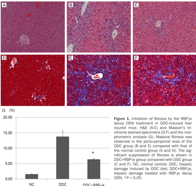
Figure 1. Inhibition of fibrosis by the RBP-jκ decoy ODN treatment in DDC-induced liver injured mice. H&E (A-C) and Masson’s tri- chrome stained specimens (D-F) and the mor- phometric analysis (G). Massive fibrosis was observed in the porto-periportal area of the DDC group (B and E) compared with that of the normal control group (A and D). The sig- nificant suppression of fibrosis is shown in DDC+RBP-jκ group compared with DDC group (C and F). NC, normal control; DDC, hepatic damage induced by DDC diet; DDC+RBP-jκ, hepatic damage treated with RBP-jκ decoy ODN. *P < 0.05.
in normal control cases (Figure 1A). Two or three bile ductules existed in one portal area.
The ductular system formed an even round to oval luminal space. No morphologic differences were detected between the NC group and the RBP-jκ decoy treated group (data not shown).
In contrast, all liver tissues obtained from DDC- fed mice showed massive fibrosis with marked bile duct proliferation in the porto-periportal area, as revealed by Masson’s trichrome stained specimens (Figure 1B, 1E, and 1G).
The proliferated bile duct cells were flattened with or without luminal space and observed in the periportal or intralobular area, which was called a ductular reaction (Figure 1B). Biliary fibrosis in DDC-fed mice was paralleled by pro- nounced proliferation of the bile duct (Figure 1E).
In contrast, the scrambled decoy treated group (DDC+Scr) reduced fibrosis and an expanded lobular areas were found in the DDC+RBP-jκ group (Figure 1C and 1F). The Masson’s tri- chrome stain results were correlated with these findings (Figure 1G). No significant microscopic changes were detected in the morphology of hepatocytes with or without decoy treatment in the DDC-fed groups (Figure 1A-C).
Expression of fibrogenic and inflammatory markers
The effects of RBP-jκ decoy ODN on liver fibro- sis and matrix accumulation were examined by Western blot analysis of TGF-β1, p-smad, vimentin, fibronectin, and α-SMA. TGF-β1, p-smad2, vimentin, fibronectin, and α-SMA were upregulated, whereas smad7 was down- regulated in the DDC-fed groups compared with Figure 2. Inhibition of fibrosis by the RBP-jκ decoy ODN treatment in DDC-induced liver injured mice. The levels of TGF-β1, p-smad2, Smad7, α-SMA and fibronectin were analyzed by Western blot. TGF-β1, p-smad2, vimentin, fibro- nectin, and α-SMA were upregulated and smad7 was downregulated in the DDC-fed groups compared to those in the NC group. In contrast, expression of TGF-β1, p-smad, vimentin, fibronectin, and α-SMA was significantly inhibited and smad7 was overexpressed in the DDC+RBP-jκ group compared with DDC and DDC+Scr groups. NC, normal control; NC+RBP-jκ, normal control treated with RBP-jκ decoy ODN; DDC, hepatic damage induced by DDC diet;
DDC+Scr, hepatic damage treated with Scr decoy ODN; DDC+RBP-jκ, hepatic damage treated with RBP-jκ decoy ODN. *P < 0.05.
those in the NC group (Figure 2). In contrast, expression of TGF-β1, p-smad, vimentin, fibro- nectin, and α-SMA was significantly inhibited and smad7 was overexpressed in the DDC+RBP- jκ group compared with those in the DDC and DDC+Scr groups (Figure 2).
The expression of tumor necrosis factor (TNF)-α and p-stat3, which are major inflammatory response markers, was examined with Western blot analysis to assess the molecular mecha- nism. The overexpressed TNF-α and p-stat3 in the DDC-fed mice were markedly downregulat-
ed after treatment with the RBP-jκ decoy ODN (Figure 3).
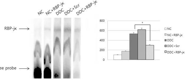
Suppressive effect of decoy ODN to RBP-jκ An EMSA was performed to analyze RBP-jκ transcriptional activity and investigate the effect of RBP-jκ decoy ODN on the target DNA binding activity in liver specimen. This tran- scription factor increased significantly in the DDC and DDC+Scr mice. In contrast, the increase was significantly suppressed by RBP- jκ decoy ODN treatment (Figure 4).
Figure 3. Inhibition of inflammation by the RBP-jκ decoy ODN treatment in DDC-induced liver injured mice. The levels of TNF-α and p-stat3, which are major inflammation-related genes, were analyzed by Western blot. The over- expressed TNF-α and p-stat3 in the DDC-fed mice were markedly downregulated after treatment with the RBP-jκ decoy ODN. NC, normal control; NC+RBP-jκ, normal control treated with RBP-jκ decoy ODN; DDC, hepatic damage induced by DDC diet; DDC+Scr, hepatic damage treated with Scr decoy ODN; DDC+RBP-jκ, hepatic damage treated with RBP-jκ decoy ODN. *P < 0.05.
Figure 4. DNA binding activity according to the electrophoretic mobility shift assay (EMSA) results. The RBP-jκ decoy oligodeoxynucleotide (ODN)-treated livers showed decreased expression of the DNA-RBP-jκ complex compared with that in the scrambled decoy ODN treated group. NC, normal control; NC+RBP-jκ, normal control treated with RBP- jκ decoy ODN; DDC, hepatic damage induced by DDC diet; DDC+Scr, hepatic damage treated with Scr decoy ODN;
DDC+RBP-jκ, hepatic damage treated with RBP-jκ decoy ODN. *P < 0.05.
Discussion
Chronic liver damage can be triggered by differ- ent mechanisms and is accompanied by chang- es in several key biochemical pathways involved in hepatic tissue homeostasis. One of the most important alterations is hepatic fibrosis, which is characterized by deposition of extracellular matrix components around the sinusoidal layer in the space of Disse, together with molecular reorganization of the matrix components result- ing in altered composition. Chronic DDC feed- ing of mice is a well-established model to induce chronic cholestatic liver disease, chol- angitis with pronounced ductular reaction, onion-skin-type periductal fibrosis, and biliary type liver fibrosis [31-33]. Therefore, we select- ed the DDC-induced cholangiopathy and biliary fibrosis mouse model as an atypical ductular reaction in patient with chronic liver diseases because it shares several specific pathological hallmarks with at least some human cholangi- opathies associated with biliary type liver fibrosis.
A variety of profibrogenic growth factors and cytokines are expressed during the ductular reaction that likely contribute to fibrosis and inflammation by promoting activation, prolifera- tion, and collagen synthesis in surrounding pro- fibrogenic cells [1]. In this study, we observed that periportal fibrosis was accompanied by bile duct proliferation in DDC-induced liver. The fibrosis was suppressed by inhibiting the relat- ed pathway with the RBP-jκ decoy ODN. RBP-jκ is a co-transcription factor of Notch signaling [14-17], suggesting that the Notch signaling pathway has an inhibitory effect on fibrosis.
Notch-1 and Notch-3 are both expressed by quiescent hepatic stellate cells (HSC) [34] and are respectively downregulated and upregulat- ed during HSC transdifferentiation into myofi- broblasts. The ligand Jag-1, has been detected on proliferating bile ductules [35, 36] and hepatocytes [34], as well as on activated HSCs [34] and is strongly upregulated in the injured liver. Notably, patients with Alagille syndrome, which is a human congenital biliary agenesis syndrome induced by mutations in the Notch ligand (Jagged-1) [37] or Notch receptor (Notch- 2) [38], show limited deposition of fibrotic tis- sue, consistent with the slow progression to cirrhosis seen in these patients [39]. Thus, Jag- 1, the defective protein in Alagille syndrome,
may signal portal myofibroblasts and induce collagen production or proliferation [13]. Notch activation and upregulation of Notch-3 in myofi- broblasts have been described in carbon tetra- chloride-induced liver fibrosis experimental rat model. In this model, pharmacologically inhibit- ing Notch reduces the extent of liver fibrosis [34]. However, the functional role of Notch in regulating epithelial/mesenchymal cross-talk related with the TGF-β1 signaling pathway dur- ing fibrogenic pathologic repair remains to be fully understood [13].
A reduced inflammatory response is needed to suppress fibrosis and for the indirect effect of RBP-jκ decoy ODN treatment to be induced. It is typically seen in liver diseases associated with increased deposition of collagenous matrices produced by activated HSCs [40]. In such liver diseases, Kupffer cells are also activated to produce various inflammatory cytokines, such as TNF-α, which is thought to play important pathogenic roles [41]. Nishikawa et al showed that inflammatory cytokines exert distinct effects on hepatocyte differentiation, indicat- ing that TNF-α is unique among these cytokines in its ability to suppress the hepatocytic pheno- type [42]. Their results indicate that TNF-α also profoundly influences the differentiation status of hepatocytes [42]. They showed that TNF-α strongly enhances bile ductular transdifferenti- ation of hepatocytes within a collagen-rich matrix, particularly by suppressing hepatocytic differentiation and enhancing ductular morpho- genesis [42]. The direct effects of TNF-α on hepatocyte differentiation might be involved in the pathogenesis of hepatic dysfunctional characteristic of the chronic liver injury associ- ated with fibrosis [42]. In this study, the increased expression of TNF-α and p-stat3 induced by the DDC feeding was markedly downregulated by the RBP-jκ decoy ODN treat- ment, suggesting that DDC-induced inflamma- tion and fibrosis is suppressed by inhibiting the Notch signaling pathway. Further studies are needed to establish the mechanisms of the interactions between Notch signaling and inflammatory responses.
In conclusion, we found that Notch signaling played an important part in the progression of the ductular reaction and fibrosis to induce dif- ferentiation and proliferation of bile duct epi- thelial cells. Further studies will be required to
unveil how ductular cells interact with other liver cell types, such as HSCs and Kupffer cells, during cholestatic liver diseases based on Notch signaling. Our result that the ductular reaction was controlled by the synthetic ring type decoy RBP-jκ ODN will help in the develop- ment of a novel therapeutic approach targeting biliary fibrosis in patients with chronic liver diseases.
Acknowledgements
This work was supported by the National Research Foundation of Korea Grant funded by the Korean Government [2014R1A1A20- 08955].
Disclosure of conflict of interest None.
Address correspondence to: Dr. Kwan-Kyu Park, Department of pathology, Catholic University of Daegu, College of Medicine, 3056-6 Daemyung 4-Dong, Nam-Gu, Daegu, 705-718, Republic of Korea. Tel: +82-53-650-4149; Fax: +82-53-650- 4834; E-mail: [email protected]
References
[1] Milani S, Herbst H, Schuppan D, Stein H and Surrenti C. Transforming growth factors beta 1 and beta 2 are differentially expressed in fi- brotic liver disease. Am J Pathol 1991; 139:
1221-1229.
[2] Pinzani M, Milani S, Herbst H, DeFranco R, Grappone C, Gentilini A, Caligiuri A, Pellegrini G, Ngo DV, Romanelli RG and Gentilini P. Ex- pression of platelet-derived growth factor and its receptors in normal human liver and during active hepatic fibrogenesis. Am J Pathol 1996;
148: 785-800.
[3] Grappone C, Pinzani M, Parola M, Pellegrini G, Caligiuri A, DeFranco R, Marra F, Herbst H, Al- pini G and Milani S. Expression of platelet-de- rived growth factor in newly formed cholangio- cytes during experimental biliary fibrosis in rats. J Hepatol 1999; 31: 100-109.
[4] Kinnman N, Hultcrantz R, Barbu V, Rey C, Wen- dum D, Poupon R and Housset C. PDGF-medi- ated chemoattraction of hepatic stellate cells by bile duct segments in cholestatic liver inju- ry. Lab Invest 2000; 80: 697-707.
[5] Pinzani M, Milani S, De Franco R, Grappone C, Caligiuri A, Gentilini A, Tosti-Guerra C, Maggi M, Failli P, Ruocco C and Gentilini P. Endothelin 1 is overexpressed in human cirrhotic liver and exerts multiple effects on activated hepatic
stellate cells. Gastroenterology 1996; 110:
534-548.
[6] Caligiuri A, Glaser S, Rodgers RE, Phinizy JL, Robertson W, Papa E, Pinzani M and Alpini G.
Endothelin-1 inhibits secretin-stimulated duc- tal secretion by interacting with ETA receptors on large cholangiocytes. Am J Physiol 1998;
275: G835-846.
[7] Marra F, DeFranco R, Grappone C, Milani S, Pastacaldi S, Pinzani M, Romanelli RG, Laffi G and Gentilini P. Increased expression of mono- cyte chemotactic protein-1 during active he- patic fibrogenesis: correlation with monocyte infiltration. Am J Pathol 1998; 152: 423-430.
[8] Eghbali-Fatourechi G, Sieck GC, Prakash YS, Maercklein P, Gores GJ and Fitzpatrick LA. Type I procollagen production and cell proliferation is mediated by transforming growth factor-beta in a model of hepatic fibrosis. Endocrinology 1996; 137: 1894-1903.
[9] Inagaki Y and Okazaki I. Emerging insights into Transforming growth factor beta Smad signal in hepatic fibrogenesis. Gut 2007; 56: 284- [10] Gressner AM, Weiskirchen R, Breitkopf K and 292.
Dooley S. Roles of TGF-beta in hepatic fibrosis.
Front Biosci 2002; 7: d793-807.
[11] Annoni G, Weiner FR and Zern MA. Increased transforming growth factor-beta 1 gene expres- sion in human liver disease. J Hepatol 1992;
14: 259-264.
[12] Zavadil J, Cermak L, Soto-Nieves N and Bot- tinger EP. Integration of TGF-beta/Smad and Jagged1/Notch signalling in epithelial-to-mes- enchymal transition. EMBO J 2004; 23: 1155- 1165.
[13] Morell CM and Strazzabosco M. Notch signal- ing and new therapeutic options in liver dis- ease. J Hepatol 2014; 60: 885-890.
[14] Artavanis-Tsakonas S, Rand MD and Lake RJ.
Notch signaling: cell fate control and signal in- tegration in development. Science 1999; 284:
770-776.
[15] Bray SJ. Notch signalling: a simple pathway be- comes complex. Nat Rev Mol Cell Biol 2006; 7:
678-689.
[16] Kopan R and Ilagan MX. The canonical Notch signaling pathway: unfolding the activation mechanism. Cell 2009; 137: 216-233.
[17] Lai EC. Notch signaling: control of cell commu- nication and cell fate. Development 2004;
131: 965-973.
[18] Sung WJ, Kim KH, Kim YJ, Chang YC, Lee IH and Park KK. Antifibrotic effect of synthetic Smad/Sp1 chimeric decoy oligodeoxynucleo- tide through the regulation of epithelial mes- enchymal transition in unilateral ureteral ob- struction model of mice. Exp Mol Pathol 2013;
95: 136-143.
[19] Morishita R, Sugimoto T, Aoki M, Kida I, Tomita N, Moriguchi A, Maeda K, Sawa Y, Kaneda Y, Higaki J and Ogihara T. In vivo transfection of cis element “decoy” against nuclear factor- kappaB binding site prevents myocardial in- farction. Nat Med 1997; 3: 894-899.
[20] Morishita R, Higaki J, Tomita N and Ogihara T.
Application of transcription factor “decoy”
strategy as means of gene therapy and study of gene expression in cardiovascular disease.
Circ Res 1998; 82: 1023-1028.
[21] Tomita N, Kashihara N and Morishita R. Tran- scription factor decoy oligonucleotide-based therapeutic strategy for renal disease. Clin Exp Nephrol 2007; 11: 7-17.
[22] Magae J, Munemura K, Ichikawa C, Osada K, Hanada T, Tsuji RF, Yamashita M, Hino A, Hori- uchi T, Uramoto M, et al. Effects of microbial products on glucose consumption and mor- phology of macrophages. Biosci Biotechnol Biochem 1993; 57: 1628-1631.
[23] Yamashita J, Yoshimasa T, Arai H, Hiraoka J, Takaya K, Miyamoto Y, Ogawa Y, Itoh H and Na- kao K. Identification of cis-elements of the hu- man endothelin-A receptor gene and inhibition of the gene expression by the decoy strategy. J Biol Chem 1998; 273: 15993-15999.
[24] Ishibashi H, Nakagawa K, Onimaru M, Castella- nous EJ, Kaneda Y, Nakashima Y, Shirasuna K and Sueishi K. Sp1 decoy transfected to carci- noma cells suppresses the expression of vas- cular endothelial growth factor, transforming growth factor beta1, and tissue factor and also cell growth and invasion activities. Cancer Res 2000; 60: 6531-6536.
[25] Kum YS, Kim KH, Park TI, Suh IS, Oh HK, Cho CH, Park JB, Chang YC, Park JH, Lee KG and Park KK. Antifibrotic effect via the regulation of transcription factor Sp1 in lung fibrosis. Bio- chem Biophys Res Commun 2007; 363: 368- [26] Kawauchi M, Suzuki J, Morishita R, Wada Y, 374.
Izawa A, Tomita N, Amano J, Kaneda Y, Ogihara T, Takamoto S and Isobe M. Gene therapy for attenuating cardiac allograft arteriopathy us- ing ex vivo E2F decoy transfection by HVJ-AVE- liposome method in mice and nonhuman pri- mates. Circ Res 2000; 87: 1063-1068.
[27] Maeshima Y, Kashihara N, Yasuda T, Sugiyama H, Sekikawa T, Okamoto K, Kanao K, Wata- nabe Y, Kanwar YS and Makino H. Inhibition of mesangial cell proliferation by E2F decoy oligo- deoxynucleotide in vitro and in vivo. J Clin In- vest 1998; 101: 2589-2597.
[28] Park JH, Jo JH, Kim KH, Kim SJ, Lee WR, Park KK and Park JB. Antifibrotic effect through the regulation of transcription factor using ring type-Sp1 decoy oligodeoxynucleotide in car- bon tetrachloride-induced liver fibrosis. J Gene Med 2009; 11: 824-833.
[29] Kim KH, Park JH, Lee WR, Park JS, Kim HC and Park KK. The inhibitory effect of chimeric de- coy oligodeoxynucleotide against NF-kappaB and Sp1 in renal interstitial fibrosis. J Mol Med (Berl) 2013; 91: 573-586.
[30] Kim KH, Lee WR, Kang YN, Chang YC and Park KK. Inhibitory effect of nuclear factor-kappaB decoy oligodeoxynucleotide on liver fibrosis through regulation of the epithelial-mesenchy- mal transition. Hum Gene Ther 2014; 25: 721- [31] Fickert P, Stoger U, Fuchsbichler A, Moustafa T, 729.
Marschall HU, Weiglein AH, Tsybrovskyy O, Jae- schke H, Zatloukal K, Denk H and Trauner M. A new xenobiotic-induced mouse model of scle- rosing cholangitis and biliary fibrosis. Am J Pathol 2007; 171: 525-536.
[32] Fickert P, Thueringer A, Moustafa T, Silbert D, Gumhold J, Tsybrovskyy O, Lebofsky M, Jae- schke H, Denk H and Trauner M. The role of osteopontin and tumor necrosis factor alpha receptor-1 in xenobiotic-induced cholangitis and biliary fibrosis in mice. Lab Invest 2010;
90: 844-852.
[33] Zatloukal K, Stumptner C, Fuchsbichler A, Fick- ert P, Lackner C, Trauner M and Denk H. The keratin cytoskeleton in liver diseases. J Pathol 2004; 204: 367-376.
[34] Chen Y, Zheng S, Qi D, Guo J, Zhang S and Weng Z. Inhibition of Notch signaling by a gam- ma-secretase inhibitor attenuates hepatic fi- brosis in rats. PLoS One 2012; 7: e46512.
[35] Boulter L, Govaere O, Bird TG, Radulescu S, Ramachandran P, Pellicoro A, Ridgway RA, Seo SS, Spee B, Van Rooijen N, Sansom OJ, Iredale JP, Lowell S, Roskams T and Forbes SJ. Macro- phage-derived Wnt opposes Notch signaling to specify hepatic progenitor cell fate in chronic liver disease. Nat Med 2012; 18: 572-579.
[36] Nijjar SS, Wallace L, Crosby HA, Hubscher SG and Strain AJ. Altered Notch ligand expression in human liver disease: further evidence for a role of the Notch signaling pathway in hepatic neovascularization and biliary ductular de- fects. Am J Pathol 2002; 160: 1695-1703.
[37] Oda T, Elkahloun AG, Pike BL, Okajima K, Krantz ID, Genin A, Piccoli DA, Meltzer PS, Spinner NB, Collins FS and Chandrasekharap- pa SC. Mutations in the human Jagged1 gene are responsible for Alagille syndrome. Nat Gen- et 1997; 16: 235-242.
[38] McDaniell R, Warthen DM, Sanchez-Lara PA, Pai A, Krantz ID, Piccoli DA and Spinner NB.
NOTCH2 mutations cause Alagille syndrome, a heterogeneous disorder of the notch signaling pathway. Am J Hum Genet 2006; 79: 169-173.
[39] Tsai S, Gurakar A, Anders R, Lam-Himlin D, Boitnott J and Pawlik TM. Management of large hepatocellular carcinoma in adult pa- tients with Alagille syndrome: a case report
and review of literature. Dig Dis Sci 2010; 55:
3052-3058.
[40] Clouston AD, Powell EE, Walsh MJ, Richardson MM, Demetris AJ and Jonsson JR. Fibrosis cor- relates with a ductular reaction in hepatitis C:
roles of impaired replication, progenitor cells and steatosis. Hepatology 2005; 41: 809-818.
[41] Bradham CA, Plumpe J, Manns MP, Brenner DA and Trautwein C. Mechanisms of hepatic toxic- ity. I. TNF-induced liver injury. Am J Physiol 1998; 275: G387-392.
[42] Nishikawa Y, Sone M, Nagahama Y, Kumagai E, Doi Y, Omori Y, Yoshioka T, Tokairin T, Yoshi- da M, Yamamoto Y, Ito A, Sugiyama T and Eno- moto K. Tumor necrosis factor-alpha promotes bile ductular transdifferentiation of mature rat hepatocytes in vitro. J Cell Biochem 2013;
114: 831-843.