J. C. Koo ( )
Division of Science Education and Institute of Science Education, Chonbuk National University, Jeonju, 561-756, Korea
e-mail: [email protected]
Isolation of an actin promoter for strong expression of transgenes in the orchid genus Dendrobium
Ja Choon Koo
Received: 14 January 2013 / Accepted: 31 January 2013
ⓒKorean Society for Plant Biotechnology
Abstract We isolated and functionally characterized a Dendrobium Actin1 (DmACT1) promoter that drives strong gene expression in the orchid genus Dendrobium. A genomic fragment containing the region 3227 bp upstream of the coding region of DmACT1 was obtained by inverse PCR.
Detailed comparison of the full-length cDNA and genomic sequences revealed that DmACT1 has a 1374 bp first intron in the 5’ UTR. However, the 5’ flanking sequences upstream of the coding region showed no obvious sequence similarities compared to those of known promoters, including plant actin promoters. Serial deletion constructs of the 5’ flanking region from the translation initiation codon were fused to the coding sequence of a GUS/luciferase fusion reporter to identify the regulatory elements necessary for promoter activity. Transient assays in the flowers of Dendrobium revealed that the 5’ UTR-intron greatly enhanced pro- moter activity. Moreover, the DmACT1 promoter with its 5’
UTR-intron yielded approximately 10-fold higher reporter activity than the rice Act1 promoter-intron. Our data suggest that the DmACT1 promoter with its 5’ UTR-intron is a useful tool for strong expression of transgenes in Dendrobium orchids.
Keywords 5’ UTR-intron, transient expression
Abbreviations CaMV, Cauliflower mosaic virus; GUS, β -Glucuronidase; X-GlcA, 5-bromo-4-chloro-3-indolyl-β-D- glucuronic acid cyclohexylammonium; DmACT1, Dendrobium moniliforme Actin1; RACE, rapid amplification of cDNA ends;
UTR, untranslated region
Introduction
Orchids in the genus Dendrobium are some of the most important commercial orchid plants in the floricultural industry because many species in this genus produce a wide variety of beautifully colored flowers with unique morphologies and diverse fragrances. Furthermore, chemical compounds from orchids in this genus are used in trad- itional Chinese medicine (Wood 2006). Extensive conven- tional breeding programs based on intra- and inter-specific hybridization have been conducted over the last decade in Dendrobium species to improve floricultural traits such as color, plant shape, and growth. However, these approaches have resulted in limited success due to the lack of genetic resources, the sterility of inter-specific hybrids, and diffi- culty obtaining a genotype with all favorable genetic alleles present in the hybrids (Shibata 2008). Recently, technical advances in genetic transformation technology in Dendrobium have made it possible to modify target traits by direct incorporation of related genes with few undesirable effects on the existing plant phenotype (Chia et al. 1994; Kuehnle and Sugii 1992; Yang et al. 2003; Yu et al. 2001).
Strong, constitutive promoters are generally required for ectopic expression of target genes in a transgenic plant.
Several promoters have been investigated as useful tools for strong expression of target genes in plants. Promoters obtained from CaMV 35S RNA (Guilley et al. 1982), the maize polyubiquitin 1 (Ubi 1) gene (Elliott et al. 1999), and the chimeric HBT promoter consisting of the CaMV 35S enhancer fused to the basal promoter and the 5′ leader sequence of the maize C4PPDK gene (Sheen et al. 1995) have been evaluated in Dendrobium by genetic transfor- mation or transient expression analysis (Chia et al. 1994;
Tee et al. 2003; Yu et al. 2001). The HBT and Ubi1 promoters showed relatively low GUS expression compared to that of the CaMV 35S promoter in Dendrobium sonia (Tee et al. 2003). Similarly, the rice Actin1 (Act1) (McElroy DOI:http://dx.doi.org/10.5010/JPB.2013.40.1.027
Research Article
et al. 1990) and maize Ubi1 promoters showed lower activity than the CaMV 35S promoter in some non-cereal monocot plants, namely Lolium, Liliaceae, and Gladiolus (Kamo et al. 1995; Wilmink et al. 1995). These results sug- gest that typical monocot promoters, such as Act1 and Ubi1, although they perform well in some monocot species, do not perform well in all monocot species. In contrast, the CaMV 35S promoter drove developmental- and tissue-specific expression of transgenes in transgenic rice and cotton plants, but was not constitutive (Sunilkumar et al. 2002; Tereda and Shimamoto 1990; Zhang and Wu 1988). Thus, for successful transformation of a target plant species, the promoter should be chosen with care.
Actin is a ubiquitous protein that plays essential roles in plant developmental processes such as cytoskeletal function, cell division, cell elongation, cellular trafficking, transportation of organelles, and pollen tube growth (Wasteneys and Galway 2003). High levels of Actin transcript were observed in all tissues and development stages in rice plants (McElroy et al. 1990). Likewise, the Actin1 (Act1) promoter directed high GUS expression in all tissues of transgenic rice plants (Zhang et al. 1991).
Recently, the rice Actin2 promoter, which drives high-level constitutive expression of GUS in transgenic rice, was also isolated for monocot transformations (He et al. 2009).
However, the feasibility of using rice actin promoters in Dendrobium remains to be evaluated.
Some 5’ UTR-introns, but not all, greatly enhance gene expression in humans (Jonsson et al. 1992), mice (Palmiter et al. 1991), Arabidopsis (Rose and Last 1997), rice (Jeon et al. 2001; Zhang et al. 1991), and Caenorhabditis elegans (Ho et al. 2001). Although 5’ UTR-intron mediated gene expression (IME) exists in a diverse range of organisms, the precise mechanism underlying IME is likely not general. The length of 5’ UTR-introns is highly variable across species, and 5’ UTR-introns tend to be similar in size or larger than intragenic introns (Bradnam and Korf 2008). Moreover, it has been shown that the most highly expressed genes in humans and C. elegans tend to have shorter introns (Castillo-Davis et al. 2002), whereas in rice and Arabidopsis, highly expressed genes tend to have longer introns (Ren et al. 2006). Furthermore, analysis of the positional effect of IME has shown that IME effects decrease with distance from the transcription start site within a single gene in transgenic Arabidopsis (Rose 2004).
Hence, the mechanisms underlying IME still remain to be elucidated.
One of the major barriers in genetic transformation of Dendrobium is the lack of promoters able to drive strong
ectopic expression of target genes. To overcome this pro- blem, we isolated the Actin1 (DmACT1) promoter from a species of Dendrobium, because DmACT1 transcripts are abundantly expressed in the roots, leaves, and flowers of species in this genus. To identify the promoter region necessary for efficient gene expression, we examined the promoter activity of DmACT1 5’ flanking sequences using a GUS/luciferase fusion reporter system. Transient assays in Dendrobium flowers revealed that the DmACT1 5’ UTR- intron is essential for transegene expression. Moreover, the DmACT1 promoter showed approximately 8- to 10-fold higher active than the rice Actl promoter. Thus, the DmACT1 promoter can be used to drive strong ectopic gene expres- sion in Dendrobium orchids.
Materials and methods Plant Materials
Dendrobium moniliforme “Jindo” (Whang et al. 2011) was cultivated under a 16-h light/8-h dark cycle at 24℃ in a greenhouse facility at Chonbuk National University. Leaf and root samples were harvested from young seedlings to isolate total RNA. Flower buds were harvested just after flowering in late May and throughout June for total RNA isolation or transient expression assays.
Northern blot analysis
Flowers, roots, and leaves were ground in liquid nitrogen using a mortar and pestle, and total RNA was isolated using Trizol reagent (Gibco BRL, NY, USA) according to the manufacturer’s instructions. To prepare a DmACT1- specificr probe, 120 bp of the 5’ UTR was amplified by radioactive PCR with the primers ActProbe-F (5’-GCCCT GCTAGTCGGCTGC-3’) and ActProbe-R (5’-CTTTTACAA GCCTAAACAGAAG-3’). Five micrograms of total RNA was denatured and separated on a 2% agarose gel containing formaldehyde. After transferring the RNA onto a Hybond-N plus membrane (Amersham Biosciences, USA), the RNA was hybridized with 32P-labeled probe for 8 h at 65℃ in Church buffer and washed as described previously (Koo et al. 2004). The blot was exposed to a Phosphor Screen (Molecular Dynamics, Amersham Bioscience, Inc.) to visu- alize the signal. The pixel intensity of each band was quantified and analyzed using TotalLab100 software (Nonlinear Dynamics, USA). The relative expression levels are shown as the ratio of the expression in the leaves to that in the other tissues.
Fig. 1 Nucleotide and deduced amino acid sequence of DmACT1 (Genbank accession No. KC171011). The lower-case letters represent the 5' and 3’ UTR sequences. Underlined sequences indicate the sequences isolated by degenerate RT-PCR
Cloning of partial Actin1 cDNA by degenerate RT-PCR
Partial DmACT1 cDNA was isolated using RT-PCR with degenerate primers. The primers dACT-F (5’-GARAARA TGACNCARATHATG-3’) as the upstream forward primer and dACT-R (5’-TCNACRTCRCAYTTCATDAT-3’) as the downstream reverse primer were designed based on a multiple alignment of orchid actin genes including those isolated from Dendrobium hybrid cultivars (EF612438.1 and GU301090.1), Phalaenopsis (AF246715.1), and Cymbidium (JQ360575.1 and JN177721.1). Five micrograms of total RNA was used to synthesize first strand cDNA using the SMARTer RACE cDNA Amplification Kit (Clontech Laboratories Inc., CA, USA) according to the manufacturer's instructions. PCR was performed with one-tenth of the synthesized cDNA, 20 μM dACT-F, and 20 μM dACT-R in 20 μl of PCR premix (Clontech Laboratories Inc., CA, USA). The PCR conditions were as follows: initial dena- turation at 95℃ for 2 min, 30 cycles of 94℃ for 30 s, 50℃ for 30 s, 72℃ for 1 min, followed by a final extension of 72℃ for 7 min. PCR products were separated on a 0.8% agarose gel (w/v). Specifically amplified DNA was recovered from the agarose gel using a PCR DNA purification Kit (GeneAll, Korea) and cloned into the pTOP TA V2 vector (Enzynomics, Inc., Korea). The nucleotide sequence of the insert was determined using vector-
derived primers and analyzed using the BlastN program of NCBI (http://blast.ncbi.nlm.nih.gov/Blast.cgi).
Isolation of the full-length DmACT1 cDNA by 5’- and 3’-RACE
Full-length DmACT1cDNA was isolated by 5’- and 3’-RACE using the SMARTer RACE cDNA Amplification Kit according to the manufacturer's instructions with minor modifications. Briefly, 5 μg of total RNA was used to synthesize first strand cDNA using the SMARTer RACE cDNA Amplification Kit (Clontech Laboratories Inc., CA, USA). One-tenth of the first strand cDNA was subjected to PCR with the DmACT1-specific reverse primer, Act- RACE-R (5’-GTCGTGCGGCCACTAGCATATAG-3’) for 5’-RACE or the forward primer, Act-RACE-F (5’-CTATT GGAGCTGGGAGATTTAGGTGC-3’), for 3’-RACE and vector derived universal primers to synthesize DmACT1, respectively. The PCR profile was as follows: initial dena- turation at 95℃ for 2 min followed by 30 cycles of 95℃
for 15 s, 60℃ for 30 s, and 72℃ for 1 min. PCR products were purified using a PCR DNA purification Kit (GeneAll, Korea) and cloned into the pTOP TA V2 vector (Enzynomics, Inc., Korea) and sequenced with vector-derived primers.
The complete DmACT1 cDNA sequence is available at Genbank (accession number KC171011, Fig. 1).
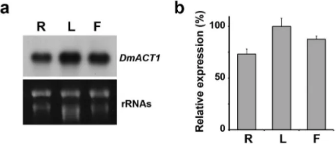
Fig. 2 Expression pattern of DmACT1 in Dendrobium. (a) RNA blot analysis of DmACT1. Total RNA was isolated from root (R), leaf (L), and flower (F) tissues. RNA was transferred to a membrane and hybridized with a 32P-labeled DmACT1 5’ UTR probe. As a loading control, rRNAs was visualized after staining with ethidium bromide. (b) Quantification of DmACT1 expression. The DmACT1 band intensity from the RNA gel blot was measured and normalized to those of the 28S rRNA bands.
Relative expression indicates the ratio of the value for the leaf to that of the other tissues. Bars indicate the SD of mean values Isolation of DmACT1 5’-flanking sequences by inverse PCR
Genomic DNA was extracted from fresh leaves using the CTAB method (Doyle and Doyle 1988). For inverse PCR, 1 μg of genomic DNA was fully digested with Eco RI at 37℃ for 4 h in a reaction volume of 100 μl. The digested DNA was extracted with a PCR DNA purification kit (GeneAll, Korea). The linearized DNA was then self- ligated to form circular DNA with T4 DNA ligase (Enzynomics Inc., Korea) in a 10 μl reaction volume at room temperature overnight. Self-ligated DNA was used as a template for inverse PCR. The PCR reaction was carried out in 40 μl PCR mix containing 1 μl of self- ligated DNA and 20 μM each of the inverse PCR primers, iACT-E-3-1 (5'-GCCATCCTTCGATTGGATCTTGCGAG CCG-3') and iACT-E-5-1 (5’-CTCTAAAACAAGCCAAA ACCCTCGCAG-3’). The PCR conditions were as follows:
one cycle of initial denaturation at 95℃ for 2 min, followed by 30 cycles of 95℃ for 15 s, 65℃ for 30 s, 72℃ for 3 min, followed by 72℃ for 5 min. PCR products were purified using a PCR DNA purification kit and used as template for a second round of nested inverse PCR. The reaction mix and PCR condition were the same as those described above except the primers, iACT-E-3-2 (5'-CTCCT TGATGAAGATCCTCACTGAGAGAGG-3') and iACT-E-5-2 (5’- AAAGTAAGTCGCCTTATACCAAGGGCG -3’) were used. Specifically amplified DNA was recovered from the agarose gel and cloned in the pTOP TA V2 vector for DNA sequencing. To obtain additional 5’ flanking sequences of DmACT1, inverse PCR was conducted with Nde I-circularized genomic DNA as described above. The first round of PCR was performed using the primers iACT-
Nd-3-1 (5’-TCAAGGGTGGATCTGGAGCTACCA-3’) and iACT-Nd-5-1 (5’-CAAAAGAACACAAACACCCAGATC TACC-3’). The second nested round of PCR was performed using the primers iACT-Nd-3-2 (5’- CCTGTTTGAATCG CGTTTGTAGCA-3’) and iACT-Nd-5-2 (5’-CAGATCTA CCAAACGGCCCACCG-3’). Specifically amplified DNA was recovered from the agarose gel and cloned into the pTOP TA V2 vector for DNA sequencing. DNA sequenc- ing was carried out using vector-derived primers and primers designed based on internal sequences, and both strands were sequenced. Nucleotide sequences were analyzed using the BlastN program of NCBI (http://blast.ncbi.nlm.
nih.gov/Blast.cgi). A database search for promoter motifs and regulatory elements was performed using PlantCARE (http://bioinformatics.psb.ugent.be/webtools/plantcare/html).
Vector construction
The DmACT1 5’ flanking sequence was deposited in Genbank (accession number KC171012, Fig. 3). A 3227 bp fragment of the DmACT1 5’ flanking sequence, containing the promoter and the first intron sequence (from -1724 to +1503 relative to the transcription start site) was amplified by LA-PCR with the primers Act1-Nd (5’-ttggatcCATAT GCAAAGGACTAAACAAATTAAGTAG-3’) and Act1- int-R (5’-ttaagcttCTTTTACAAGCCTGAAGAATAACTG ACATG-3’). The underlined sequences indicate Bam HI and Hind III sites, respectively. The PCR product was digested with Bam HI and Hind III and cloned into the same sites in the GUS/Luciferase3300 vector, which encodes the GUS/Luciferase fusion reporter gene (Koo et al. 2007), to create pDmACT1Nd. In the same manner, the 1833 bp PCR product (from -1724 to +109) lacking the first intron sequence was amplified by PCR with the primers Act1-Nd and Act1-R (5’- ttaagcttAGAATCTGAC GAGGATCGATTCAAAGC-3’), and cloned in the GUS/
Luciferase3300 vector to create pDmACT1NdΔint. A 2555 bp fragment of the DmACT1 5’ flanking sequence (from -1052 to +1503) was amplified by PCR with the primers Act1-Xb (5’-ttggatcctTCTAGAAAAACTCGGTACAACT CTGTATTACC-3’) and Act1-int-R, and cloned into the GUS/Luciferase3300 vector to create pDmACT1Xb. A 1602 bp fragment of the DmACT1 5’ flanking sequence (from -99 to +1503) was amplified by PCR with the primers, Act1-St (5’-ttggatccAGGCCTAAGCCCGTCTTGCTCAA -3’) and Act1-int-R, and cloned into the GUS/Luciferase 3300 vector to create pDmACT1St. To compare with other monocot actin promoter, the Pst I and Hind III fragment of pCAMYS02 containing rice Actin1 promoter (a kind
a
b
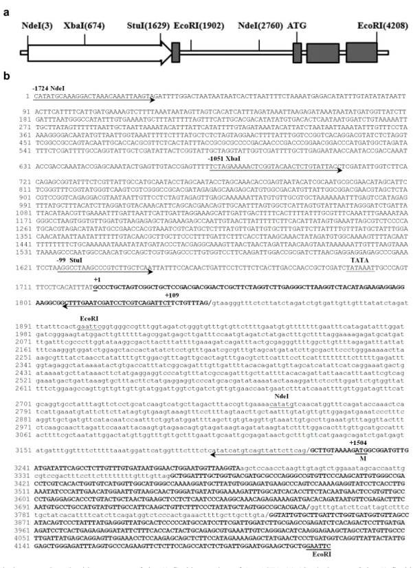
Fig. 3 Physical map and nucleotide sequence of the 5’ flanking region of DmACT1. (a) Physical map of the 5’ flanking region of DmACT1. Open arrow and gray boxes indicate the promoter region and exons of DmACT1, respectively. ATG indicates the translation initiation site. (b) Nucleotide sequence of the 5’ flanking region of DmACT1 (Genbank accession No. KC171012). Primers used for subsequent construction of promoter truncation vectors are indicated by arrows. The restriction sites used in inverse PCR are underlined. The transcription initiation site is designated as +1 and is underlined. The translation initiation site is indicated by an underlined M. The TATA box is underlined. Bold upper-case and lower-case letters represent exon and intron sequences, respectively.
Slashed lines indicate exon-intron boundaries. Note that the first intron resides in the 5’ UTR
gift from In-Sun Yoon, RDA, Korea) with its first intron was cloned in the same sites of GUS/Luciferase3300 vector to create pAct1. The DNA sequences of all constructs
were confirmed by DNA sequencing analysis. These constructs were used for transient luciferase reporter assays in Dendrobium flower cells.
Microprojectile bombardment
Promoter activity of constructs containing various lengths of DmACT1 5’ flanking sequence were examined by a microprojectile bombardment-mediated transient assay (Whang et al. 2011). Approximately 3.5 μg of gold particles (0.6 μm, Bio-Rad Laboratory, USA) were coated with 5 μg of DNA construct. As an internal control, 1 μg of CaMV35S promoter-Renilla luciferase construct (a kind gift from H.
G. Nam, DGIST, Korea) was co-bombarded. Flowers were bombarded with the DNA construct-coated gold particles at a helium gas pressure of 1,000 Psi and a target distance of 6 cm.
Analysis of Gus/luciferase activity
The reporter activity of the GUS/luciferase fusion protein was examined as described previously (Koo et al. 2007).
Briefly, the bombarded samples were transferred to a 24- well plate containing 0.5 mM D-luciferin or coelenterazine in MS medium, and bioluminescence was measured using a high-performance CCD camera (VersArray system, Roper Scientific Ltd., UK). Images were analyzed by MetaVue version 5.2 software (Universal Imaging Corporation, USA).
To normalize the actin promoter activity, the ratio of firefly luciferase activity was calculated and represented by comparison with that of the control Renilla luciferase.
Data are shown as mean values obtained from three independent microprojectile bombardment experiments.
Histochemical staining of GUS activity was performed by overnight incubation in X-GlcA (Duchefa, The Netherlands) substrate solution as described previously (Koo et al. 2007).
When necessary, samples were bleached with 70% ethanol.
Results
Molecular cloning of full-length DmATC1 cDNA
To isolate an actin gene from Dendrobium, we used a degenerate PCR approach using primers designed to DNA sequences conserved among orchid actin genes (See Materials and Methods). We cloned a 513 bp partial cDNA fragment, which we named DmACT1 (Fig. 1). Then, we isolated and characterized the 1573 bp full-length DmACT1 cDNA by 5’ and 3’ RACE PCR. RACE PCR was conducted using the SMARTer RACE cDNA amplification kit that allows the cloning of the full-length sequence of a target tran- script. Sequence analysis indicated that DmACT1 contains a 129 bp 5’ UTR, a 1131 bp coding sequence, and a 288
bp 3’ UTR (Fig. 1). A database search revealed that the nucleotide and deduced amino acid sequences of DmACT1 were the most similar to the nucleotide and amino acid sequences of Phalaenopsis ACT2 (Accession No. AF246715.1) (87% and 99%, respectively).
Expression of DmACT1 in Dendrobium
To determine whether DmACT1 expression was ubiquitous in Dendrobium, we performed RNA gel blot analysis using total RNA isolated from roots, leaves, and flowers.
Actins are ubiquitous proteins in plant cells and are encoded by a multigene family (Šlajcherová et al. 2012).
Because coding sequences are highly conserved among actin genes, the 5’ UTR of DmACT1 was amplified for use as a probe in RNA gel blot analysis. As shown in Fig. 2, DmACT1 transcripts were abundantly expressed in all organs that we examined. Although transcript levels were slightly higher in leaf than the other tissues, our results never- theless indicate that DmACT1is ubiquitously expressed in Dendrobium.
Isolation of genomic fragments containing the 5’ flanking region of DmACT1
The 5’ flanking sequences of DmACT1 were isolated by two round of inverse PCR. Circularized genomic DNA digested with Eco RI was used as a template in the first round of inverse PCR. However, a sequence comparison with the full-length DmACT1 cDNA revealed that the isolated genomic clone contained only the exon-intron boundaries and the truncated form of the intron without the sequence corresponding to the 5’ UTR (Fig. 3). Thus, we isolated upstream sequences by a second round of inverse PCR using circularized genomic DNA digested with Nde I. Figure 3 shows the full-length sequence of the isolated genomic clone. Sequence analysis revealed that the genomic clone contained a 3216 bp region upstream of the coding sequence of DmACT1. Because the nucleotide sequence of the full-length DmACT1 cDNA matched the base at position 1725 exactly, this was designated +1 as the putative transcription start site. In a motif search, a TATA box sequence was found between bases -29 and -23. The translation initiation codon was found at position +1504 in the second exon. Interestingly, the 5’ UTR of DmACT1 contains a 1374 bp long intron from +119 to +1492. However, the 5’ UTR-intron of DmACT1 is some- what longer than that reported in other plant species (Bradnam and Korf 2008), and shared no sequence similarity with known sequences in a database search.
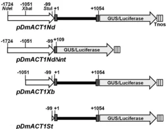
Fig. 4 Physical map of the DmACT1 5’ flanking sequence-GUS/
luciferase fusion constructs. Various portions of the 5'-flanking and/or 5'-noncoding UTRs of the DmACT1 gene were fused to the coding sequence of a GUS/luciferase fusion reporter gene.
White arrows show the promoter region. Gray and black boxes represent the exon and 5’UTR intron, respectively
DmACT1 promoter analysis by GUS/luciferase reporter transient assays
5’ UTR-introns can enhance promoter activity in animals and plants (Bradnam and Korf 2008). Thus, we generated four promoter constructs with or without the 5’ UTR-intron to examine the promoter activity of the 5’ flanking sequence of DmACT1.
For analysis of promoter activity, choice of reporter system is very important to avoid artifacts arising from the stability or multimerization of reporter protein. We previ- ously demonstrated that the GUS/luciferase fusion protein has the advantages of both GUS and luciferase proteins, namely high resolution histochemical GUS staining and real-time bioluminescence imaging with a luciferin-dependent rapid turn-over rate (Koo et al. 2007). Hence, we used the GUS/luciferase fusion reporter system for analysis of 5’
flanking sequence of DmACT1. As shown in Figure 4, a 2778 bp fragment of the flanking sequence of DmACT1 5’
(from -1724 to +1054) was fused to the coding sequence of the GUS/luciferase reporter gene to create pDmACT1Nd.
To examine the effect of the 5’ UTR-intron on gene ex- pression, we fused the 1833 bp fragment (-1724 to +105) lacking the 5’ UTR-intron to the coding sequence of the reporter gene to create pDmACT1Nd△int. Similarly, pDmACT1Xb and pDmACT1St constructs containing 2105 bp (-1051 to +1054) and 1153 bp (-99 to +1054) fragments of the 5’ flanking sequence of DmACT1, respectively, were generated by fusion with the coding sequence of Gus/
luciferase.
The flower is a very important plant characteristic in the floricultural industry and genetic manipulation of orchids.
Therefore, many researches primarily target the improve- ment of flower traits. RNA blot analysis indicated that DmACT1 is also highly expressed in flowers (Fig. 2).
Thus, we analyzed the promoter activity of the 5’ flanking sequence of DmACT1 in flowers of Dendrobium by transient expression assays.
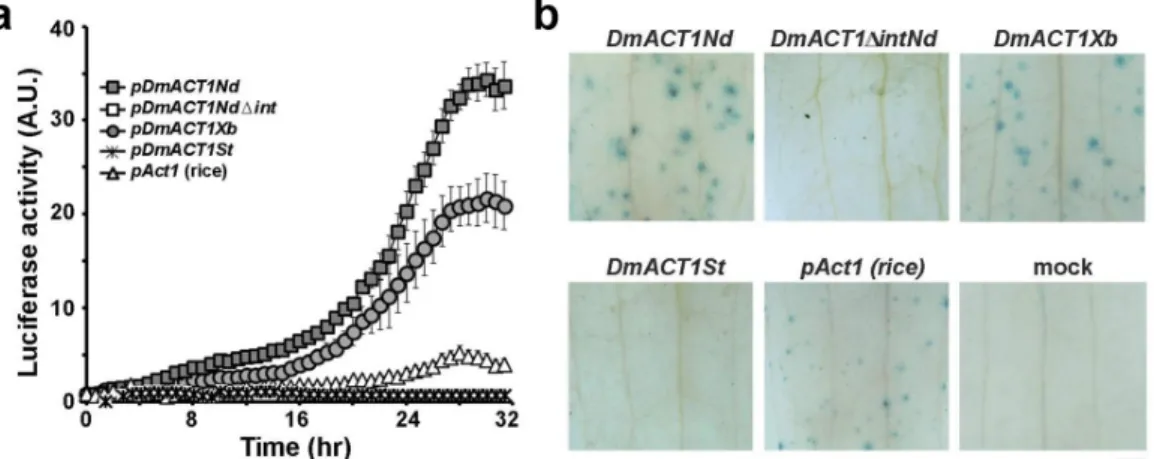
Since transient expression system was highly affected by DNA concentration, efficiency of bombardment and conditions of plant samples, CaMV35S promoter-Renilla luciferase construct was used in the experiment as an internal control to normalize the promoter activity. An unbombarded sample was used as a negative control. The rice actin promoter (pAct1), which is widely used in monocot plants, was also included in the experiment to compare promoter activities. The promoter activity of each bombarded sample was monitored by the time-lapse biolu- minescence imaging using a high-performance CCD camera.
Then, the value of firefly luciferase activity driven by actin promoter constructs was normalized with that of Renilla luciferase driven by CaMV35S promoter. As shown in Figure 5, flowers bombarded with pDmACT1Nd and pDmACT1Xb containing the 5’ UTR-intron showed high levels of luciferase activity, although pDmACT1Nd had 1.5-fold higher activity than pDmACT1Xb. Luciferase activity driven by pDmACT1Nd and pDmACT1Xb increased gradually after bombardment, and steady-state levels were reached after 28 to 30 h of incubation. Furthermore, pDmACT1Nd showed 8- to 10-fold higher luciferase activity than the rice actin promoter (pAct1). However, flowers bombarded with pDmACT1Nd△int displayed undetectable levels of luciferase activity. These results suggest that the 5’ UTR-intron is necessary for efficient promoter activity of DmACT1. Flowers bombarded with pDmACT1St, which lacks the large portion of promoter region, showed near basal-levels of luciferase activity.
The promoter activity of the 5’ flanking sequence of DmACT1 was further confirmed by histochemical GUS staining. Similar with luciferase activity, the strong his- tochemical GUS stains were observed in flowers bombarded with pDmACT1Nd and pDmACT1Xb, whereas no detect- able activity was observed in flowers bombarded with pDmACT1Nd△int and pDmACT1St. These results confirm that the 5’ UTR-intron is necessary for efficient promoter activity of DmACT1. As expected, the rice Act1 promoter (pAct1) displayed weaker GUS stains than the DmACT1 5’
flanking sequence. Taken together, our results demonstrate that the 5’-flanking sequence of DmACT1 can promote strong gene expression in Dendrobium.
Fig. 5 Analysis of transient GUS/luciferase activity driven by various portions of the DmACT1 5’-flanking sequence in Dendrobium flowers. Each construct was co-bombarded with pCaMV35S-Renilla luciferase. Expression of the promoter from the rice actin promoter (pAct1) in Dendrobium was also evaluated for comparison purposes. (a) Relative luciferase activity in bombarded flowers.
Bombarded sample was divided into two parts and transferred to medium containing luciferin and coelenterazine in a 24-well plate.
Bioluminescence was measured using a high-performance cool CCD every 40 min for 32 hr. The values were subtracted from those of unbombarded samples (mock) and normalized relative to the Renilla luciferase activity. Note that pDmACT1Nd△Int and pDmACT1St showed background levels of bioluminescence. Bars indicate SD of the mean value (n=3). (b) GUS histochemical staining obtained after 32 hr of incubation. Representative images from three independent experiments are shown. The scale bar indicates 1 mm
Discussion
Promoters that drive strong and constitutive gene expression are important for plant biotechnology applications. Several promoters have been developed for use in monocot transformation including maize Ubi1 (Elliott et al. 1999) and rice Act1 (McElroy et al. 1990). Although these promoters drive strong expression of genes in rice, they show lower activity than the CaMV 35S promoter in some monocot plants (Kamo et al. 1995; Wilmink et al. 1995).
This is also the case in Dendrobium. GUS expression driven by the Ubi1 promoter was lower than that of GUS expression driven by the CaMV 35S promoter in Dendrobium sonia (Tee et al. 2003). Furthermore, the efficiency of the Act1 promoter in orchids, including Dendrobium, has not been evaluated. Actin genes belong to a multigene family in plants, with some actin isoforms constitutively and strongly expressed in plants (An et al. 1996; McElroy et al.
1990; Šlajcherová et al. 2012). In this study, we demonstrated that DmACT1 expression is ubiquitous and abundant in the flowers, leaves, and roots of Dendrobium by RNA blot analysis (Fig. 1). Thus, we isolated the DmACT1 promoter to determine if it could drive strong expression of a reporter gene in Dendrobium.
We isolated the 5’-flanking sequence of DmACT1 by two round of inverse PCR. Sequence analysis indicated that it contained a 1374 bp intron in the 5’ UTR region.
To examine if this region had promoter activity, we fused various lengths of DmACT1 5’ flanking sequence to a GUS/
luciferase fusion reporter (Koo et al. 2007) to monitor
promoter activity by real-time luciferase imaging and to assess GUS activity by histochemical staining. We also utilized a transient expression system for promoter analysis.
Although cellular expression patterns can be determined in stable transgenic plants in contrast to transiently transfected plants, undesirable effects may occur in measurement of promoter activity due to the presence of flanking sequences in the T-DNA insertion site, multiple gene copies, and silencing or co-suppression of genes. Transient GUS/
luciferase expression analysis revealed that the DmACT1 5’ flanking sequence had 8- to 10-fold higher promoter activity in the flower organs of Dendrobium orchids than the rice actin promoter (pAct1). Hence, the DmACT1 pro- moter can be used to drive strong ectopic expression of transgenes in Dendrobium.
It has been shown that some 5’ UTR-introns contribute to high levels of transgene expression in both dicot and monocot transgenic plans including Arabidopsis, maize, and rice (Bradnam and Korf 2008; Jeon et al. 2001; Lu et al. 2008; Zhang et al. 1991; Rose and Last 1997). Although the underlying mechanism remains unclear, it has been shown that the effect of the 5’ UTR-intron decreases grad- ually as its distance from the translation initiation site increases (Rose 2004). Furthermore, the 5’ UTR-intron does not drive gene expression when located in the 3’ UTR.
Second, the IME effect of the 5’ UTR-intron is not general to all organisms. The rice Act1 intron and the maize Ubi1 intron greatly enhances gene expression in rice, but have negligible effects in non-cereal monocot plants (Kamo et al. 1995; Tee et al. 2003; Wilmink et al.
1995). Moreover, the combination of the CaMV35S pro- moter and maize Adh15’ UTR-intron resulted in decreased GUS expression in the protoplasts of sugarcane (Wilmink et al. 1995), whereas addition of the maize shrunken-1 locus 5’ UTR-intron to the same promoter resulted in greatly increased CAT gene expression in the protoplasts of various grass species (Vasil et al. 1989). Third, IME differs between plant and animal cells. Highly expressed genes have a shorter 5’ UTR-intron in animal cells than plant cells, and vice versa (Castillo-Davis et al. 2002; Ren et al. 2006). Fourth, the effect of IME may be due to trans- criptional or post-transcriptional events. In some cases, IME has been shown to be related to increased enzyme activity or protein accumulation rather than increased mRNA levels (Bourdon et al. 2001; Kamo et al. 2012;
Mascarenhas et al. 1990; Samadder et al. 2008; Rose 2004). Moreover, the effect of the 5’ UTR-intron of rice ubiquitin (rub3) on promoter activity has been shown to be tissue- and cell-type specific (Lu et al. 2008). Thus, it would be interesting to explore the IME mechanism of the 5’ UTR-intron in DmACT1 in future studies. Taken together, our data suggest that the DmACT1 promoter will be of great use in orchid biotechnology as it drives high levels of transgene expression, and represents a good genetic resource for the development of even more efficient orchid promoters.
Acknowledgements
The author wish to thank Drs. Hong Gil Nam, Ju-Kon Kim and In Sun Yoon for generously providing Renilla luciferase construct and rice actin promoter. The author would like to thank Dr. Moon-Sik Yang, Hyo-Yeon Moon and Ki-Nam Kim for kindly providing equipment and technical assistance in the experiment of microprojectile bombardment. This work was supported by grants from Korea National Arboretum (the development and conser- vation of useful plant resources of in and outside the country, 2012).
References
An YQ, McDowell JM, Huang S, McKinney EC, Chambliss S, Meagher RB (1996) Strong, constitutive expression of the Arabidopsis ACT2/ACT8 actin subclass in vegetative tissues.
Plant J 10:107-121
Bourdon V, Harvey A, Lonsdale DM (2001) Introns and their positions affect the translational activity of mRNA in plant cells. EMBO Rep 2: 394-398
Bradnam KR, Korf I (2008) Longer first introns are a general property of eukaryotic gene structure. PloS one 3: e3093.
Castillo-Davis CI, Mekhedov SL, Hartl DL, Koonin EV, Kondrashov FA (2002) Selection for short introns in highly expressed genes. Nat Genet 31:415-418
Chia TF, Chan YS, Chua NH (1994) The firefly luciferase gene as a non-invasive reporter for Dendrobium transformation. Plant J 6:441-446
Doyle JJ, Doyle L (1990) Isolation of plant DNA from fresh tissue.
Focus 12:13-15
Elliott AR, Campbell JA, Dugdale B, Brettell, RIS, Grof CPL (1999) Green fluorescent protein facilitates rapid in vivo detection of genetically transformed plant cells. Plant Cell Rep 18:707-714
Guilley H, Dudley RK, Jonard G, Balfizs E, Richards KE (1982) Transcription of cauliflower mosaic virus DNA: Detection of promoter sequences, and characterization of transcripts. Cell 30:763-773
He C, Lin Z, McElroy D, Wu R (2009) Identification of a rice actin2 gene regulatory region for high-level expression of transgenes in monocots. Plant Biotechnol J 7:227-239 Ho SH, So GM, Chow KL (2001) Postembryonic expression of
Caenorhabditis elegans mab-21 and its requirement in sensory ray differentiation. Dev Dyn 221: 422-430
Jeon JS, Lee S, Jung KH, Jun SH, Kim C, An G (2000) Tissue- preferential expression of a rice alpha-tubulin gene, OsTubA1, mediated by the first intron. Plant Physiol 123: 1005-1014 Jonsson JJ, Foresman MD, Wilson N, McIvor RS (1992) Intron
requirement for expression of the human purine nucleoside phosphorylase gene. Nucleic Acids Res 20: 3191-3198 Kamo K, Blowers A, Smith F, Van Eck J, Lawson R (1995) Stable
transformation of Gladiolus using suspension cells and callus.
J Am Soc Hort Sci 120:347-352
Kamo K, Kim AY, Park SH, Joung YH (2012) The 5' UTR-intron of the Gladiolus polyubiquitin promoter GUBQ1 enhances translation efficiency in Gladiolus and Arabidopsis. BMC Plant Biol 12:79
Koo J, Kim Y, Kim J, Yeom M, Lee IC, Nam HG (2007) A GUS/luciferase fusion reporter for plant gene trapping and for assay of promoter activity with luciferin-dependent control of the reporter protein stability. Plant Cell Physiol 48:1121-1131 Koo JC, Asurmendi S, Bick J, Woodford-Thomas T, Beachy
RN (2004) Ecdysone agonist-inducible expression of a coat protein gene from tobacco mosaic virus confers viral resistance in transgenic Arabidopsis. Plant J 37:439-448
Kuehnle AR, Sugii N (1992) Transformation of Dendrobium orchid using particle bombardment of protocorms. Plant Cell Rep 11:484-488
Lu J, Sivamani E, Li X, Qu R (2008) Activity of the 5' regulatory regions of the rice polyubiquitin rubi3 gene in transgenic rice plants as analyzed by both GUS and GFP reporter genes. Plant Cell Rep 27:1587-1600
Mascarenhas D, Mettler IJ, Pierce DA, Lowe HW (1990) Intron- mediated enhancement of heterologous gene expression in maize. Plant Mol Biol 15: 913-920
McElroy D, Rothenberg M, Reece KS, Wu R (1990) Charac- terization of the rice actin gene family. Plant Mol Biol 15:
257-268
Palmiter RD, Sandgren EP, Avarbock MR, Allen DD, Brinster RL (1991) Heterologous introns can enhance expression of transgenes in mice. Proc Natl Acad Sci U S A 88: 478-482 Ren X-Y, Vorst O, Fiers MWEJ, Stiekema WJ, Nap J-P (2006) In
plants, highly expressed genes are the least compact. Trends Genet 22:528-532
Rose AB (2004) The effect of intron location on intron-mediated enhancement of gene expression in Arabidopsis. Plant J 40:744-751
Rose AB, Last RL (1997) Introns act post-transcriptionally to increase expression of the Arabidopsis thaliana tryptophan pathway gene PAT1. Plant J 11: 455-464
Samadder P, Sivamani E, Lu J, Li X, Qu R (2008) Transcriptional and post-transcriptional enhancement of gene expression by the 5' UTR intron of rice rubi3 gene in transgenic rice cells.
Mol Genet Genomics 279:429-439
Sheen J, Hwang S, Niwa Y, Kobayashi H, Galbraith DW (1995) Green fluorescent protein as a new vital marker in plant cells.
Plant J 8:777-784
Shibata, M. (2008) Importance of genetic transformation in ornamental plant breeding. Plant Biotech 25:3-8
Šlajcherová K, Fišerová J,Fischer L,Schwarzerová K (2012) Multiple actin isotypes in plants: diverse genes for diverse roles? Front Plant Sci. 3:226
Sunilkumar G., Mohr LA, Lopata-Finch E, Emani C, Rathore KS (2002) Developmental and tissue-specific expression of CaMV 35S promoter in cotton as revealed by GFP. Plant Mol Biol 50: 463-474
Tee CS, Marziah M, Tan CS, Abdullah MP (2003) Evaluation of
different promoters driving the GFP reporter gene and select- ed target tissues for particle bombardment of Dendrobium Sonia 17. Plant Cell Rep 21: 452-458
Tereda R, Shimamoto K (1990) Expression of CaMV 35S-GUS gene in transgenic rice plants. Mol Gen Genet 220:389-392 Vasil V, Clancy M, Ferl RJ, Vasil IK, Hannah LC (1989) Increased
gene expression by the first intron of maize shrunken-1 locus in grass species. Plant Physiol 91:1571-1579
Wasteneys GO, Galway ME (2003) Remodeling the cytoskeleton for growth and form: an overview with some new views. Ann Rev Plant Biol 54:691-722
Wilmink A, Van de Ven BCE, Dons JJM (1995) Activity of constitutive promoters in various species of the Liliaceae.
Plant Mol Biol 28:949-955
Whang SS, Um WS, Song IJ, Lim PO, Choi K, Park KW, Kang KW, Choi MS, Koo JC (2011) Molecular analysis of anthocyanin biosynthetic genes and control of flower coloration by Fla- vonoid 3′,5′-Hydroxylase (F3′5′H) in Dendrobium moniliforme.
J Plant Biol 54: 209-218
Wood HP (2006) The Dendrobiums. AR. G. Gantner Verlag, Ruggell, Leichenstein.
Yang SH, Yu H, Goh CJ (2003) Functional characterisation of a cytokinin oxidase gene DSCKX1 in Dendrobium orchid. Plant Mol Biol 51:237-248
Yu H, Yang SH, Goh CJ (2001) Agrobacterium-mediated trans- formation of a Dendrobium orchid with the class 1 knox gene DOH1. Plant Cell Rep 20:301-305
Zhang W, Wu R (1988) Efficient regeneration of transgenic plants from rice protoplasts and correctly regulated expression of the foreign gene in the plants. Theor Appl Genet 76: 835-840 Zhang W, McElroy D, Wu R (1991) Analysis of rice Act 1 5' region
activity in transgenic rice plants. Plant Cell 3:1155-1165