24
접수일: 2009년 10월 22일, 수정일: 2009년 12월 14일 승인일: 2009년 12월 15일
교신저자: 홍대식
Tel: 032-621-5186, Fax: 032-621-5018 E-mail: [email protected]
우리나라 일개 병원 암 환자에서 중심정맥관 합병증에 관한 후향적 조사
김은정ㆍ김현정ㆍ김한조ㆍ김경하ㆍ김세형ㆍ이상철ㆍ배상병 김찬규ㆍ이남수ㆍ이규택ㆍ박성규ㆍ원종호ㆍ박희숙ㆍ홍대식
순천향대학교 의과대학 혈액종양내과학교실
Retrospective Analysis for Complications of the Central Venous Catheter in Patients with Cancer at a Single Center in Korea
Eun-Jung Kim, M.D., Hyun Jung Kim, M.D., Han Jo Kim, M.D., Kyoung Ha Kim, M.D., Se Hyung Kim, M.D., Sang-Cheol Lee, M.D., Sang Byung Bae, M.D., Chan Kyu Kim, M.D., Nam Su Lee, M.D., Ph.D., Kyu Taek Lee, M.D., Ph.D., Sung Kyu Park, M.D., Ph.D., Jong-Ho Won, M.D., Ph.D., Hee Sook Park, M.D., Ph.D. and Dae Sik Hong, M.D., Ph.D.
Division of Hematology and Oncology, Department of Internal Medicine, Soonchunhyang University College of Medicine, Bucheon, Korea
Purpose: A central venous catheterization (CVC) is frequently used for delivering anti-cancer chemotherapeutic
agents, blood products, parenteral nutrition, and other intravenous therapy in patients with cancer. Major com- plications of CVC use are thrombosis, infection, and mechanical complications. The aim of this study was to evaluate the frequency of CVC complications and related factors. Methods: The records of cancer patients who received a CVC at our university hospital from March 2001 to October 2006 were retrospectively investigated. Chi square test was used to determine whether there was a related factor for thrombosis or infection, and Kaplan-Meier analysis for univariate analysis, or Cox-regression analysis for multivariate analysis was used for catheter life span. Results:Three hundred and ten CVCs (235 nontunneled, 75 tunneled) were inserted in 310 patients (157 males, 153 females). Among them, 104 had hematologic cancers and 206 had solid cancers. The mean age of the patients was 52 years (range, 19∼82 years). CVC complications occurred in 60 cases (19%). CVC-related thrombosis occurred frequently in patients with infection (P=0.003), whereas diagnosis, catheter type, transfusion, and TPN history did not affect infection or thrombosis. The mean duration of the catheter was 102 days (range, 2∼1,330 days), and the duration was prolonged in patients with tunneled catheters (P=0.000), or without transfusion through CVC (P=0.030). Conclusion: The major complications for long-term use of a CVC were infectionand thrombosis. Tunneled catheter was effective tool for long term use, especially in cases without transfusion through CVC. The studies on the prevention or treatment ofthrombosis and infection are, therefore, warranted by using CVC for an extended period of time. (Korean J Hosp Palliat Care 2010;13:24-31)
Key Words: Catheterization, Central venous, Hematologic neoplasms/complications, Neoplasms
서 론
중심정맥관 삽입은 일반적으로 환자들의 생징후가 불 안정하여 다량의 수액 및 약물 투여를 해야할 경우나, 말초 혈관의 혈관 확보가 어려운 경우, 응급 혈액 투석
이 필요한 경우 시행되며, 암환자들에게 있어서는 항암 화학 요법, 수혈, 영양 공급 등의 목적으로 유용하게 사 용되는 시술이며, 특히 호스피스환자에서는 정맥관삽입 이 치료의 첫 단계가 되기도 한다. 크게 비터널식 정맥 관과 터널식 정맥관으로 나뉘며 일반적인 경우는 주로 비터널식 정맥관을 삽입하고, 면역성이 떨어져 있는 암 환자들에서는 감염 예방 및 장기 사용을 위해 터널식 정맥관 삽입을 고려하는 경우가 많다(1,2). 중심 정맥관 삽입 시 15% 이상에서 합병증이 발생한다는 보고가 있 으며(3-5), 그 종류로는 혈전(2∼26%), 감염(5∼26%), 물 리적 합병증(5∼19%) 등이 있다(3-7). 이는 환자의 사망 률과 입원 일수 및 의료 비용이 증가하는 문제를 야기 한다(1,2). 이러한 합병증 발생을 막기 위해서는 삽입 시 시술자의 경험이 중요하며, 예방적 항응고제 및 항생제 를 도관 내에 주입하는 방법이 사용되기도 한다.
우리나라에서 중심정맥관 시술에 따른 합병증에 대 한 연구는 적은 편으로(8,9), 이 연구는 암환자에서 중심 정맥관의 삽입과 관련된 위험인자와 효용인자를 밝혀 내고, 합병증에 대한 삽입관의 관리와 예방, 암환자에 있어 적절한 중심 정맥관 사용을 알기 위해 시행되었 다.
대상 및 방법 1. 연구대상
혈액암 혹은 고형암을 진단받고 부천 순천향대학교병 원 혈액종양내과에서 치료받는 환자 중 2001년 3월부터 2006년 8월까지 중심정맥관을 삽입한 환자들의 경과기 록 분석을 통한 후향적 연구를 실시하였다.
2. 연구방법
삽입관은 터널식과 비터널식으로 구분하였으며, 비터 널식에는 쇄골하 정맥관(subclavian catheter), 내경 정맥관 (Intrajugular vein catheter), 터널식에는 히크만 도관(Hick- mann catheter)와 Chemoport가 포함되었고, 삽입 장소는 혈관 조영실과 병실로 구분하였다. 관의 사용 기간은 관 삽입에서부터 어떤 이유로든 관을 제거한 시기까지 로 정의하였다. 그리고 삽입관의 종류 및 환자의 연령 과 성별, 수혈과 비경구 영양(Total parenteral nutrition) 시 행력에 따른 관의 사용기간 및 중심 정맥관으로 인한 여러 합병증에 대해 조사하였다.
중심 정맥관 합병증을 크게 감염과 혈전, 물리적 합 병증으로 나누었으며 물리적 합병증에는 기흉, 혈흉, 정
맥관 첨부의 위치이상, 정맥관 파손, 환자가 자가 제거 한 경우, 관의 기능이상, 관으로 인한 통증 등을 포함하 였고, 감염의 경우, 정맥관의 군락, 삽입 부위의 감염, 관 통과부위(터널) 감염 혹은 혈중 감염을 포함하였다.
또한 감염은 추정감염 및 확진 감염으로 나누었는데, 추정 감염은 패혈증의 증후, 패혈성 쇼크, 삽입부위 감 염을 포함하여 발열, 백혈구와 C-reactive protein 등 염증 수치의 증가, 삽입부위의 발진 및 통증, 농양의 생성 등 감염이 의심되고 정맥관 감염 외 다른 감염 원인을 찾 을 수 없으나 균이 증명 안된 경우로 정의하였고, 확진 감염은 말초 및 도관을 통해 채혈한 혈액 배양 혹은 도 관 제거 후 도관 첨부의 균 배양을 통해 균이 증명된 경우로 정의하였다. 혈전은 사지나 얼굴의 부종, 통증, 호흡곤란 등 급성 심부 정맥 혈전증이나 폐색전증의 증 상을 나타내는 환자 및 관을 통한 수액 흐름의 장애, 증 상과 상관없이 정맥 혈관 조영술 및 도플러 초음파 등 영상학적 진단기법으로 확진이 이루어진 경우로 정의 하였다.
혈전에 대하여 예방적 항혈전제는 투여하지 않았으 며, 혈전 발생 시는 도관 내에 우로키나아제(Urokinase) 를 채워 처치하였고, 감염이 의심될 경우는 2주간의 전 신적 항생제 투여 및 도관 내 항생제 주입 방법(Locking therapy)을, 도관 삽입부위 감염이나 합병증 발생시, 진 균이나 그람 음성균 감염이 확진된 경우는 도관을 제거 하였다.
3. 분석방법
환자군의 특성을 파악하기 위해서, 범주형 변수에 따 른 차이를 분석하기 위해 chi-square test를 사용하였고, 연속형 변수에 대해서는 independent t-test를 사용하였다.
또한 중심 정맥관 합병증과 관련된 인자가 있는지 알기 위해 교차분석을 사용하였다.
더불어 삽입관의 수명과 이에 대한 예측인자가 있는 지 알기 위해 단변량 분석에는 log-rank test를 이용한 Kaplan-meier 방법을 사용하였고, 다변량분석시에는 Cox regression analysis test를 사용해 분석하였다.
결 과 1. 환자의 특성
2001년 3월부터 2006년 8월까지 총 310명의 환자가 후향적으로 등록되었으며, 그 중 235건의 비터널식 및 75건의 터널식 중심 정맥관 시술이 시행되었다. 이 중
Table 1. Characteristics of Patients.
Nontunneled cath.
Tunneled
cath. Total P value*
Sex
Male 118 39 157 0.793
Female 117 36 153
Insertion state
Angio-room 215 75 290
0.005
Bedside 20 0 20Disease
Solid cancer 142 64 206
0.000
Blood cancer 93 11 104Duration of cath.
Use day, 28±23 322±317 99±201
0.000
mean±SD (range) (2∼144) (6∼1,330) (2∼1,330) Age yr, mean±SD, 52±17 52±14 52±16 0.905(range) (15∼82) (20∼78) (15∼82)
Cath: catheter, SD: standard deviation, Yr: year. *This values were analyzed by the chi-square test or independent t-test.
Table 2. Complications Related to the Central Venous Catheter in 310 Patients.
Complications n %
Infection
Suspicious 12 3.9
Definite 22 7.1
Thrombosis 11 3.5
Mechanical complications
Self-removal 2 0.6
Malfunction 6 1.9
Pain 5 1.7
Mal-position 1 0.3
Breakage of catheter 1 0.3
Total 60 19.3
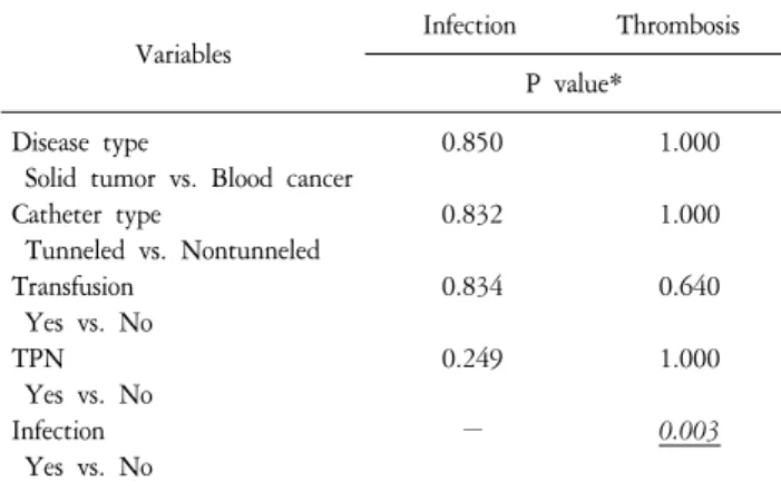
Table 3. The Affecting Factors for Complications.
Variables Infection Thrombosis
P value*
Disease type 0.850 1.000
Solid tumor vs. Blood cancer
Catheter type 0.832 1.000
Tunneled vs. Nontunneled
Transfusion 0.834 0.640
Yes vs. No
TPN 0.249 1.000
Yes vs. No
Infection −
0.003
Yes vs. No
TPN: total parenteral nutrition. *This values were analyzed by chi-square test.
남자는 157명, 여자는 153명이었고, 혈액암 환자가 132 명, 고형암 환자 178명이었으며, 혈관 조영실에서 삽입 한 경우가 290명, 병실에서 삽입한 경우가 20명이었다.
병실에서보다 혈관 조영실에서(P=0.005), 혈액암보다 고형암에서(P=0.000) 터널식 중심 정맥관 삽입이 유의하 게 많았으며, 성별과는 크게 상관이 없었다(P=0.793).
평균 연령은 52세였고(range: 15∼82) 삽입관의 평균 수 명은 99일이었다(range: 2∼1,330)(Table 1).
2. 중심정맥관과 관련된 합병증의 빈도와 연관인자
삽입관으로 인한 합병증은 총 60예(19%)에서 발생하 였는데, 이 중 감염이 34예(10.9%)이었고 추정감염 12예 (3.9%), 확진 감염 22예(7.1%)였다. 혈전은 11예(3.5%), 통 증이나 관의 손상, 위치 및 기능이상, 자가제거 같은 물 리적 합병증은 15예(5%)로 나타났다(Table 2).
발생한 여러 합병증 중 감염과 혈전은 밀접한 연관이 있는 것으로 나타났는데, 감염이 발생한 34예 중 5예에 서 혈전이 발생하였으며(P=0.003), 암(고형암 vs. 혈액암) 과 삽입관의 종류(터널식 vs 비터널식), 수혈 및 비경구 영양 시행 여부와 합병증 발생과는 큰 연관성이 없었다 (Table 3).
3. 삽입관의 수명과 제거이유
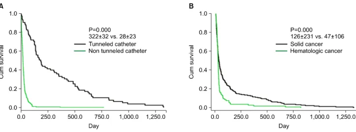
삽입관의 평균수명은 99일(range: 2∼1,330)이었는데, 고형암인 경우와 혈액암인 경우를 비교했을 때 각각 126일과 47일로 고형암인 경우 수명이 더 길었으며(P=
0.000), 터널식 정맥관의 경우과 비터널식 정맥관의 경 우 각각 322일과 28일로 터널식에서 수명이 연장된 것 으로 나타났다(P=0.000). 또한 중심 정맥관을 통해 수혈 을 한 경우가 88일, 수혈을 하지 않은 경우가 172일로 수명이 연장되었다(P=0.044). 이에 대한 인자들을 다시 다변량분석으로 확인했을 때 고형암은 의미 있는 인자 가 아니었고(P=0.146), 터널식 정맥관과(P=0.000) 수혈 여부(P=0.030)는 삽입관 수명에 영향을 미치는 독립적 인자로 확인되었다. 이에 비해 정맥관의 수명과 연령, 성별, 비경구 영양 시행여부, 감염, 혈전, 물리적 합병증 과는 유의한 연관성이 없었다(Table 4, Figure 1).
조사기간 중 전체 310명 중 49명(13%)이 합병증으로 인해 관을 제거하였고, 가장 많은 원인은 감염이었다 (7.7%).
Table 4. The Affecting Factors for Catheter Life Span.
Variables Life span, days, median (95% CI) Univariate analysis Multivariate analysis P value†
Age <55 vs. ≥55 29 (25∼33) vs. 24 (19∼29) 0.373 −
Male vs. Female 29 (24∼34) vs. 26 (21∼31) 0.521 −
Solid cancer vs. Blood cancer 33 (25∼41) vs. 24 (21∼27)
0.000
0.146Tunneled vs. Nontunneled catheter 187 (134∼240) vs. 22 (20∼24)
0.000 0.000
Infection 26 (23∼29) vs. 53 (30∼76) 0.697 −
Thrombosis 28 (25∼31) vs. 38 (16∼60) 0.491 −
Mechanical complications* 28 (25∼31) vs. 66 (7∼125) 0.356 −
Transfusion 37 (0∼79) vs. 28 (25∼31)
0.044 0.030
TPN 30 (23∼37) vs. 26 (22∼30) 0.131 −
CI: confidence interval, TPN: total parenteral nutrition. *Mechanical complications included in cases of self-removal, malfunction, pain, malposition and breakage of catheter. †This value were analyzed by log rank test in univariate anlaysis or Cox regression analysis in multivariate anlaysis.
Figure 1. Differences of the life span of catheter according to type of catheter (A) and cancer (B).
고 찰
중심 정맥관 삽입은 암환자에서 여러 목적으로 흔히 사용되는 술기이며, 보고된 합병증으로는 삽입하는 동 안 혹은 삽입 직후 직접적으로 발생하는 기흉, 혈흉, 관 의 위치 및 기능 이상, 통증, 자가 제거, 정맥관 파손과 같은 물리적 합병증 및 장기적 합병증으로 혈전과 감염 이 있다(1,2). 중심 정맥관 삽입 시 15% 이상에서 합병 증이 발생한다는 보고가 있으며(3-5), 각각의 발생률을 연구한 결과들을 보면 혈전 2∼26%, 감염 5∼26%, 물 리적 합병증 5∼19% 정도이다(3-7). 물리적 합병증을 줄 이기 위해서는 경험이 있는 의사가 주의 깊게 시술해야 하며(10,11) 만약 3번의 시도에도 삽입이 되지 않았다면, 다른 의사의 도움을 받아야 한다. 3번의 시도 이후에 발
생하는 물리적 합병증의 발생률은 한 번의 시도로 성공 했을 경우에 비해 6배가 높다고 알려져 있다(7). 초음파 를 이용하여 정맥의 위치와 깊이를 측정 후 관을 삽입 하는 것도 삽입 도중 합병증 발생의 위험을 낮추는 데 도움이 될 수 있으나 쇄골 하 중심 정맥관 삽입 시에는 쇄골 하 정맥과 쇄골의 고정된 해부학적 위치 때문에 피부의 해부학적 표지를 기준으로 삽입하는 것보다 더 어려울 수 있다. 그래서 초음파를 이용하여 중심 정맥 관을 삽입하는 것은 대개 내경 정맥관 삽입을 시도할 때 고려된다(12,13). 본 연구에서는 총 310명의 환자들 중 15예(4.8%)에서 물리적 합병증이 발생하여 이전 연 구들의 결과와 큰 차이가 없었다. 또한 혈관 조영실에 서 정맥관을 삽입하는 경우는 초음파와 정맥 혈관 조영 술을 이용하여 시술하였고, 병실에서 시행한 경우는 해 부학적 표지를 이용하여 정맥관을 삽입하였는데, 삽입
장소에 따른 물리적 합병증 발생률은 유의한 차이가 없 었다. 하지만 병실에서 삽입한 사례가 혈관 조영실에서 삽입한 경우보다 매우 적음을 생각할 때 이러한 결과는 통계적 오류의 가능성이 있다.
다음으로, 중심 정맥관으로 인한 혈전은 무증상이면 서 선별적 영상검사를 통해 발견되기도 하고, 팔, 목, 머 리 통증이나 부종, 사지의 저림과 홍반, 정맥 확장 및 턱의 통증 등 증상으로 의심해 볼 수 있다(2,14,15). 중심 정맥관 관련 혈전의 발생 기전에는 다양한 인자가 관여 하는데, 비르코우 3주징(Virshow’s triad)의 축을 이루는 혈관의 손상이 중요한 역할을 한다고 알려져 있다(16).
혈관 손상은 중심 정맥관을 삽입 시 정맥 내피의 물 리적 손상, 정맥 천자의 횟수, 항암제에 의한 혈관벽의 자극 등에 의해 일어날 수 있는데(16,17), 중심 정맥관 삽입 후에는 섬유소와 교원질이 관을 둘러싸게 되고, 이러한 피복막은 그 자체로는 큰 문제가 없는 합병증이 지만 관의 기능이상을 일으킬 수 있고, 감염을 촉진시 키거나 관 내 혈전을 유발하는 요인이 된다(18,19).
중심 정맥관으로 인한 혈전은 심각한 합병증들을 야 기할 수 있는데, 관이 종격동 깊이 위치 함으로써, 혈전 이 심각한 상태에 이르기까지 임상적으로는 이상 증상 이 나타나지 않을 수 있다. 폐색전증이 증상을 가진 중 심 정맥관 혈전 환자의 15∼25%에서 일어나며(20,21), 상지 심부 정맥 혈전을 가진 환자의 14.8%에서 정맥염 후 증후군이 발생한다는 보고가 있다(22). 또한 정맥관 혈전을 가진 환자에서 감염의 위험성이 높아진다는 보 고들이 있다(23,24). 본 연구의 결과에서 중심 정맥관으 로 인한 혈전의 발생은 11예로(3.5%) 이전 연구 결과들 에 비해 적은 편이었으나 그 중 5예에서 감염이 동반되 어(P=0.003), 혈전의 발생과 감염이 서로 밀접한 관련이 있음이 증명되었다.
중심정맥관 관련 혈전에 대한 치료는 정해진 지침은 없으나 혈전 용해 요법 및 전신 항혈전 요법의 시행, 관 의 제거 등을 포함한다. 고형암을 가진 환자에서는 저 용량 헤파린과 함께 경구 항혈전제를 투여하고 최소 3
∼6개월을 유지하는 것을 우선으로 하나 혈액암이면서 특히 혈소판이 감소된 환자에서는 출혈의 위험성이 있 어 상대적인 금기이다(25-27). 고식적인 치료법은 저용 량의 우로키나아제(Urokinase)나 스트렙토키나아제(Strep- tokinase) 혹은 조직 플라스미노겐 활성제(Tissue plasmi- nogen activator)를 단발성 혹은 반복적으로 정맥관에 주 입하여 첨부 혹은 피막의 폐색을 막는 방법으로, 이는 대부분의 환자에서 정맥관의 개통성이 복원되며 관의
위치를 잘 유지할 수 있게 한다(28,29).
정맥관 혈전을 막기 위한 예방적 항혈전제 투여는 출 혈 위험성 때문에 혈액암 환자에서는 그 사용이 제한적 이고, 주로 고형암을 가진 환자에서 하게 되는데 그 방 법으로는 주로 하루 와파린 1 mg을 복용하는 경구 요법 을 사용한다(30,31). 그러나 최근의 여러 연구결과에서 저용량 와파린의 예방효과는 없는 것으로 나타나 현재 는 특별한 예방적 항응고제 사용은 추천되지 않는다 (32,33). 본 연구에서는 예방적 항혈전제 투여는 하지 않 았으며, 혈전 발생 시에는 저용량 우로키나아제를 사용 하여 정맥관에 주입하는 치료 방법을 사용하였고, 1예 에서 이러한 방법으로 혈전 발생시 정맥관을 제거하지 않고 지속적으로 사용할 수 있었다.
중심정맥관 관련 감염은 관의 균 군락형성, 삽입 부 위의 감염, 터널 감염 및 혈류감염 등으로 나눌 수 있는 데(34), 특히 자가 조혈 모세포 이식에 따른 항암요법을 받았거나 중심 정맥관으로 인한 혈전을 가진 환자에서 그 위험성이 높아진다고 알려져 있다(23,35).
균의 가능한 침투 경로로는 관 삽입부위의 피부오염, 도관 연결부위(hub)의 오염, 감염이 있는 원위부로터의 혈류성 전파, 혹은 오염된 수액이나 주사제의 주입 등 이며(34,36), 터널식 중심정맥관의 경우 도관 연결부위 의 오염이, 비터널식의 경우 피부의 오염이 가장 흔한 감염의 원인으로 보고되었다(36). 흔한 원인균으로는 그 람음성 포도알균(Coagulase-negative Staphylococcus), 황색 포도알균(Staphylococcus aureus), 그람음성 막대균(Gram negative bacilli)-ex. 장내세균(Enterobacteriaceae), 대장균(Es- cherichia coli)- 녹농균(Pseudomonas spp.), 칸디다(Candida spp.) 등이 있다(34).
진단에는 미생물학적 기준이 필수적으로, 도관을 제 거하여 첨부 균배양을 하거나, 말초 및 도관을 통한 혈 액 채취로 균배양을 하는 것, 혹은 도관 내를 닦아내어 균배양 등을 하게 된다(37-39).
중심 정맥관 관련 감염을 예방하기 위해서는 관을 삽 입하고 다루는 과정에서 무균적으로 처리하고 조작하 는 과정이 필수적으로 선행되어야 한다(40). 도관 내에 반코마이신과 같은 항생제를 주입하고 채움으로써 감 염을 예방하는 방법이 많이 사용되나 내성균의 발현에 주의해야 하며, 백혈구 감소증을 가진 환자에서 감염 예방을 위한 전신 항생제 요법이 효과가 있었다는 보고 도 있었다(41,42).
중심 정맥관 관련 감염을 가진 환자가 발생했을 때에 는 패혈성 혈전증, 심내막염, 골수염 및 암의 전이 가능
성 등과 같은 합병증성 감염과 감별진단 해야하는데, 합병증성 감염이거나 황색 포도알균, 그람음성 막대균, 칸디다에 의한 감염이면 도관제거를 비합병증성 도관 감염의 경우 항균 치료를 10∼14일 유지해야 하고, 도 관 내 항생제 주입 요법을 전신 항균 치료와 함께 사용 할 수 있다(34).
관의 제거 후에도 재발되거나, 지속되는 발열 혹은 균혈증이 있다면 지속적인 감염의 병인을 고려하여 4∼
6주의 장기 항균요법 혹은 변형된 항균요법을 시행하고 전이성 농양 혹은 패혈성 정맥염, 심내막염 등을 적극 적으로 감별해야 한다(34,40,41). 본 연구에서 감염 합병 증은 310명 중 34예에서 발생하였는데(10.9%) 이는 이전 연구의 결과들과 큰 차이가 없었으나, 감염으로 관을 제거한 경우는 7.7%로 전체 관 제거 이유 중 가장 큰 비중을 차지했다.
삽입관의 평균 수명은 99일(range: 2∼1,330)이었고, 고 형암인 경우가 126일, 혈액암인 경우 47일로 고형암을 가진 환자에서 수명이 더 길었으며(P=0.000), 터널식 정 맥관의 경우 322일, 비터널식 정맥관의 경우 28일로 터 널식에서 수명이 연장된 것으로 나타났다(P=0.000). 다 변량 분석에서 터널식 정맥관은 삽입관 수명에 대한 의 미 있는 인자였으나 고형암은 의미 있는 인자가 아닌 것으로 나타났는데, 이는 고형암을 가진 환자에서 터널 식 삽입관을 더 많이 시술하였고(P=0.000), 이것이 고형 암 환자의 삽입관 수명에 영향을 끼쳤으리라 여겨진다.
또한 중심 정맥관을 통해 수혈을 하지 않은 경우가 172 일로 수혈을 한 경우보다 연장되었는데(P=0.030), 이에 대한 기전은 분명치 않으며, 전향적 연구를 통해 확인 이 필요할 것으로 여겨진다.
정리하면, 본 연구에서는 310명의 환자에서 중심 정 맥관에 관한 합병증과 그 요인들을 조사하였고, 약 19%
에서 합병증이 나타났으며 이는 이전 연구들의 통계와 큰 차이는 없었다. 또한 적절한 처치를 함으로써 평균 99일 장기 사용이 가능하였다. 그러나 본 연구는 후향 적 연구로 부작용의 빈도에 대한 조사 및 중복삽입한 환자가 포함되어 이에 대한 제한점이 있다.
중심 정맥관의 삽입이 많이 사용되는 중요한 시술이 므로, 합병증을 줄이기 위해 적절한 시술과 관리를 요 하며, 또한 혈전과 감염 등 합병증에 대한 예방 및 치료 에 관한 전향적 연구가 더 이루어져야할 것으로 여겨진 다.
요 약
목적: 암환자에게 있어 중심 정맥관 삽입은 항암치료 나 수혈, 비경구 영양공급 목적으로 흔히 시행된다. 중 심정맥관 삽입으로 인해 발생하는 합병증으로는 감염, 혈전, 물리적 합병증이 있는데, 이 연구는 합병증의 빈 도와 관련 인자들을 규명하기 위해 계획되었다.
방법: 2001년 3월부터 2006년 8월까지 중심 정맥관을 삽입한 고형암과 혈액암 환자를 대상으로 경과기록 분 석을 통한 후향적 연구를 시행하였다. 감염, 혈전, 물리 적 합병증 각각의 빈도를 조사하였고, 감염과 혈전의 발생에 영향을 미치는 여러 인자들과 그 연관성을 분석 하기 위해 교차 분석을 사용하였다. 또한, 삽입관 수명 에 영향을 미치는 인자들을 알기 위해 단변량 분석에는 log-rank test를 이용한 Kaplan-Meier 방법과 다변량 분석 에는 Cox regression analysis 를 사용하였다.
결과: 310명의 암환자들에게 310개의 중심 정맥관 삽 입이 시술되었고, 남자 157명, 여자 153명이었고, 혈액 암 환자가 132명, 고형암 환자 178명이었으며, 평균 연 령은 52세였다(range: 15∼82). 60예(19%)의 환자에서 합 병증이 나타났으며, 혈전을 가진 환자에서 감염이 더 빈번하게 일어났고(P=0.003), 암이나 삽입관의 종류, 수 혈 및 비경구 영양 시행 여부와 합병증 발생과는 큰 연 관성이 없었다. 삽입관의 평균수명은 99일(range: 2∼
1,330)이었는데 삽입관의 수명은 터널식 삽입관(P=0.000) 을 가진 경우에, 그리고 중심정맥관을 통해 수혈을 하 지 않은 경우(P=0.030) 더 연장되었다.
결론: 중심 정맥관 삽입의 주요 합병증은 혈전과 감염 이었다. 터널식 정맥관은 장기적인 사용에 효과적인 방 법이며, 특히 중심정맥관을 통해 수혈을 받지 않는 경 우에 그러하다. 중심 정맥관 장기 사용을 위해 혈전과 감염의 예방 및 치료에 대한 연구가 더욱 이루어져야할 것으로 여겨진다.
중심단어: 중심정맥관, 합병증, 암 참 고 문 헌
1. Gallieni M, Pittiruti M, Biffi R. Vascular access in oncology pa- tients. CA Cancer J Clin 2008;58(6):323-46.
2. Boersma RS, Jie KS, Verbon A, van Pampus EC, Schouten HC.
Thrombotic and infectious complications of central venenous ca- theters in patients with hematological malignancies. Ann Oncol
2008;19(3):433-42.
3. Merrer J, De Jonghe B, Golliot F, Lefrant JY, Raffy B, Barre E, et al. Complications of femoral and subclavian venous catheteri- zation in critically ill patients: a randomized controlled trial.
JAMA 2001;286(6):700-7.
4. Vescia S, Baumgärtner AK, Jacobs VR, Kiechle-Bahat M, Rody A, Loibl S, et al. Management of venous port systems in onco- logy: a review of current evidence. Ann Oncol 2008;19(1):9-15.
5. Veenstra DL, Saint S, Saha S, Lumley T, Sullivan SD. Efficacy of antiseptic-impregnated central venous catheters in preventing catheter-related bloodstream infection: a meta-analysis. JAMA 1999;281(3):261-7.
6. Mansfield PF, Hohn DC, Fornage BD, Gregurich MA, Ota DM.
Complications and failures of subclavian-vein catheterization. N Engl J Med 1994;331(26):1735-8.
7. Raad I, Darouiche R, Dupuis J, Abi-Said D, Gabrielli A, Hachem R, et al. Central venous catheters coated with Minocycline and rifampin for the prevention of catheter related colonization and bloodstream infections: a randomized, double-blind trial. The Texas Medical Center Catheter Study Group. Ann Intern Med 1997;127(4):267-74.
8. Kim JT, Oh TY. Clinical review of totally implantable venous catheter. Korean J Thorac Cardiovasc Surg 2007;40(10):691-5.
9. Cho SG, Kim SH, Song HH, Song SH, Lee KH, Chung DY, et al. Radiologic placement of subcutaneous infusion ports in cancer patients: analysis of 45 cases. J Korean Cancer Assoc 2000;32(6):
1115-21.
10. Mansfield PF, Hohn DC, Fornage BD, Gregurich MA, Ota DM.
Complications and failures of subclavian-vein catheterization. N Engl J Med 1994;331(26):1735-8.
11. Fares LG II, Block PH, Feldman SD. Improved house staff results with subclavian cannulation. Am Surg 1986;52(2):108-11.
12. Lefrant JY, Cuvillon P, Bénézet JF, Dauzat M, Peray P, Saïssi G, et al. Pulsed Doppler ultrasonography guidance for catheterization of the subclavian vein: a randomized study. Anesthesiology 1998;
88(5):1195-201.
13. Bold RJ, Winchester DJ, Madary AR, Gregurich MA, Mansfield PF. Prospective, randomized trial of Doppler-assisted subclavian vein catheterization. Arch Surg 1998;133(10):1089-93.
14. Cortelezzi A, Moia M, Falanga A, Pogliani EM, Agnelli G, Bonizzoni E, et al. CATHEM Study Group. Incidence of throm- botic complications in patients with haematological malignancies with central venous catheters: a prospective multicentre study. Br J Haematol 2005;129(6):811-7.
15. van Rooden CJ, Rosendaal FR, Barge RM, van Oostayen JA, van der Meer FJ, Meinders AE, et al. Central venous catheter related thrombosis in haematology patients and prediction of risk by screening with Doppler-ultrasound. Br J Haematol 2003;123(3):
507-12.
16. Lee AY, Levine MN, Butler G, Webb C, Costantini L, Gu C, et al. Incidence, risk factors, and outcomes of catheter-related throm-
bosis in adult patients with cancer. J Clin Oncol 2006;24(9):
1404-8.
17. Cortelezzia A, Fracchiolla NS, Maisonneuve P, Moia M, Luchesini C, Ranzi ML, et al. Central venous catheter related complications in patients with hematological malignancies: a retrospective analy- sis of risk factors and prophylactic measures. Leuk Lymphoma 2003;44(9):1495-501.
18. Martin C, Viviand X, Saux P, Gouin F. Upper-extremity deep vein thrombosis after central venous catheterization via the axillary vein. Crit Care Med 1999;27(12):2626-9.
19. Xiang DZ, Verbeken EK, Van Lommel AT, Stas M, De Wever I.
Composition and formation of the sleeve enveloping a central venous catheter. J Vasc Surg 1998;28(2):260-71.
20. Monreal M, Lafoz E, Ruiz J, Valls R, Alastrue A. Upper-ex- tremity deep venous thrombosis and pulmonary embolism. A prospective study. Chest 1991;99(2):280-3.
21. Monreal M, Raventos A, Lerma R, Ruiz J, Lafoz E, Alastrue A, et al. Pulmonary embolism in patients with upper extremity DVT associated to venous central lines-a prospective study. Thromb Haemost 1994;72(4):548-50.
22. Prandoni P, Polistena P, Bernardi E, Cogo A, Casara D, Verlato F, et al. Upper-extremity deep vein thrombosis. Risk factors, diagnosis, and complications. Arch Intern Med 1997;157(1):57- 62.
23. Lordick F, Hentrich M, Decker T, Hennig M, Pohlmann H, Hartenstein R, et al. Ultrasound screening for internal jugular vein thrombosis aids the detection of central venous catheter- related infections in patients with haemato-oncological diseases: a prospective observational study. Br J Haematol 2003;120(6):
1073-8.
24. Journeycake JM, Buchanan GR. Catheter-related deep venous thrombosis and other catheter complications in children with cancer. J Clin Oncol 2006;24(28):4575-80.
25. Büller HR, Agnelli G, Hull RD, Hyers TM, Prins MH, Raskob GE. Antithrombotic therapy for venous thromboembolic disease:
the Seventh ACCP Conference on Antithrombotic and Throm- bolytic Therapy. Chest 2004;126(3 Suppl):401S-28S.
26. Karabay O, Yetkin U, Onol H. Upper extremity deep vein thrombosis: clinical and treatment characteristics. J Int Med Res 2004;32(4):429-35.
27. Frank DA, Meuse J, Hirsch D, Ibrahim JG, van den Abbeele AD. The treatment and outcome of cancer patients with thromboses on central venous catheters. J Thromb Thrombolysis 2000;10(3):271-5.
28. Ponec D, Irwin D, Haire WD, Hill PA, Li X, McCluskey ER.
COOL Investigators. Recombinant tissue plasminogen activator (alteplase) for restoration of flow in occluded central venous access devices: a double-blind placebo-controlled trial-the Cardiovascular Thrombolytic to Open Occluded Lines (COOL) efficacy trial. J Vasc Interv Radiol 2001;12(8):951-5.
29. Monturo CA, Dickerson RN, Mullen JL. Efficacy of thrombolytic
therapy for occlusion of long-term catheters. JPEN J Parenter Enteral Nutr 1990;14(3):312-4.
30. Van Rooden CJ, Monraats PS, Kettenis IM, Rosendaal FR, Huisman MV. Low physician compliance of prescribing anticoa- gulant prophylaxis in patients with solid tumor or hematological malignancies and central vein catheters. J Thromb Haemost 2003;
1(8):1842-3.
31. Carr KM, Rabinowitz I. Physician compliance with warfarin pro- phylaxis for central venous catheters in patients with solid tumors.
J Clin Oncol 2000;18(21):3665-7.
32. Baskin JL, Pui CH, Reiss U, Wilimas JA, Metzger ML, Ribeiro RC, et al. Management of occlusion and thrombosis associated with long-term indwelling central venous catheters. Lancet 2009;
374(9684):159-69.
33. Young AM, Billingham LJ, Begum G, Kerr DJ, Hughes AI, Rea DW, et al. WARP Collaborative Group, UK. Warfarin throm- boprophylaxis in cancer patients with central venous catheters (WARP): an open label randomised trial. Lancet 2009;373(9663):
567-74.
34. Mermel LA, Farr BM, Sherertz RJ, Raad II, O’Grady N, Harris JS, et al. Infectious Diseases Society of America; American College of Critical Care Medicine; Society for Healthcare Epidemiology of America. Guidelines for the management of intravascular catheter- related infections. Clin Infect Dis 2001;32(9):1249-72.
35. Nouwen JL, Wielenga JJ, van Overhagen H, Laméris JS, Kluyt- mans JA, Behrendt MD, et al. Hickman catheter-related infec- tions in neutropenic patients: insertion in the operating theater
versus insertion in the radiology suite. J Clin Oncol 1999;17(4):
1304.
36. Trautner BW, Darouiche RO. Catheter-associated infections: pa- thogenesis affects prevention. Arch Intern Med 2004;164(8):842- 50.
37. Brun-Buisson C, Abrouk F, Legrand P, Huet Y, Larabi S, Rapin M. Diagnosis of central venous catheterrelated sepsis. Critical level of quantitative tip cultures. Arch Intern Med 1987;147(5):873-7.
38. Catton JA, Dobbins BM, Kite P, Wood JM, Eastwood K, Sugden S, et al. In situ diagnosis of intravascular catheter-related blood- stream infection: a comparison of quantitative culture, differential time to positivity, and endoluminal brushing. Crit Care Med 2005;33(4):787-91.
39. Safdar N, Fine JP, Maki DG. Meta-analysis: methods for diag- nosing intravascular device-related bloodstream infection. Ann Intern Med 2005;142(6):451-66.
40. Eggimann P, Hugonnet S, Sax H, Harbarth S, Chevrolet JC, Pittet D. Long-term reduction of vascular access-associated blood- stream infection. Ann Intern Med 2005;142(10):875-6.
41. Hospital Infection Control Practices Advisory Committee (HICPAC).
Recommendations for preventing the spread of vancomycin resi- stance. Infect Control Hosp Epidemiol 1995;16(2):105-13.
42. Ljungman P, Hägglund H, Björkstrand B, Lönnqvist B, Ringdén O. Peroperative teicoplanin for prevention of Gram-positive in- fections in neutropenic patients with indwelling central venous catheters: a randomized, controlled study. Support Care Cancer 1997;5(6):485-8.