• 검색 결과가 없습니다.

Impact of Hospital Volume and the Experience of Endoscopist on Adverse Events Related to Endoscopic Retrograde Cholangiopancreatography: A Prospective Observational Study

N/A
N/A
Protected

Academic year: 2021

Share "Impact of Hospital Volume and the Experience of Endoscopist on Adverse Events Related to Endoscopic Retrograde Cholangiopancreatography: A Prospective Observational Study"

Copied!
8
0
0

로드 중.... (전체 텍스트 보기)

전체 글

(1)

Background/Aims: Few studies have addressed the rela- tionship between the occurrence of adverse events (AEs) in endoscopic retrograde cholangiopancreatography (ERCP) and hospital case volume or endoscopist’s experience with inconsistent results. The aim of our study was to investigate the impact of hospital case volume and endoscopist’s expe- rience on the AEs associated with ERCP and to analyze pa- tient- and procedure-related risk factors for post-ERCP AEs.

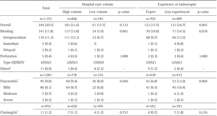
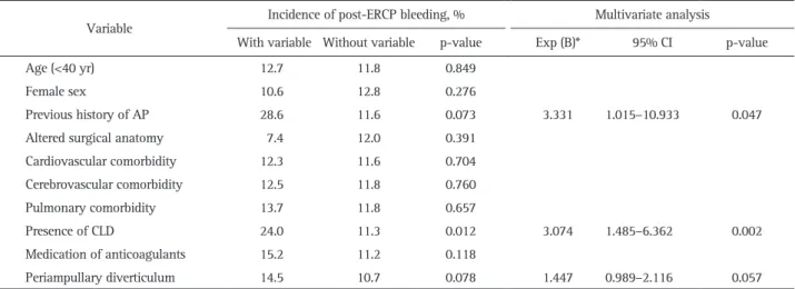
Methods: From January 2015 to December 2015, we pro- spectively enrolled patients with naïve papilla who underwent ERCP at six centers. Patient- and procedure-related variables were recorded on data collection sheets at the time of and after ERCP. Results: A total of 1,191 patients (median age, 71 years) were consecutively enrolled. The overall success rate of biliary cannulation was 96.6%. Overall, 244 patients (20.5%) experienced post-ERCP AEs, including pancreatitis (9.0%), bleeding (11.8%), perforation (0.4%), cholangitis (1.2%), and others (0.9%). While post-ERCP pancreatitis (PEP) was more common when the procedure was performed by less experienced endoscopists, bleeding was more common in high-volume centers and by less experienced endosco- pists. Multivariate analysis showed that a less experience in ERCP was significantly associated with PEP (odds ratio [OR], 1.630; 95% confidence interval [CI], 1.050 to 2.531;

p=0.030) and post-ERCP bleeding (OR, 1.439; 95% CI, 1.003 to 2.062; p=0.048). Conclusions: Our study demonstrated that overall AEs following ERCP were associated with the experience of the endoscopist. To minimize post-ERCP AEs, rigorous training with a sufficient case volume is required, and treatment strategies should be modified according to the

endoscopist’s expertise. (Gut Liver 2020;14:257-264) Key Words: Cholangiopancreatography, endoscopic retro- grade; Adverse events; Hospital volume; Endoscopic experi- ence

INTRODUCTION

Since the introduction of endoscopic sphincterotomy in 1974,

1,2

endoscopic retrograde cholangiopancreatography (ERCP) has evolved from a diagnostic to a therapeutic procedure for the management of various pancreatobiliary disorders.

3

How- ever, ERCP is a relatively invasive procedure associated with potential adverse events (AEs) that range from trivial incidents to major life-threatening crises requiring additional hospital stays or interventional procedures. The major AEs after ERCP are well recognized and the reported incidence varies widely across different studies ranging for 5% to 10% in pancreatitis, 1% to 4% in hemorrhage, 1% to 5% in cholangitis, and 1% to 2% in perforation. The magnitude and independence of the as- sociated risk factors varies widely and are uncertain, but the procedure-related mortality was about 1%.

4-6

Although ERCP procedures are becoming increasingly safer owing to techni- cal advancements and the growing experience of endoscopists, ERCP-related AEs cannot be completely avoided. There may be an association between ERCP case volume and endoscopist’s expertise with outcomes and post-ERCP AEs. Several previous univariate and multivariate analyses including large series of patients have identified either patient- or procedure-related risk factors for ERCP-related AEs.

7-12

However, controversy remains

This is an Open Access article distributed under the terms of the Creative Commons Attribution Non-Commercial License (http://creativecommons.org/licenses/by-nc/4.0) which permits unrestricted non-commercial use, distribution, and reproduction in any medium, provided the original work is properly cited.

Impact of Hospital Volume and the Experience of Endoscopist on Adverse Events Related to Endoscopic Retrograde Cholangiopancreatography:

A Prospective Observational Study

Hyun Jik Lee

1

, Chang Min Cho

2,3

, Jun Heo

3

, Min Kyu Jung

3

, Tae Nyeun Kim

4

, Kook Hyun Kim

4

, Hyunsoo Kim

5

, Kwang Bum Cho

1

, Ho Gak Kim

6

, Jimin Han

6

, Dong Wook Lee

6

, and Yoon Suk Lee

7

1

Department of Internal Medicine, Keimyung University Dongsan Medical Center,

2

Center for Pancreatobiliary Tumor, Kyungpook National University Chilgok Hospital,

3

Department of Internal Medicine, School of Medicine, Kyungpook National University,

4

Department of Internal Medicine, Yeungnam University Medical Center,

5

Department of Internal Medicine, Daegu Fatima Hospital,

6

Department of Internal Medicine, Daegu Catholic University Medical Center, Daegu, and

7

Department of Internal Medicine, Inje University College of Medicine, Busan, Korea

Correspondence to: Chang Min Cho

Center for Pancreatobiliary Tumor, Kyungpook National University Chilgok Hospital, 807 Hokuk-ro, Buk-gu, Daegu 41404, Korea Tel: +82-53-200-2608, Fax: +82-53-200-2028, E-mail: cmcho@knu.ac.kr

Received on December 7, 2018. Revised on March 17, 2019. Accepted on March 24, 2019. Published online January 7, 2020.

pISSN 1976-2283 eISSN 2005-1212 https://doi.org/10.5009/gnl18537

(2)

regarding the impact of the expertise of the endoscopist and the case volume of the center on the outcome of ERCP procedures.

This study aimed to investigate whether hospital case volume and endoscopist’s experience affect the incidence of ERCP- related AEs and to identify patient- and procedure-related risk factors for ERCP-related AEs using univariate and multivariate analyses.

MATERIALS AND METHODS 1. Patients

This study was conducted as a prospective multicenter obser- vational study in six centers of the Daegu-Gyeongbuk province, South Korea. Among these centers, more than 200 therapeutic ERCP procedures per year were performed in four centers (high- volume centers) and less than 200 procedures per year were performed in two centers (low-volume centers). From January 2015 to December 2015, participating centers enrolled patients with naïve papilla who required diagnostic or therapeutic ERCP procedures. This study was approved by the Medicity Daegu Joint Institutional Review Board (IRB number: DGIRB 2014-07- 003-004) and registered in the Clinical Research Information Service (KCT0002993). Informed consent was routinely obtained from all patients prior to performing ERCP.

2. Study protocol and data collection

Patients with any of the following criteria were excluded from the study: aged less than 18 years; pregnancy; previous history of sphincterotomy; underlying chronic pancreatitis;

main pancreatic duct dilation of more than 5 mm; medically unstable condition including severe cardiopulmonary disease for conscious sedation; or uncontrolled coagulopathy (i.e., inter- national normalized ratio of >1.5 or platelet count of <50,000/

mm

3

). Data were recorded on the data collection sheets before ERCP. Details of the ERCP procedures were recorded at the time of the procedure (Supplementary Material 1). Amylase and/or lipase levels were assessed at 6 hours and 24 hours after ERCP.

In patients with persistently high 24-hour amylase levels, the enzymatic profile was re-assessed at 48 hours. All patients underwent follow-up until hospital discharge to monitor their clinical conditions and the occurrence of procedure-related AEs.

Patients who developed AEs continued to be hospitalized and followed up until their clinical conditions improved.

3. Definitions of observation variables and AEs

Cannulation time was measured from the time when the catheter or papillotome was advanced out of the scope channel in front of the papilla to the time when successful deep biliary cannulation was accomplished under fluoroscopy. Difficult can- nulation was defined by the occurrence of any of the following prior to deep biliary cannulation: more than 10 attempts of catheter or guidewire insertion into the pancreatic duct and a

cannulation time of more than 5 minutes.

13

Definitions of individual AEs after ERCP were similar to those reported by Cotton et al.,

11

with some modifications made to the severity and classifications. Post-ERCP pancreatitis (PEP) was defined as the occurrence of new-onset or worsened abdominal pain lasting for more than 24 hours that was associated with an increase in serum amylase level of at least 3 times the up- per limit of normal at 24 hours after the procedure. We used a modified definition of severity for PEP.

14

Instead of the duration of hospital stay, we evaluated the degree of severity using the number of days before normal feeding resumed. ERCP-related bleeding was defined as intraprocedural when bleeding occurred during the procedure and an additional procedure was required for hemostasis, as immediate when bleeding or a hemoglobin drop of at least 2 g/dL was observed in the first 24 hours, and as delayed when bleeding occurred within 15 days. Perforation was defined when an air or contrast leak into the peritoneal or retroperitoneal cavity was recognized at the time of ERCP or on subsequent chest and/or abdomen plain imaging. Perforation was classified into four types according to the perforation site.

15

As an operator variable, less experience was defined as less than 200 procedures based on the cumulative number of ERCP procedures. The threshold for defining a low-volume center was fewer than 200 annual ERCPs per center.

16,17

4. Study outcomes

The primary outcome of this study was to investigate overall AEs and to compare the rate of AEs based on hospital case vol- ume and endoscopist’s experience. The secondary outcome was to identify patient- and procedure-related risk factors predispos- ing to the development of AEs.

5. Statistical analysis

Data were described as absolute and relative frequencies for

categorical variables and mean with standard deviation or me-

dian with range for continuous variables. Differences in the in-

cidence of post-ERCP AEs among groups were compared using

the chi-square or Fisher exact test for categorical variables. To

identify the independent risk factors for post-ERCP AEs, patient-

and procedure-related variables were examined in univariate

and multivariate analyses. All variables with a p-value of <0.1

in the univariate analysis were included in a forward stepwise

regression multivariate analysis and multiple logistic regres-

sion models. The results are shown as odds ratios (ORs) with

95% confidence intervals (CIs). All reported probability values

were two-sided, and p-values of less than 0.05 were considered

statistically significant. Data were analyzed using SPSS version

22.0 (IBM Corp., Armonk, NY, USA).

(3)

RESULTS

1. Baseline characteristics

From January 2015 to December 2015, a total of 1,201 pa- tients with naïve papilla underwent ERCP, and data regarding the patients and procedures were collected from six centers (Table 1). After the exclusion of 10 patients owing to chronic pancreatitis (n=3), previous history of sphincterotomy (n=1), pancreatic duct dilation of >5 mm (n=3), and incomplete pro- tocol completion (n=3), 1,191 patients (median age, 71 years;

male:female ratio, 1.38:1) were included in the study; 846 pro- cedures (71%) were performed in the four high-volume centers and 345 (29%) in the two low-volume centers. Among the participating endoscopists, 10 had performed fewer than 200 therapeutic ERCPs and eight had performed more than 200 pro- cedures. Overall, 702 ERCPs (58.9%) were performed by eight expert endoscopists and 489 (41.1%) by 10 less experienced ones. Patient demographics, including comorbidities, previous history of surgery, clinical manifestations at the time of ERCP, indications for ERCP, current history of medication, and pro- phylactic management, are described in Table 1. Prophylactic medication for the prevention of PEP was administered at the discretion of the attending endoscopist.

2. ERCP-related AEs according to hospital case volume and endoscopist’s experience

Overall, ERCP-related AEs developed in 244 patients (20.5%) (Table 2). The occurrence of ERCP-related AEs was statistically higher with less experienced operators than with expert opera- tors (24.7% vs 17.5%, p=0.003), and no difference was observed in the occurrence of these complications between the high- volume and low-volume centers (21.6% vs 17.7%, p=0.133).

After the exclusion of 130 patients with acute pancreatitis at enrollment, PEP occurred in 95 (9.0%). It was classified as mild in 86 cases (8.1%), moderate in seven (0.7%), and severe in two (0.2%). ERCP-related bleeding occurred in 141 cases (11.8%); it was classified as intraprocedural in 134 (11.3%), immediate in five (0.4%), and delayed in two (0.2%). The mean incidence of PEP was 9.4% (69/736 cases) in high-volume centers and 8.0%

(26/325 cases) in low-volume centers; the difference was not significant (p=0.560). However, in terms of endoscopist’s ex- perience, less experienced operators showed statistically higher rates of PEP compared with experts (12.0% vs 6.8%, p=0.004).

This result was attributed to higher rates of difficult cannulation among less experienced operators than among experts (215/433 [49.7%] vs 234/628 [37.3%], p<0.001).

The occurrence of ERCP-related bleeding was more common in high-volume centers (13.8% vs 7.0%, p=0.001) and among less experienced operators (14.5% vs 10.0%, p=0.018). Other AEs included perforation in five patients (0.4%), cholangitis in 11 (1.2%) following the exclusion of 236 patients with acute cholangitis at enrollment, and others in 11 (0.9%).

Table 1. Baseline Characteristics of the Enrolled Patients (n=1,191)

Characteristic Patients

Age, year 71 (19–101)

Sex, male/female 690/501

Comorbidities

Cardiovascular disorders 397 (33.3)

Cerebrovascular disorders 112 (9.4)

Pulmonary disorders 51 (4.3)

Diabetes 163 (13.7)

Chronic liver diseases 50 (4.2)

Altered surgical anatomy 54 (4.5)

STG with B-II anastomosis 21 (1.8)

STG with B-I anastomosis 17 (1.4)

Liver transplantation 14 (1.2)

Hepatectomy 2 (0.2)

Clinical presentation at ERCP

Acute pancreatitis 130 (10.9)

Acute cholangitis 236 (19.8)

Acute cholecystitis 81 (6.8)

Reason for ERCP

Removal of choledocholithiasis and/or sludges 856 (71.9) Tissue acquisition for histologic diagnosis 219 (18.4)

Drainage procedure 392 (32.9)

Current history of medication

None 993 (83.4)

Aspirin 128 (10.7)

Antiplatelet agents 83 (7.0)

Warfarin 16 (1.3)

NSAIDs 8 (0.7)

Prophylactic medication

None 355 (29.8)

Nafamostate mesilate 342 (28.7)

Ulinastin 243 (20.4)

Gabexate mesilate 223 (18.7)

Indomethacin 28 (2.4)

Presence of diverticulum 359 (30.1)

Type 1/2/3/4 53/164/142/0

Attempts of catheter insertion into main PD

None / <5 / 5–10 / >10 times 962/189/24/16 Attempts of guidewire insertion into main PD

None / <5 / 5–10 / >10 times 864/278/35/14 Attempts of contrast injection into main PD

None / <5 / 5–10 / >10 times 1015/164/12/0 Contact of ampulla before deep biliary cannulation

None / <5 / 5–10 / >10 times 668/390/89/44

Precut sphincterotomy 94 (7.9)

Infundibulotomy 100 (8.4)

Data are presented as median (range) or number (%).

STG with B-II, subtotal gastrectomy with Billroth-II anastomosis; STG

with B-I, subtotal gastrectomy with Billroth-I anastomosis; ERCP, en-

doscopic retrograde cholangiopancreatography; NSAIDs, nonsteroidal

anti-inflammatory drugs; PD, pancreatic duct.

(4)

The observed overall mortality rates were 0.4% (3/846) and 0.9% (3/345) in the high- and low-volume centers (p=0.364), respectively. However, there was only one case of ERCP-related mortality in a high-volume center. The other causes of mortality were septic shock or hepatic failure; according to endoscopist’s experience, there was no significant difference in the mortal- ity rate (0.4% in the less experienced group, 0.6% in the expert group, p=1.000), with one case of ERCP-related mortality in the expert group.

3. Univariate and multivariate analysis for PEP and bleeding Analysis of risk factors was performed only for PEP and ERCP-related bleeding because the occurrence of the other AEs was too low to be further analyzed in our study. Of the nine pa- tient-related risk factors considered in the study protocol (Table 3), the development of PEP was significantly associated with fe- male sex alone in univariate analysis (p=0.029) and with female sex and cardiovascular comorbidity in multivariate analysis (OR, Table 2. Frequencies of ERCP-Related Adverse Events According to Hospital Case Volume and Endoscopist’s Experience

Total Hospital case volume Experience of endoscopist

High volume Low volume p-value Expert Less experienced p-value

n=1,191 n=846 n=345 n=702 n=489

Overall 244 (20.5) 183 (21.6) 61 (17.7) 0.133 123 (17.5) 121 (24.7) 0.003

Bleeding 141 (11.8) 117 (13.8) 24 (7.0) 0.001 70 (10.0) 71 (14.5) 0.018

Intraprocedural 134 (11.3) 111 (13.1) 23 (6.7) 68 (9.7) 66 (13.5)

Immediate 5 (0.4) 5 (0.6) 0 1 (0.1) 4 (0.8)

Delayed 2 (0.2) 1 (0.1) 1 (0.3) 1 (0.1) 1 (0.2)

Perforation 5 (0.4) 4 (0.5) 1 (0.3) 1.000 2 (0.3) 3 (0.6) 1.000

Type I/II/III/IV 3/0/0/2 2/0/0/2 1/0/0/0 1/0/0/1 2/0/0/1

Others* 11 (0.9) 3 (0.4) 8 (2.3) - 9 (1.3) 2 (0.4) -

n=1,061 n=736 n=325 n=628 n=433

Pancreatitis

95 (9.0) 69 (9.4) 26 (8.0) 0.560 43 (6.8) 52 (12.0) 0.004

Mild 86 (8.1) 64 (8.7) 22 (6.8) 41 (6.5) 45 (10.4)

Moderate 7 (0.7) 4 (0.5) 3 (0.9) 1 (0.2) 6 (1.4)

Severe 2 (0.2) 1 (0.1) 1 (0.3) 1 (0.2) 1 (0.2)

n=955 n=650 n=305 n=562 n=393

Cholangitis

11 (1.2) 7 (1.1) 4 (1.3) 0.751 4 (0.7) 7 (1.8) 0.216

Data are presented as number (%).

ERCP, endoscopic retrograde cholangiopancreatography.

*Others included acute cholecystitis (n=4), cerebrovascular accident (n=2), transient fever (n=1), hypoxia (n=1), hypotension (n=1), abnormality of liver battery profiles (n=1), and nausea (n=1).

Data were analyzed after excluding 130 patients with acute pancreatitis at enrollment.

Data were analyzed after excluding 236 patients with acute cholangitis at enrollment.

Table 3. Results of Univariate and Multivariate Analyses of Patient-Related Risk Factors for Post-ERCP Pancreatitis (n=1,061)

Variable Incidence of post-ERCP pancreatitis, % Multivariate analysis

With variable Without variable p-value Exp (B)* 95% CI p-value

Age (<70 yr) 8.9 9.0 1.000

Female sex 11.3 7.3 0.029 1.604 1.041–2.470 0.032

Previous history of AP 11.1 8.9 0.572

Altered surgical anatomy 10.4 8.9 0.611

Cardiovascular comorbidity 6.6 10.1 0.067 0.588 0.356–0.972 0.038

Cerebrovascular comorbidity 13.5 8.5 0.130

Pulmonary comorbidity 4.3 9.2 0.428

Presence of CLD 8.5 9.0 1.000

Periampullary diverticulum 9.4 8.7 0.726

ERCP, endoscopic retrograde cholangiopancreatography; CI, confidence interval; AP, acute pancreatitis; CLD, chronic liver disease.

*Exp (B), odds ratios.

(5)

1.604; 95% CI, 1.041 to 2.470; p=0.032; OR, 0.588; 95% CI, 0.356 to 0.972; p=0.038, respectively). Among eight procedure- related risk factors, difficult cannulation, acquisition of pancrea- togram, pancreatic sphincterotomy, and less experienced opera- tor were significantly associated with PEP in univariate and multivariate analyses (Table 4). Although precut sphincterotomy was identified as a statistically significant risk factor for PEP (p=0.003) in the univariate analysis, the multivariate analysis did not show significance (p=0.082).

The results of the forward stepwise binary logistic regression analysis from the pool of 17 potential risk factors for ERCP- related bleeding identified the following four risk factors by multivariate analysis, one patient- and three procedure-related, all of which were independently associated with ERCP-related bleeding: comorbidity of chronic liver disease, pancreatic sphincterotomy, less experienced operator, and high hospital case volume (Tables 5 and 6).

DISCUSSION

ERCP has been widely practiced as a diagnostic and thera- peutic procedure for pancreatobiliary disease. The risk of ERCP- related AEs was recognized early and has been the focus of many studies.

8-12,18-20

Although technical advances and the expanding experience of endoscopists have made the ERCP procedure increasingly safe, it is still associated with a high po- tential for AEs. Several prospective multicenter studies have de- scribed ERCP success rates and related AEs, and these findings have been useful in understanding the patient- and procedure- related risk factors.

6,10,12,21-26

However, most of these studies are not recent and may not reflect the current clinical picture, as ERCP is now routinely performed to treat complex pancreato- biliary diseases and conditions, such as post-liver transplanta- tion complications, and is a technically demanding procedure.

These applications increase the risk of ERCP-related AEs. In this study, we aimed to determine whether hospital case volume and endoscopist’s experience correlated with ERCP-related AEs.

Table 4. Results of Univariate and Multivariate Analyses of Procedure-Related Risk Factors for Post-ERCP Pancreatitis (n=1,061)

Variable Incidence of post-ERCP pancreatitis, % Multivariate analysis

With variable Without variable p-value Exp (B)* 95% CI p-value

Difficult cannulation 14.0 5.2 <0.001 2.341 1.430–3.833 0.001

Pancreatogram 21.3 7.1 <0.001 2.132 1.251–3.633 0.005

Precut incision 19.0 8.1 0.003 1.784 0.928–3.430 0.082

Infundibulotomy 13.2 8.6 0.175

Biliary balloon dilation 6.8 9.3 0.418

Pancreatic sphincterotomy 18.9 7.4 <0.001 1.867 1.103–3.160 0.020

Less experienced operator 12.0 6.8 0.004 1.630 1.050–2.531 0.030

Low hospital case volume 8.0 9.4 0.560

ERCP, endoscopic retrograde cholangiopancreatography; CI, confidence interval.

*Exp (B), odds ratios.

Table 5. Results of Univariate and Multivariate Analyses of Patient-Related Risk Factors for Post-ERCP Bleeding (n=1,191)

Variable Incidence of post-ERCP bleeding, % Multivariate analysis

With variable Without variable p-value Exp (B)* 95% CI p-value

Age (<40 yr) 12.7 11.8 0.849

Female sex 10.6 12.8 0.276

Previous history of AP 28.6 11.6 0.073 3.331 1.015–10.933 0.047

Altered surgical anatomy 7.4 12.0 0.391

Cardiovascular comorbidity 12.3 11.6 0.704

Cerebrovascular comorbidity 12.5 11.8 0.760

Pulmonary comorbidity 13.7 11.8 0.657

Presence of CLD 24.0 11.3 0.012 3.074 1.485–6.362 0.002

Medication of anticoagulants 15.2 11.2 0.118

Periampullary diverticulum 14.5 10.7 0.078 1.447 0.989–2.116 0.057

ERCP, endoscopic retrograde cholangiopancreatography; CI, confidence interval; AP, acute pancreatitis; CLD, chronic liver disease.

*Exp (B), odds ratios.

(6)

In addition, we identified a number of patient and procedural correlates with complications in 6 centers in Daegu-Gyeongbuk province, South Korea.

According to previous reports, the incidence of ERCP-related AEs varies from 5% to 12%.

7-10,12,26-29

The discrepancies in the reported rate may be attributed to heterogeneity of the defini- tion and inclusion criteria of ERCP-related AEs, inclusion of diagnostic ERCP, and differences in the experience of the en- doscopists. The present study provides up-to-date findings with regard to the differences in post-ERCP AEs according to hospital case volume and endoscopist’s experience. The overall success rate for deep bile duct cannulation was 96.6%, with a cannula- tion success rate of 96.1% (813/846) in patients in high-volume centers compared to 98.0% (338/345) in low-volume centers (p=0.104). The cannulation success rate was higher with expert endoscopists (97.4%) than with less experienced endoscopists (95.5%, p=0.068) and was slightly lower in high-volume centers, but there were no statistically significant differences according to hospital case volume. These results reveal mixed findings between the hospital groups. Similar to the results of a recent systematic review,

20

endoscopist’s experience was evaluated in this study and found to be a significant risk factor associated with overall AEs, though there was no significant difference in the overall AEs according to hospital case volume. Owing to sources of bias such as the complexity of case mixes, referral patterns, patient factors, and differences in endoscopist’s experi- ence and educational programs for ERCP training, the interpre- tation of AEs according to hospital volume should be performed with caution.

6,30

In our study, the AEs were generally of minor severity, with additional hospital stay followed by full recovery, and were rarely severe with permanent disability. Low-volume centers were not associated with increased mortality or poor outcomes.

Bleeding is a serious AE of ERCP that is most commonly related to endoscopic sphincterotomy. The exact incidence of clinically significant ERCP-related bleeding is variable and diffi- cult to define in the consensus criteria. The reported occurrence

at the time of sphincterotomy of bleeding ranging from oozing to severe bleeding is as high as 10% to 30%, while the rate of post-sphincterotomy bleeding after ERCP is estimated to be 0.1%

to 2%.

3,7,10,12,28,31,32

In the present study, the bleeding rate was 11.8% and most cases of bleeding were intraprocedural or immediate events (95.0% and 3.5%, respectively), which included continuous bleeding for several minutes after the sphincterotomy. These types of bleeding are generally considered mild AEs if signifi- cant blood loss, transfusion requirement, or changes in vital signs do not occur. Identified risk factors for post-sphincteroto- my bleeding include coagulopathy, thrombocytopenia, antico- agulant treatment, presence of active cholangitis, and low case volume on the part of the endoscopist (performance of not more than 1 sphincterotomy per week).

3,7,31

In the present study, mul- tivariate analysis identified three independent procedure-related risk factors for post-ERCP bleeding: pancreatic sphincterotomy (OR, 1.766; p=0.021), less experienced endoscopist (OR, 1.439;

p=0.048), and high hospital volume (OR, 2.016; p=0.005). Pres- ence of chronic liver disease was the only patient-related factor (OR, 3.074; p=0.002). These findings indicate that less expe- rienced endoscopists may control the bleeding less precisely or less effectively and that the volume of hospital cases is not associated with a reduced rate of bleeding. This may reflect the fact that high-volume centers have more patients at clinical high risk of bleeding, with poor general conditions that entail a higher degree of difficulty. These conflicting data regarding the negative impact of high-volume centers on the post-ERCP bleeding rate warrant further well-designed studies.

PEP is one of the most common AEs of ERCP, and it is asso- ciated with both patient- and procedure-related factors. Patient- related factors are younger age, female sex, history of previous PEP, non-dilated ducts, normal bilirubin level, and suspected sphincter of Oddi dysfunction. Procedure-related factors are difficult cannulation, multiple pancreatic injections, pancreatic sphincterotomy, precut sphincterotomy, and pancreatic sam- pling. These factors have been shown to increase the risk of Table 6. Results of Univariate and Multivariate Analyses of Procedure-Related Risk Factors for Post-ERCP Bleeding (n=1,191)

Variable Incidence of post-ERCP bleeding, % Multivariate analysis

With variable Without variable p-value Exp (B)* 95% CI p-value

Difficult cannulation 11.8 11.9 1.000

Precut incision 6.4 12.3 0.097 0.491 0.208–1.162 0.105

Infundibulotomy 20.0 11.1 0.014 1.685 0.954–2.978 0.072

Biliary balloon dilation 8.7 12.3 0.263

Pancreatic sphincterotomy 18.8 10.8 0.007 1.766 1.091–2.861 0.021

Less experienced operator 14.5 10.0 0.018 1.439 1.003–2.062 0.048

High hospital case volume 13.8 7.0 0.001 2.016 1.232–3.298 0.005

ERCP, endoscopic retrograde cholangiopancreatography; CI, confidence interval.

*Exp (B), odds ratios.

(7)

PEP.

3,8,10,12,18,26-28,31,32

In the present study, the incidence of PEP was similar to that reported in previous studies.

33

In the present study, among the patient-related risk factors, only female sex and cardiovascular comorbidity were significantly associated with PEP in the multivariate analysis. In terms of procedure- related risk factors, difficult cannulation, acquisition of pancrea- togram, pancreatic sphincterotomy, and less experienced endos- copist were significantly associated with PEP in univariate and multivariate analyses. These results indicate that endoscopist’s experience is a relatively important risk factor for AEs of ERCP (OR, 1.630) from a technical standpoint.

In one large Italian multicenter prospective study, a compari- son of high-volume (>200 ERCPs/year) and low-volume centers (<200 ERCPs/year) showed that the risk of PEP was significantly higher in low-volume centers in univariate analysis (relative risk, 2.8).

12

In another recent multicenter study, PEP was not associated with the case volume of either the single endosco- pist (3.8% vs 5.5%; expert vs nonexpert, p=0.345) or the center (3.9% vs 3.1%; high-volume centers vs low-volume centers, p=0.379).

25

The effect of endoscopist’s experience is quite difficult to evaluate and interpret. Continuous training and an adequate case volume should be required for the performance of ERCP in practice to reduce AEs. Furthermore, endoscopists must consider if they have an adequate case volume to maintain their ERCP skills and minimize AEs before performing ERCP. The success and outcome of ERCP procedures is critically important, and minimizing AEs has equal significance. Therefore, endoscopist’s experience is an essential factor in the success of ERCP, along with a skilled ERCP team consisting of nurses and radiogra- phers. The number of purely diagnostic ERCPs has decreased, and the number of interventional ERCPs, with high risk and complexity, has increased. Endoscopists in high-volume centers are continuously undergoing training and performing ERCP.

As a result, endoscopists in low-volume centers or those who are less experienced are performing fewer total procedures an- nually. Our data showed that endoscopists with less experience have higher rates of AEs. Although increasing endoscopist’s ex- perience was associated with low PEP rates, this association was not observed with regard to center volume. Given the greater frequency of more complex and difficult procedures in high- volume centers, these centers are more likely to have a large proportion of patients with a high risk of pancreatitis. Therefore, endoscopist’s experience and careful patient selection play an important role in preventing PEP. Rigorous training and a suf- ficient case volume along with an adequate system of referral to experts for complex cases are required to reduce the incidence of PEP in practice. Appropriate modification of the treatment approach according to the individual endoscopist’s experience may be the most important factor.

This prospective multicenter study revealed the incidence of ERCP-related AEs according to different endoscopists’ experi-

ence and center case volumes. The experience of the endoscopist is associated with the overall incidence rate of AEs. In addition, the current data reveal the patient- and procedure-related risk factors for PEP and bleeding. To minimize the AEs of ERCP, en- doscopists should endeavor to ensure an adequate case volume to maintain their technical skills. Before considering performing an ERCP, the endoscopist should be attentive to the selection of patients with appropriate indications, perform individual risk- benefit analyses, and understand how to effectively manage any potential AEs.

CONFLICTS OF INTEREST

No potential conflict of interest relevant to this article was reported.

ACKNOWLEDGEMENTS

This work was supported by a Clinical Research Institute grant from the Daegu-Gyeongbuk Society of Gastroenterology (2014).

AUTHOR CONTRIBUTIONS

Study concept and design: C.M.C., K.B.C., Y.S.L., H.J.L. Defini- tion of intellectual content: C.M.C. Literature search and clinical studies: C.M.C., K.B.C., Y.S.L., H.J.L. Data and statistical analysis, manuscript preparation: C.M.C., K.B.C., Y.S.L., H.J.L. Data acqui- sition: C.M.C., M.K.J., J. Heo, T.N.K., K.H.K., H.K., K.B.C., Y.S.L., H.J.L., H.G.K., J. Han, D.W.L. Manuscript editing and review: all authors.

ORCID

Hyun Jik Lee https://orcid.org/0000-0002-3929-9180 Chang Min Cho https://orcid.org/0000-0002-9903-1282 Jun Heo https://orcid.org/0000-0003-3180-1655 Min Kyu Jung https://orcid.org/0000-0001-8749-408X Tae Nyeun Kim https://orcid.org/0000-0003-4178-2056 Kook Hyun Kim https://orcid.org/0000-0001-7786-7882 Hyunsoo Kim https://orcid.org/0000-0001-5572-5453 Kwang Bum Cho https://orcid.org/0000-0003-2203-102X Ho Gak Kim https://orcid.org/0000-0003-3365-1662 Jimin Han https://orcid.org/0000-0001-8674-370X Dong Wook Lee https://orcid.org/0000-0002-1029-9064 Yoon Suk Lee https://orcid.org/0000-0002-5835-9417

REFERENCES

1. Classen M, Demling L. Endoscopic sphincterotomy of the papilla

of Vater and extraction of stones from the choledochal duct (au-

thor’s transl). Dtsch Med Wochenschr 1974;99:496-497.

(8)

2. Kawai K, Akasaka Y, Murakami K, Tada M, Koli Y. Endoscopic sphincterotomy of the ampulla of Vater. Gastrointest Endosc 1974;20:148-151.

3. Freeman ML. Complications of endoscopic retrograde cholangio- pancreatography: avoidance and management. Gastrointest En- dosc Clin N Am 2012;22:567-586.

4. Siiki A, Tamminen A, Tomminen T, Kuusanmäki P. ERCP proce- dures in a Finnish community hospital: a retrospective analysis of 1207 cases. Scand J Surg 2012;101:45-50.

5. Kapral C, Mühlberger A, Wewalka F, et al. Quality assessment of endoscopic retrograde cholangiopancreatography: results of a run- ning nationwide Austrian benchmarking project after 5 years of implementation. Eur J Gastroenterol Hepatol 2012;24:1447-1454.

6. Glomsaker T, Hoff G, Kvaløy JT, et al. Patterns and predictive fac- tors of complications after endoscopic retrograde cholangiopan- creatography. Br J Surg 2013;100:373-380.

7. Freeman ML, Nelson DB, Sherman S, et al. Complications of endo- scopic biliary sphincterotomy. N Engl J Med 1996;335:909-918.

8. Masci E, Toti G, Mariani A, et al. Complications of diagnostic and therapeutic ERCP: a prospective multicenter study. Am J Gastro- enterol 2001;96:417-423.

9. Vandervoort J, Soetikno RM, Tham TC, et al. Risk factors for complications after performance of ERCP. Gastrointest Endosc 2002;56:652-656.

10. Wang P, Li ZS, Liu F, et al. Risk factors for ERCP-related com- plications: a prospective multicenter study. Am J Gastroenterol 2009;104:31-40.

11. Cotton PB, Garrow DA, Gallagher J, Romagnuolo J. Risk factors for complications after ERCP: a multivariate analysis of 11,497 procedures over 12 years. Gastrointest Endosc 2009;70:80-88.

12. Loperfido S, Angelini G, Benedetti G, et al. Major early complica- tions from diagnostic and therapeutic ERCP: a prospective multi- center study. Gastrointest Endosc 1998;48:1-10.

13. Udd M, Kylänpää L, Halttunen J. Management of difficult bile duct cannulation in ERCP. World J Gastrointest Endosc 2010;2:97-103.

14. Kawaguchi Y, Ogawa M, Omata F, Ito H, Shimosegawa T, Mine T.

Randomized controlled trial of pancreatic stenting to prevent pan- creatitis after endoscopic retrograde cholangiopancreatography.

World J Gastroenterol 2012;18:1635-1641.

15. Stapfer M, Selby RR, Stain SC, et al. Management of duodenal perforation after endoscopic retrograde cholangiopancreatography and sphincterotomy. Ann Surg 2000;232:191-198.

16. ASGE Training Committee, Adler DG, Bakis G, et al. Principles of training in GI endoscopy. Gastrointest Endosc 2012;75:231-235.

17. Moon HS, Choi EK, Seo JH, et al. Education and training guide- lines for the board of the Korean society of gastrointestinal endos- copy. Clin Endosc 2017;50:345-356.

18. Masci E, Mariani A, Curioni S, Testoni PA. Risk factors for pancre- atitis following endoscopic retrograde cholangiopancreatography:

a meta-analysis. Endoscopy 2003;35:830-834.

19. Andriulli A, Loperfido S, Napolitano G, et al. Incidence rates of post-ERCP complications: a systematic survey of prospective stud-

ies. Am J Gastroenterol 2007;102:1781-1788.

20. Keswani RN, Qumseya BJ, O’Dwyer LC, Wani S. Association be- tween endoscopist and center endoscopic retrograde cholangio- pancreatography volume with procedure success and adverse out- comes: a systematic review and meta-analysis. Clin Gastroenterol Hepatol 2017;15:1866-1875.

21. Kapral C, Duller C, Wewalka F, Kerstan E, Vogel W, Schreiber F.

Case volume and outcome of endoscopic retrograde cholangio- pancreatography: results of a nationwide Austrian benchmarking project. Endoscopy 2008;40:625-630.

22. Vitte RL, Morfoisse JJ; Investigator Group of Association Natio- nale des Gastroentérologues des Hôpitaux Généraux. Evaluation of endoscopic retrograde cholangiopancreatography procedures performed in general hospitals in France. Gastroenterol Clin Biol 2007;31:740-749.

23. Masci E, Minoli G, Rossi M, et al. Prospective multicenter quality assessment of endotherapy of biliary stones: does center volume matter? Endoscopy 2007;39:1076-1081.

24. Enochsson L, Swahn F, Arnelo U, Nilsson M, Löhr M, Persson G.

Nationwide, population-based data from 11,074 ERCP procedures from the Swedish Registry for Gallstone Surgery and ERCP. Gas- trointest Endosc 2010;72:1175-1184.

25. Testoni PA, Mariani A, Giussani A, et al. Risk factors for post- ERCP pancreatitis in high- and low-volume centers and among expert and non-expert operators: a prospective multicenter study.

Am J Gastroenterol 2010;105:1753-1761.

26. Freeman ML, DiSario JA, Nelson DB, et al. Risk factors for post- ERCP pancreatitis: a prospective, multicenter study. Gastrointest Endosc 2001;54:425-434.

27. Cheng CL, Sherman S, Watkins JL, et al. Risk factors for post- ERCP pancreatitis: a prospective multicenter study. Am J Gastro- enterol 2006;101:139-147.

28. Williams EJ, Taylor S, Fairclough P, et al. Risk factors for compli- cation following ERCP; results of a large-scale, prospective multi- center study. Endoscopy 2007;39:793-801.

29. Galeazzi M, Mazzola P, Valcarcel B, et al. Endoscopic retrograde cholangiopancreatography in the elderly: results of a retrospec- tive study and a geriatricians’ point of view. BMC Gastroenterol 2018;18:38.

30. Petersen BT. ERCP outcomes: defining the operators, experience, and environments. Gastrointest Endosc 2002;55:953-958.

31. Rustagi T, Jamidar PA. Endoscopic retrograde cholangiopancre- atography-related adverse events: general overview. Gastrointest Endosc Clin N Am 2015;25:97-106.

32. Mergener K. Complications of endoscopic and radiologic in- vestigation of biliary tract disorders. Curr Gastroenterol Rep 2011;13:173-181.

33. Kochar B, Akshintala VS, Afghani E, et al. Incidence, severity, and

mortality of post-ERCP pancreatitis: a systematic review by using

randomized, controlled trials. Gastrointest Endosc 2015;81:143-

149.

수치

Table 1. Baseline Characteristics of the Enrolled Patients (n=1,191)
Table 3. Results of Univariate and Multivariate Analyses of Patient-Related Risk Factors for Post-ERCP Pancreatitis (n=1,061)
Table 5. Results of Univariate and Multivariate Analyses of Patient-Related Risk Factors for Post-ERCP Bleeding (n=1,191)

참조

관련 문서

6. Acute biliary pancreatitis: the roles of endoscopic ultrasonography and endoscopic retrograde cholangiopancreatography.. endoscopic ultrasonography in the

In the chapters of Journey of Renewal that describe the Mission Accountability Framework there will be flashbacks to the story of Richmond Corps, showing clearly how

It considers the energy use of the different components that are involved in the distribution and viewing of video content: data centres and content delivery networks

1 John Owen, Justification by Faith Alone, in The Works of John Owen, ed. John Bolt, trans. Scott Clark, &#34;Do This and Live: Christ's Active Obedience as the

The related factors with clustering healthy behaviors by multinominal logistic regression analysis were higher for “females, the elderly, people with higher level of

The purpose of this study was to identify the frequency and related factors of advanced airway management for patients with cardiac arrest by the

A Retrospective Study of Prevalence and Risk factors for Neonatal Occipital

• 기회주의에 기반한 이론이 높은 수준의 거래 특유의 투자를 이유로 수직적 통합의 필요성을 주장할 때. – 능력에 기반한 이론이 수직적 통합에 필요한 자원과