Comparing the Immediate Effectiveness of Lumbar Flexion and Extension Exercise With Regards to Pain, Range of Motion,
Pelvic Tilt, and Functional Gait Ability in Patients With Lumbar Spinal Stenosis
Hyun-ho Do1, MSc, PT, Seung-chul Chon2, PhD, PT
1Dept. of Rehabilitation Medicine, Konyang University Hospital
2Dept. of Physical Therapy, College of Medical Science, Konyang University
Abstract
1)Background: In patients with lumbar spinal stenosis (LSS), lumbar flexion exercise (LFE) is considered a standard therapeutic exercise that widens the space between the spinal canal and intervertebral foramen. However, some researchers have reported that lumbar extension exercise (LEE) may improve lumbar pain and functional ability in patients with LSS. Although exercise intervention methods for patients with LSS have been widely applied in clinical settings, few studies have conducted comparative analysis of these exercise methods.
Objects: This study aimed to compare the effects of LFE, LEE, and lumbar flexion combined with lumbar flexion-extension exercise (LFEE) on pain, range of motion (ROM), pelvic tilt angle, and functional gait ability in patients with LSS.
Methods: A total of 30 patients with LSS, LFE (n1=10), LEE (n2=10), and LFEE (n3=10) were assigned to each of the three exercise groups. The numerical pain rating scale (NPRS), modified-modified schober test (MMST)-flexion, MMST-extension, pelvic tilt inclinometer, and 6-minute walking test (6MWT) were measured.
Results: After the intervention, statistically significant differences were observed in the NPRS (p=.043), MMST-flexion (p<.001), MMST-extension (p<.001), and 6MWT (p=.005) between groups. According to the post hoc test, the NPRS was statistically significant difference between the LFEE and LEE groups (p=.034). The MMST-flexion was statistically significantly different between the LFE and LEE (p=.000), LFE and LFEE (p=.001), and LEE and LFEE (p=.001) groups. The MMST-extension was statistically significantly different between the LFE and LEE (p<.001), LFE and LFEE (p=.002), and LEE and LFEE (p=.008) groups. The 6MWT was statistically significantly different between the LFE and LFEE (p=.042) and the LEE and LFEE (p=.004) groups.
Conclusion: This study suggested that LFEE was the most effective exercise for pain and functional gait ability in patients with LSS, LFE was the most effective exercise for lumbar flexion ROM, and LEE was the most effective exercise for lumbar extension ROM.
Key Words: Lumbar extension exercise; Lumbar flexion exercise; Lumbar spinal stenosis.
Introduction
Lumbar spinal stenosis (LSS) is a degenerative disease that is frequently observed in the elderly. It presents with pain in the lumbar spine and legs due
to a narrowing space in the nerve and blood vessel distribution in the lumbar spinal canal. (Lafian and Torralba, 2018). Degenerative changes in the spinal area can lead to the deterioration of the spinal canal space and degeneration of the intervertebral disc, Corresponding author: Seung-chul Chon [email protected]
This research was supported by Basic Science Research Program through the National Research Foundation of Korea (NRF) funded by the Ministry of Education (2017R1D1A3B03031876).
causing instability and hypermobility of the facet joints, resulting in an enlargement of the superior articular process of the facet joints and nerve and blood vessel compression (Costandi et al, 2015).
Common symptoms of LSS include features of neu- rogenic intermittent claudication (NIC) (Suzuki et al, 2010), which increases pain during walking and lumbar extension and decreases pain during lumbar flexion and rest and pain, numbness, and muscle weakness in the lumbar spine and leg (Lafian and Torralba, 2018). Pain expression during lumbar extension causes abnormal postural changes due to limited range of motion (ROM) and inadequate weight-bearing ability in the direction of extension (Costandi et al, 2015). Additionally, the major cause of LSS is degenerative change due to old age (Chen et al, 2017). Turner et al. (1992) reported that NIC was observed in approximately 62% of pa- tients with LSS, which reduced functional gait and overall physical conditions (Tomkins-Lane et al, 2014).
Suzuki et al. (2010) reported that positive sagittal bal- ance and pelvic tilt were increased in patients with LSS complaining of NIC.
Therapeutic intervention for LSS is largely divided into surgical and conservative methods (Inoue et al, 2016). Surgical methods typically use laminectomy and foraminotomy for lumbar decompression if there is no improvement in conservative treatment, severe NICs are identified, and severe pain is experienced in daily life (Binder et al, 2002). In surgical methods, despite the reduction of pain and NIC, postoperative functional disorders still remain an unresolved problem (Inoue et al, 2016). Therefore, in consideration of postoperative functional impairment of LSS, most patients with LSS have undergone rehabilitation and physical therapy 3∼
6 months before surgery (Chen et al, 2017).
Conservative treatment includes a variety of ex- ercise treatments such as drug administration, acu- puncture, strength training, stretching, and joint mo- bility (Zaina et al, 2016). Professional exercise thera- pies have been reported to have positive outcomes on pain and functional motor capacity of patients with LSS (Mu et al, 2018). Fritz et al. (1997) found
that treadmill walking exercise improves pain reduc- tion and walking ability in patients with LSS. Mu et al. (2018) reported that exercise therapy combined with core stabilization exercises such as plank and bridge exercises improve walking and functional mo- tor performance in patients with LSS. Goren et al.
(2010) found that lumbar flexion and extension mus- cle strengthening combined with low-intensity bi- cycle exercise were effective in reducing lumbar and leg pain in patients with LSS.
The most recommended exercise therapy for pa- tients with LSS is to limit the lumbar extension that can cause pain (Creighton et al, 2006). Because lum- bar extension narrows the spinal and nerve root ca- nal, lumbar flexion is recommended only (Fast, 1988).
Creighton et al. (2006) reported that lumbar flexion exercise (LFE) improved pain and functional motor and walking ability in patients with LSS. Whitman et al. (2006) found that LFE combined with treadmill walking can improve pain and walking ability in pa- tients with LSS. However, one researcher insisted the combination of lumbar flexion and extension ex- ercises (Onel et al, 1993). Atlas et al. (2000) found that lumbar extension exercise (LEE) was effective for pain, functional motor capacity, and patient sat- isfaction in patients with LSS.
Hence, the results of the studies on exercise methods to improve pain and motor function of pa- tients with LSS are controversial. This study aimed to compare the effectiveness of LFE, LEE, and lum- bar flexion-extension exercise (LFEE) in pain, lum- bar ROM, pelvic tilt angle (PTA), and functional gait ability in patients with LSS and to determine the most effective exercise method.
Methods
Instruments and measurement
Sustained lumbar extension provocation test
The subjects performed lumbar extension (10∼30
degrees) after placing both hands in the hips in a stand- ing position, and lumbar pain was noted if there was pain while maintaining this position. Subsequently, symp- toms observed in this position were assessed. Complaints of lumbar and leg pain due to prolonged lumbar ex- tension posture have been associated with LSS (Katz et al, 1995). The sensitivity and specificity of SLEPT were 51% (95% confidence interval (CI), 36%-66%) and 69%
(95% CI, 53%-85%), respectively (Katz et al, 1995).
Modified lumbar extension test
The subjects extended their lumbar spine for 1 minute in a standing position. If symptoms did not appear for 1 minute, lumbar extension was performed again after 1 minute of rest. During this time, the lateral flexion was performed to the left and right lumbar spine, and if pain was felt in the ipsilateral side, it was positive in the ipsilateral direction and negative when the pain was felt in the opposite side.
The sensitivity and specificity were 92% (95% CI, 72%-99%) and 40% (95% CI, 7%-82%), respectively (Dobbs et al, 2016).
Numerical pain rating scale
The NPRS subjectively measures pain. It consists of 11 items scored from 0 to 10, with 0 for “no pain” and 10 for “most severe pain.” Subjects were asked about the degree of pain and were instructed to verbalize or indicate how they felt with the fol- lowing classifications: 1 to 3 points were classified as mild pain, 4 to 6 points as moderate pain, and 7 to 10 points as severe pain. In clinical practice, a score of 4 points is usually defined as moderate pain (Krebs et al, 2007). The intraclass correlation co- efficient (ICC) was .95 (Alghadir et al, 2018).
Modified modified schober test-flexion The MMST-flexion was used to measure ROM for lumbar flexion. The first landmark was marked at the spinal intersection of the left and right posterior supe- rior iliac spine (PSIS), and the second landmark was 15 ㎝ above the first landmark. The examiner aligned
the tape measure between the two skin marks with zero at the inferior skin mark and 15 ㎝ at the superi- or skin mark. The measuring tape was kept firmly against the subject’s skin, while the subject was asked to bend forward with the following instruction: “Bend forward as far as you can while keeping the knee straight.” At the end of flexion ROM, the distance be- tween the two marks was noted. The ROM was the difference between 15 ㎝ and length measured at the end of motion. This test has a high correlation with radiographic measurements (Battie et al, 1987), In this study, the ICC and test-retest reliability were .72 and .78∼.89, respectively (Williams et al, 1993).
Modified modified schober test- extension
The same landmarks and procedure described for the flexion technique were used for measuring lum- bar extension. The examiner gave the following in- struction: “Place the palms of your hands on your buttock and bend backward as far as you can.”
When the subject bent backward into full lumbar extension, the new distance between the superior and inferior skin markings was measured using the tape, and the change in the distance between the marks was used to indicate the amount of ROM of lumbar extension. The ICC and test-retest reliability were .76 and .69∼.91, respectively (Williams et al, 1993).
Pelvic tilt angle test
The subjects distributed their weight evenly on both feet in a barefoot standing position. Using the pelvic inclinometer (Baseline® AcuAngle Inclinometer, Fabrication Enterprises Inc., NY, USA) for the measurement, the examiner placed the tip of both calipers on the anterior superior iliac spine and PSIS and marked the indicated angles. The test-retest re- liability of the pelvic inclinometer ranged from .96 to .99 (Youdas et al, 2000).
6-minute walking test
The 6-minute walking test (6MWT) is a sub-
maximal exercise test used to assess aerobic ca- pacity, endurance, and gait performance capacity.
The distance covered over a time of 6 minutes is used as the outcome to compare the changes in per- formance capacity. The best distance walked in me- ters is recorded. At least 30-minute rest was allowed between tests. The test-retest reliability of the 6MWT was .88∼.94. (Rikli and Jones, 1998).
Intervention
The LFE applied William’s exercise, LEE applied McKenzie exercise, and LFEE applied William and McKenzie exercise evenly. The three groups con- sisted of six exercise methods, and the exercise method of each group was performed equally in 10 seconds (5 times in 3 sets). Each of the three groups equally performed abdominal draw in maneuver for core stabilization. All three groups performed this maneuver for 40 minutes and were evaluated imme- diately before and after the intervention.
Lumbar flexion exercise
1) Hip flexor stretching: To relax the hip flexor muscles, the subjects held the ankle with one hand in a side-lying and extended their hip flexor muscles for 30 seconds. During this time, the subjects kept their pelvis tilted back and were extremely careful not to overextend their lumbar spine. 2) Double knee-to-chest: To relax the paraspinal muscles around the lumbar spine, the subjects held the thigh with both hands while in a supine and pulled it to- ward the chest while exhaling. 3) Lumbar flexion in long sitting: To restore and relax the lumbosacral muscle, erector spinae muscle, and hamstring muscle, the subjects extended their legs in a long sitting with their hands pointed at their toes and bent their lumbar spine. 4) Pelvic posterior tilt exercise: To prevent anterior tilting of the pelvis and reduce for- ward bending of the lumbar spine, the subjects tilted their pelvis backward in a hook-lying, allowing their lumbar spine to reach the floor. During this time, the subjects did not reach their legs on the floor. 5)
Partial sit-up exercise: To strengthen the abdominal muscles, the subjects, while in a hook-lying, put both of their hands behind the head and slowly raised their upper body until the scapula reached the floor. 6) Bending trunk in sitting: To improve stretching ability of the lumbosacral soft tissue, the subjects sat in a chair with both hands behind their heads, and they bent their trunk forward as far as possible without pain.
Lumbar extension exercise
1) Hold in prone: To relax the full body, the sub- jects put their arms in parallel to their body while breathing comfortably, slowly turning their head to both sides. 2) Lumbar extension on the elbow while in a prone: For lumbar extension, the subjects sup- ported both of their elbows to the floor in prone and maintained lumbar extension within painless range.
3) Lumbar extension on the hand while in prone: For a larger range of lumbar extension, the subjects kept their lumbar extension with both arms extended while supporting the floor in a prone. 4) Superman exercise: For lumbar extensor muscle strengthening, the subjects lifted both of their hands slowly and si- multaneously with extension of both elbows in a prone. 5) Bird-dog exercise: For lumbar extensor muscle strengthening and balancing left and right movements, the subjects’ one hand and the other leg were simultaneously stretched parallel to the ground in one quadruped position and hold for 10 seconds and alternately as long as the subjects did not lose balance. 6) Lumbar extension in standing: To im- prove functional lumbar extension in standing with lumbar weight-bearing applied, the subjects extended their lumbar spine as far as possible without pain after placing both hands on both hips.
Lumbar flexion combined extension exercise
The LFEE was combined with LFE and LEE 1) Hip flexor stretching. 2) Lumbar extension on the el- bow while in prone. 3) Lumbar flexion in long
Variables LFEa (n1=10) LEEb (n2=10) LFEEc (n3=10) p1
NPRSe (score)
Pre 2.70±.48d 2.80±.42 2.80±.42 .845
Post 2.00±.47 2.40±.51 1.70±.67 .032*
p2 .001* .037* .001*
Change -.70±.48 -0.40±.51 -1.10±.73 .043*
MMST-flexionf (㎝)
Pre 5.20±1.40 5.67±1.34 6.95±2.02 .060
Post 5.90±1.32 5.58±1.31 7.24±2.04 .065
p2 <.001* .324 <.001*
Change .70±.24 -.09±.27 .29±.08 <.001*
MMST-extensiong (㎝)
Pre 2.00±.69 2.14±.70 2.13±.62 .875
Post 1.99±.66 2.41±.65 2.27±.64 .354
p2 .758 <.001* .001*
Change -.01±.99 .27±.08 .14±.08 <.001*
Pre 13.20±2.09 13.60±2.36 14.10±2.96 .725
PTAh (°) Post 12.70±1.82 13.50±2.27 14.20±2.57 .342
p2 .015* .726 .726
Change -.50±.52 -.10±.87 .10±.87 .231
Pre 165.20±29.20 155.80±21.33 162.90±30.50 .728
6MWTi (m) Post 167.50±29.91 156.20±19.83 170.30±28.74 .464
p2 .199 .768 <.001*
Change 2.30±5.25 .40±4.16 7.40±3.83 .005*
alumbar flexion exercise, blumbar extension exercise, clumbar flexion combined extension exercise, dmean±standard deviation, enumerical pain rating scale, fmodified modified schober test-flexion, gmodified modified schober test-extension, hpelvic tilt angle,i6 minute walking test, *p<.05.
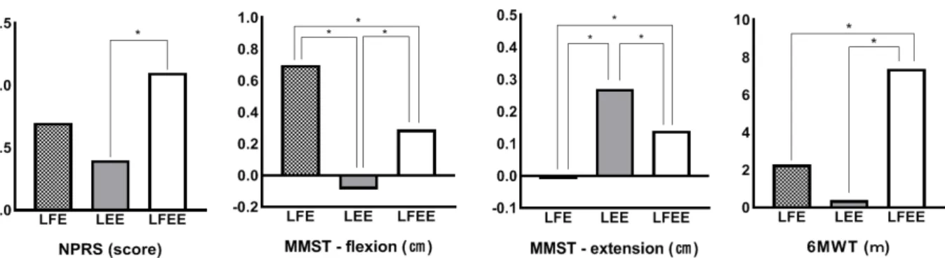
Table 2. Comparisons of LFE, LEE, LFEE on NPRS, MMST flexion, MMST extension, PTA, 6MWT Variables (unit) LFEa (n1=10) LEEb (n2=10) LFEEc (n3=10) p
Gender Male 6 (20%) 4 (13%) 5 (17%)
Female 4 (13%) 6 (20%) 5 (17%)
Age (year) 78.30±3.97d 76.60±5.44 74.00±6.01 .197
Height (㎝) 161.30±8.57 159.00±7.13 160.90±6.50 .764
Weight (㎏) 59.10±8.53 58.70±8.05 56.80±6.52 .780
alumbar flexion exercise, blumbar extension exercise, clumbar flexion combined extension exercise,
dmean±standard deviation.
Table 1. General characteristics of subjects
sitting. 4) Superman exercise. 5) Partial sit-up exercise. 6) Lumbar extension in standing.
Statistical analysis
Data were analyzed using the Statistical Package for the Social Sciences (SPSS) ver. 20.0 (IBM corp,
Armouk, NY, USA). The data are described as the mean±standard deviation. One-way analysis of var- iance (ANOVA) with the subjects’ general character- istics was used to compare among the groups. A one-way ANOVA was used to identify the differ- ences and changes in the NPRS, MMST-flexion,
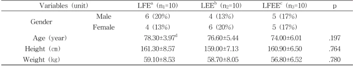
Figure 1. Post-Hoc comparisons of LFE, LEE, LFEE on changes value of NPRS, MMST flexion, MMST extension, 6MWT (LFE: lumbar flexion exercise, LEE: lumbar extension exercise, LFEE:
lumbar flexion combined extension exercise, NPRS: numerical pain rating scale, MMST-flexion:
modified modified schober test-flexion, MMST-extension: modified modified schober test-extension, 6MWT: 6-minute walking test).
MMST-extension, PTA, and 6MWT among the three groups before and after the intervention. A paired t-test was performed to compare the outcome changes before and after the intervention in each group. If the analysis revealed statistical significance, post hoc Turkey was performed to determine the differences in pairwise comparison. The level of sig- nificance was set at p <.05.
Results
General characteristics of study subjects
The general characteristics of the study subjects are presented in Table 1. There were no significant differences in age, height, and weight of the subjects among the groups (p>.05).
Comparisons of LFE, LEE, LFEE on NPRS, MMST flexion, MMST extension, PTA, 6MWT
According to the comparison among the groups by change value, significant differences were ob- served for NPRS (p=.043), MMST-flexion (p<.001), MMST-extension (p<.001), and 6MWT (p=.005) but not PTA. LFE demonstrated significant differences in NPRS (p=.001), MMST-flexion (p<.001), and PTA (p=.015), but not for MMST-extension and 6MWT,
before and after the intervention. LEE demonstrated significant differences in NPRS (p=.037) and MMST-extension (p<.001), but not for MMST-flex- ion, PTA, and 6MWT, before and after the intervention. LFEE demonstrated significant differ- ences in NPRS (p=.001), MMST-extension (p=.001), and 6MWT (p<.001), but not for PTA, before and after the intervention (Table 2).
Post-Hoc comparisons of LFE, LEE, LFEE on changes value of NPRS, MMST flexion, MMST extension, 6MWT
LFEE was significantly more effective than LEE at improving NPRS (p=.043), LFE was more effective than LEE and LFEE at improving MMST-flexion (p<.001), LEE was more effective than LFE and LFEE at improving MMST-extension (p<.001), and LFEE was more effective than LFE and LEE at im- proving 6MWT (p=.005) (Figure 1).
Discussion
LFE, LEE, and LFEE were applied to patients with LSS to compare the effectiveness of these ex- ercises by measuring pain, lumbar ROM, PTA to measure postural abnormalities, and functional gait ability. As a result, LFE showed a significant differ- ence in pain, lumbar flexion ROM, and PTA, LEE
showed significant difference in pain and lumbar ex- tension ROM, and LFEE showed significant differ- ence in pain, lumbar flexion ROM, lumbar extension ROM, and walking ability. Additionally, LFEE was most effective in pain and gait performance, LFE was most effective in lumbar flexion ROM, and LEE was most effective in lumbar extension ROM.
We used a safe and efficient MMST-flexion and MMST-extension measure in standing to measure the lumbar ROM of patients experiencing pain and difficulty in the measurement process (Tousignant et al, 2005). Additionally, PTA, which can predict pos- tural abnormalities, was immediately applied in standing (Youdas et al, 2000), and a simple and uni- versal 6MWT was used to evaluate walking ability (Bohannon and Crouch, 2017). Therefore, the meas- urements used in this study are considered clinically and highly efficient tools. These tools were similarly used in patients with LSS in the study of Tomkins-Lane et al. (2014).
The lumbar flexion and extension directions were performed to improve lumbar pain and functional motor ability of patients with LSS. LFE was based on the William exercise method, and LEE was ap- plied based on the McKenzie exercise method. The William exercise method induces lumbar flexion to widen the intervertebral disk space by mechanically inducing lumbar flexion to reduce structure com- pression (Williams, 1955). The McKenzie method in- duces lumbar extension, which reduces the loading of the posterior longitudinal ligament of the interverte- bral disc (Clare et al, 2004). Elnaggar et al. (1991) compared lumbar flexion and extension in chronic mechanical LBP patients; they found that both groups were effective in reducing pain and improving thoracolumbar spinal mobility. However, there was no difference between the groups. Hammerich (2014) reported that LEE was negatively associated with pain relief in patients with LSS. Moreover, Nwuga (1985) reported that LEE was more effective in re- ducing pain than flexion exercise. As such, the pre- vious studies that determined the most effective ex-
ercise are insufficient, with varying findings.
Universally, patients with LSS often benefit from conservative treatment and participation in a physical therapy program. However, the North American Spine Society guideline states that there is in- sufficient evidence to support the effectiveness of physical therapy (Kreiner et al, 2013). LEE should be avoided in this population as spinal extension and increased lumbar lordosis are known to worsen LSS.
Flexion exercises for the lumbar spine should be emphasized as they reduce lumbar lordosis and de- crease stress on the spine. Spinal flexion exercises increase the spinal canal dimension, thus reducing NIC. Williams’ flexion-based exercises increase lum- bar lordosis, paraspinal and hamstring inflexibility, and abdominal muscle weakness. These exercises in- corporate knee-to-chest maneuvers, pelvic tilts, wall-standing lumbar flexion, and avoidance of lum- bar extension.
However, the LEE method that can induce lumbar extensor muscle strengthening effect improves pain and motor function in patients with LSS and has been applied clinically. In this study, it was found that the LEE is effective in improving lumbar ex- tension ROM and functional exercise ability in pa- tients with LSS. The LEE method has been excluded from the structural point of view because of the nar- rowing of the spinal canal in patients with LSS.
However, neuromuscularly, the improvement of lum- bar extension ROM was shown by the improvement of mutual control between the agonist and antagonist muscles around the lumbar spine by strengthening lumbar extension muscle in patients with LSS (Steele et al, 2013). Additionally, the LEE should be included in patients with LSS as the LFEE is the most effec- tive exercise method in improving pain, lumbar ROM, and walking ability, because the advantages of LFE and LEE have been combined together.
Lumbar pain and postural abnormalities observed in elderly patients with LSS are highly associated with lumbar extensor muscle weakness. Moreover, extensor muscle (such as the multifidus and erector spinae
muscle) weakness (with fat accumulation) is consid- ered the major problem for patients with LSS (Jiang et al, 2017). Nevertheless, studies on exercise meth- ods to improve mobility and restore extensor muscle strengthening in patients with LSS are significantly insufficient. In this study, several representative methods such as the LEE were applied, and related effects were observed. In particular, the six exercise methods for strengthening the lumbar extensor mus- cles and lower extremity muscles while in a prone position were significantly associated with motor function in the lumbar spine and lower extremity.
There are some limitations in interpreting the re- sults of this study. First, the small sample size lacks the power of this study. Second, the training effect was unknown because the short intervention time was applied due to the pain and various variables of the subjects. Third, it was not an intervention method considering the differences of individuals with various problems in pain degree, site, duration, and motor function. Lastly, the results of this study cannot be generalized because subjects with LSS with mild pain less than 4 points of NPRS participated in this study.
Therefore, further studies should be conducted to sev- eral patients with LSS who have a NPRS 4 or more by applying an intervention method that specifically considers the functional level of each patient.
Conclusion
This study aimed to evaluate which exercise meth- od is more effective through pain, lumbar ROM, PTA, and functional gait ability by applying LFE, LEE, and LFEE in patients with LSS. The results showed that LFEE was the most effective exercise for pain and functional gait ability, LFE was the most effective exercise for lumbar flexion ROM, and LEE was the most effective exercise for lumbar extension ROM.
This study suggests that LFEE exercises for lumbar extensor muscle recovery may be effective in improv- ing pain and motor function in patients with LSS.
References
Alghadir AH, Anwer S, Iqbal A, et al. Test-retest reliability, validity, and minimum detectable change of visual analog, numerical rating, and verbal rating scales for measurement of osteo- arthritic knee pain. J Pain Res. 2018;(11):
851-856. https://doi.org/10.2147/JPR.S158847 Atlas SJ, Keller RB, Robson D, et al. Surgical and
nonsurgical management of lumbar spinal steno- sis: four-year outcomes from the maine lumbar spine study. Spine (Phila Pa 1976). 2000;25(5):
556-562.
Battie MC, Bigos SJ, Sheehy A, et al. Spinal flexi- bility and individual factors that influence it.
Phys Ther. 1987;67(5):653-658.
Binder DK, Schmidt MH, Weinstein PR. Lumbar spi- nal stenosis. Semin Neurol. 2002;22(2):157-166.
Bohannon RW, Crouch R. Minimal clinically im- portant difference for change in 6-minute walk test distance of adults with pathology: A sys- tematic review. J Eval Clin Pract. 2017;23(2):
377-381. https://doi.org/10.1111/jep.12629
Chen C, Lin Z, Zhang Y, et al. Does the effective- ness of core stability exercises correlate with the severity of spinal stenosis in patients with lumbar spinal stenosis? Pak J Med Sci. 2017;
33(3):631-634. https://doi.org/10.12669/pjms.333.
12123
Clare HA, Adams R, Maher CG. A systematic re- view of efficacy of McKenzie therapy for spinal pain. Aust J Physiother. 2004;50(4):209-216.
Costandi S, Chopko B, Mekhail M, et al. Lumbar spinal stenosis: Therapeutic options review. Pain Pract. 2015;15(1):68-81. https://doi.org/10.1111/
papr.12188
Creighton D, Krass J, Marcoux B. Management of lumbar spinal stenosis through the use of trans- latoric manipulation and lumbar flexion ex- ercises: A case series. J Manual Manip Ther.
2006;14(1):E1-E10.
Dobbs R, May S, Hope P. The validity of a clinical
test for the diagnosis of lumbar spinal stenosis.
Man Ther. 2016;25:27-34. https://doi.org/10.1016/
j.math.2016.05.332
Elnaggar IM, Nordin M, Sheikhzadeh A, et al.
Effects of spinal flexion and extension exercises on low-back pain and spinal mobility in chronic mechanical low-back pain patients. Spine (Phila Pa 1976). 1991;16(8):967-972.
Fast A. Low back disorders: conservative management.
Arch Phys Med Rehabil. 1988;69(10):880-891.
Fritz JM, Erhard RE, Delitto A, et al. Preliminary results of the use of a two-stage treadmill test as a clinical diagnostic tool in the differential diagnosis of lumbar spinal stenosis. J Spinal Disord. 1997;10(5):410-416.
Goren A, Yildiz N, Topuz O, et al. Efficacy of ex- ercise and ultrasound in patients with lumbar spinal stenosis: A prospective randomized con- trolled trial. Clin Rehabil. 2010;24(7):623-631.
https://doi.org/10.1177/0269215510367539
Hammerich AS. Lumbar spinal stenosis and exercise prescription. Top Geriatr Rehabil. 2014;30(2):
108-116.
Inoue G, Miyagi M, Takaso M. Surgical and non- surgical treatments for lumbar spinal stenosis.
Eur J Orthop Surg Traumatol. 2016;26(7):695- 704. https://doi.org/10.1007/s00590-016-1818-3 Jiang J, Wang H, Wang L, et al. Multifidus degener-
ation, a new risk factor for lumbar spinal steno- sis: A case-control Study. World Neurosurg.
2017;99:226-231. https://doi.org/10.1016/j.wneu.
2016.11.142
Katz JN, Dalgas M, Stucki G, et al. Degenerative lumbar spinal stenosis: Diagnostic value of the history and physical examination. Arthritis Rheum. 1995;38(9):1236-1241.
Krebs EE, Carey TS, Weinberger M. Accuracy of the pain numeric rating scale as a screening test in primary care. J Gen Intern Med.
2007;22(10):1453-1458.
Kreiner DS, Shaffer WO, Baisden JL, et al. An evi- dence-based clinical guideline for the diagnosis and
treatment of degenerative lumbar spinal stenosis.
Spine J. 2013;13(7):734-743. https://doi.org/10.1016/
j.spinee.2012.11.059
Lafian AM, Torralba KD. Lumbar spinal stenosis in older adults. Rheum Dis Clin North Am.
2018;44(3):501-512. https://doi.org/10.1016/j.rdc.
2018.03.008
Mu W, Shang Y, Mo Z, et al. Comparison of two types of exercises in the treatment of lumbar spinal stenosis. Pak J Med Sci. 2018;34(4):
897-900. https://doi.org/10.12669/pjms.344.15296 Nwuga G, Nwuga V. Relative therapeutic efficacy of
the Williams and McKenzie protocols in back pain management. Physiother Pract. 1985;1(2):
99-105.
Onel D, Sari H, Dönmez C. Lumbar spinal stenosis:
Clinical/radiologic therapeutic evaluation in 145 patients. Conservative treatment or surgical in- tervention? Spine (Phila Pa 1976). 1993;18(2):
291-298.
Rikli RE, Jones CJ. The reliability and validity of a 6-minute walk test as a measure of physical endurance in older adults. J Aging Phys Act.
1998;6(4):363-375.
Steele J, Bruce-Low S, Smith D, et al. A randomized controlled trial of limited range of motion lum- bar extension exercise in chronic low back pain.
Spine (Phila Pa 1976). 2013;38(15):1245-1252.
https://doi.org/10.1097/BRS.0b013e318291b526 Suzuki H, Endo K, Kobayashi H, et al. Total sagittal
spinal alignment in patients with lumbar canal stenosis accompanied by intermittent claudication.
Spine (Phila Pa 1976). 2010;35(9):E344-E346.
https://doi.org/10.1097/BRS.0b013e3181c91121 Tomkins-Lane CC, Battié MC, Macedo LG.
Longitudinal construct validity and responsive- ness of measures of walking capacity in in- dividuals with lumbar spinal stenosis. Spine J.
2014;14(9):1936-1943. https://doi.org/10.1016/j.
spinee.2013.11.030
Tousignant M, Poulin L, Marchand S, et al. The modified-modified Schober test for range of mo-
This article was received September 18, 2019, was re- viewed September 18, 2019, and was accepted November 8, 2019.
tion assessment of lumbar flexion in patients with low back pain: a study of criterion validity, intra- and inter-rater reliability and minimum metrically detectable change. Disabil Rehabil.
2005;27(10):553-559.
Turner JA, Ersek M, Herron L, et al. Surgery for lumbar spinal stenosis. Attempted meta-analysis of the literature. Spine (Phila Pa 1976). 1992;
17(1):1-8.
Whitman JM, Flynn TW, Childs JD, et al. A com- parison between two physical therapy treatment programs for patients with lumbar spinal steno- sis: A randomized clinical trial. Spine (Phila Pa 1976). 2006;31(22):2541-2549.
Williams PC. Examination and conservative treatment for disc lesions of the lower spine. Clin Orthop.
1955;5:28-40.
Williams R, Binkley J, Bloch R, et al. Reliability of the modified-modified Schöber and double in-
clinometer methods for measuring lumbar flexion and extension. Phys Ther. 1993;73(1):33-44.
Youdas JW, Garrett TR, Egan KS, et al. Lumbar lordosis and pelvic inclination in adults with chronic low back pain. Phys Ther. 2000;80(3):
261-275.
Zaina F, Tomkins-Lane C, Carragee E, et al.
Surgical versus non-surgical treatment for lum- bar spinal stenosis. Cochrane Database Syst Rev. 2016;29(1):CD010264. https://doi.org/10.1002/
14651858.CD010264.pub2